
POSTN Antibodies
Background
The periostin protein encoded by the POSTN gene is an extracellular matrix protein mainly secreted by fibroblasts. It is highly expressed in organs with abundant connective tissue, such as the heart, bones, lungs, and skin. This protein participates in regulating cell adhesion and migration processes by binding to integrin receptors and plays a crucial role in tissue development, injury repair, and fibrosis. Periostin was initially discovered in mouse osteoblast cell lines in 1993 and was named osteoblast-specific factor-2. Later, it was renamed POSTN due to its high expression in the heart and periodontal tissues. Studies have found that POSTN is significantly upregulated in various tumor microenvironments, affecting the epithelial-mesenchymal transition and metastasis ability of tumor cells, and has become an important biomarker for tumor progression and prognosis assessment in myocardial infarction.
Structure of POSTN
The Periostin protein encoded by the POSTN gene has a molecular weight of approximately 93 kDa. Its size varies slightly among different species. This protein contains a typical signal peptide and four tandemly repeated FAS1 domains. These domains can generate multiple isoforms through alternative splicing.
| Species | Human | Mouse | Rat | Pig |
| Molecular Weight (kDa) | 93 | 92.8 | 93.1 | 92.9 |
| Primary Structural Differences | Contains the classic EMI domain and FAS1 repeat sequence | Distributions of splicing variants are different | Slight differences in glycosylation sites | High sequence homology |
The Periostin protein encoded by the POSTN gene consists of 836 amino acids. Its three-dimensional structure is mainly composed of the N-terminal EMI domain and four consecutive FAS1 domains, forming a characteristic elongated conformation. The core region of this protein structure contains multiple cysteine residues, which stabilize the overall conformation of the protein through the formation of disulfide bonds. The Periostin protein exhibits typical structural characteristics of secreted extracellular matrix proteins. Its FAS1 domains are arranged in a similar β-triangular folding pattern, forming a functional interface for binding to integrins. The EMI domain contains a conserved cysteine pattern, mediated by disulfide bonds for protein polymerization. Multiple loop regions within the FAS1 domain participate in protein-protein interactions, regulating the binding ability of Periostin to other matrix components such as fibronectin.
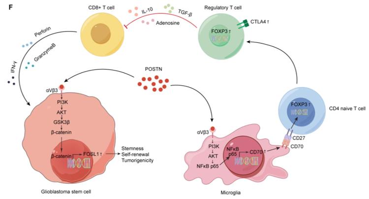 Fig. 1 POSTN is a key mediator of GSC–microglia crosstalk in GBM.1
Key structural properties of POSTN:
- Four consecutive FAS1 domains form the core scaffold
- The EMI domain mediates protein polymerization
- Multiple cysteine residues form disulfide bonds to stabilize the conformation
- Glycosylation sites modification affects cell adhesion function
Functions of POSTN
The Periostin protein encoded by the POSTN gene mainly participates in tissue remodeling and cell adhesion processes, and also plays a regulatory role under various physiological and pathological conditions.
| Function | Description |
| Cell Adhesion | Binding with ligands and integrin receptors, mediating the interaction between cells and extracellular matrix |
| Tissue Development | Promoting the normal formation of connective tissues such as cardiac muscle, bones, and valves during embryonic development |
| Injury Repair | Expressed at higher levels after tissue damage, participating in wound healing and matrix reconstruction processes |
| Fibrosis Regulation | Promoting collagen deposition by activating the TGF-β signaling pathway, playing a key role in organ fibrosis |
| Tumor Progression | Highly expressed in various tumor microenvironments, affecting the migration and invasion abilities of cancer cells |
The expression regulation of the POSTN gene exhibits a tissue-specific pattern. Unlike the constitutive expression of most matrix proteins, its expression is significantly upregulated in fibroblasts in response to TGF-β and mechanical stress stimulation. This induction property enables it to play a marker role in the process of injury repair and fibrosis.
Applications of POSTN and POSTN Antibody in Literature
1. Wang, Hao, et al. "The dual role of POSTN in maintaining glioblastoma stem cells and the immunosuppressive phenotype of microglia in glioblastoma." Journal of Experimental & Clinical Cancer Research 43.1 (2024): 252. https://doi.org/10.1186/s13046-024-03175-9
The article indicates that POSTN is secreted by glioma stem cells and, by activating the αVβ3/PI3K/AKT pathway, it not only maintains the self-renewal of stem cells but also induces microglia cells to form an immunosuppressive phenotype, thereby promoting the malignant progression of glioblastoma.
2. Chen, Chao, et al. "Single‐cell and spatial transcriptomics reveal POSTN+ cancer‐associated fibroblasts correlated with immune suppression and tumour progression in non‐small cell lung cancer." Clinical and translational medicine 13.12 (2023): e1515. https://doi.org/10.1002/ctm2.1515
The article indicates that in lung cancer, a positive POSTN fibroblast subpopulation is found, which co-localizes with SPP1-positive macrophages, promoting extracellular matrix remodeling and immunosuppression, and is associated with T cell exhaustion, low infiltration, and poor prognosis of patients.
3. Wu, Yifan, et al. "Integrative single-cell and spatial transcriptomics analysis reveals ECM-remodeling cancer-associated fibroblast-derived POSTN as a key mediator in pancreatic ductal adenocarcinoma progression." International Journal of Biological Sciences 21.8 (2025): 3573. https://doi.org/10.7150/ijbs.108618
The article indicates that in pancreatic cancer, the ECM remodeling leads to high expression of POSTN in fibroblasts. This, through the activation of the ITGAV/ITGB5 receptors, triggers the PI3K/AKT pathway in tumor cells, promoting epithelial-mesenchymal transition, and is closely related to the poor prognosis of patients.
4. Huang, Miller, et al. "ALK upregulates POSTN and WNT signaling to drive neuroblastoma." Cell reports 43.3 (2024). https://doi.org/10.1016/j.celrep.2024.113927
The article indicates that ALK mutations in neuroblastoma promote tumor cell adhesion and proliferation by upregulating POSTN expression, activating the WNT signaling pathway, and forming a positive feedback loop, in collaboration with MYCN, thereby driving tumor formation.
5. Wu, Shufei, et al. "The gene expression of CALD1, CDH2, and POSTN in fibroblast are related to idiopathic pulmonary fibrosis." Frontiers in Immunology 15 (2024): 1275064. https://doi.org/10.3389/fimmu.2024.1275064
The article indicates that hub genes such as POSTN and CALD1 are significantly upregulated in idiopathic pulmonary fibrosis. Knockdown of these genes can inhibit collagen synthesis, suggesting that these molecules could serve as potential therapeutic targets.
Creative Biolabs: POSTN Antibodies for Research
Creative Biolabs specializes in the production of high-quality POSTN antibodies for research and industrial applications. Our portfolio includes monoclonal and polyclonal antibodies tailored for ELISA, Flow Cytometry, Western blot, immunohistochemistry, and other diagnostic methodologies.
- Custom POSTN Antibody Development: Tailor-made solutions to meet specific research requirements.
- Bulk Production: Large-scale antibody manufacturing for industry partners.
- Technical Support: Expert consultation for protocol optimization and troubleshooting.
- Aliquoting Services: Conveniently sized aliquots for long-term storage and consistent experimental outcomes.
For more details on our POSTN antibodies, custom preparations, or technical support, contact us at email.
Reference
- Wang, Hao, et al. "The dual role of POSTN in maintaining glioblastoma stem cells and the immunosuppressive phenotype of microglia in glioblastoma." Journal of Experimental & Clinical Cancer Research 43.1 (2024): 252. Distributed under Open Access license CC BY 4.0, and cropped from the original figure. https://doi.org/10.1186/s13046-024-03175-9
Anti-POSTN antibodies
 Loading...
 Compare




Hot products 
- AActivation
- AGAgonist
- APApoptosis
- BBlocking
- BABioassay
- BIBioimaging
- CImmunohistochemistry-Frozen Sections
- CIChromatin Immunoprecipitation
- CTCytotoxicity
- CSCostimulation
- DDepletion
- DBDot Blot
- EELISA
- ECELISA(Cap)
- EDELISA(Det)
- ESELISpot
- EMElectron Microscopy
- FFlow Cytometry
- FNFunction Assay
- GSGel Supershift
- IInhibition
- IAEnzyme Immunoassay
- ICImmunocytochemistry
- IDImmunodiffusion
- IEImmunoelectrophoresis
- IFImmunofluorescence
- IGImmunochromatography
- IHImmunohistochemistry
- IMImmunomicroscopy
- IOImmunoassay
- IPImmunoprecipitation
- ISIntracellular Staining for Flow Cytometry
- LALuminex Assay
- LFLateral Flow Immunoassay
- MMicroarray
- MCMass Cytometry/CyTOF
- MDMeDIP
- MSElectrophoretic Mobility Shift Assay
- NNeutralization
- PImmunohistologyp-Paraffin Sections
- PAPeptide Array
- PEPeptide ELISA
- PLProximity Ligation Assay
- RRadioimmunoassay
- SStimulation
- SESandwich ELISA
- SHIn situ hybridization
- TCTissue Culture
- WBWestern Blot
