RACK1 Antibodies
Background
RACK1, as a linker protein containing seven WD40 repeat domains, is widely expressed in various tissues of eukaryotic organisms. This protein was initially identified as the anchoring receptor of protein kinase C and participates in regulating important biological processes such as gene transcription, protein translation, and cell migration by mediating the assembly of various signaling molecules. Studies have shown that RACK1 is abnormally highly expressed in various tumors such as oral squamous cell carcinoma and ovarian cancer, and its expression level is significantly correlated with poor prognosis of patients. Moreover, this protein plays a key regulatory role in macrophage pyroptosis induced by Streptococcus suis infection, mediating the inflammatory response through interaction with the NEK7-NLRP3 complex. Due to its central position in the signal transduction network, RACK1 has become an important model for studying protein interactions and disease molecular mechanisms.
Structure of RACK1
RACK1 is a scaffold protein composed of seven WD40 repeat domains, with a molecular weight of approximately 36 kDa. This protein is highly conserved in mammals, and the structural differences mainly manifest in the subtle variations of amino acid sequences among different species.
| Species | Human | Mouse | Rat | Cow | Zebrafish |
| Molecular Weight (kDa) | 36.1 | 36.1 | 36.1 | 36.1 | 36.2 |
| Primary Structural Differences | Classic WD40 Structure | 99% Homologous to Human Source | 99% Homologous to Human Source | Highly Conserved | Homology about 95% |
The RACK1 protein contains approximately 317 amino acids and adopts a typical seven-leaf propeller-like three-dimensional conformation. Its secondary structure is mainly composed of β-sheets, with each WD repeat sequence forming a β-sheet layer. This unique structure provides a binding platform for various signaling molecules (such as PKC, Src, and PDE4D). The hydrophobic cavity at the protein center stabilizes the overall conformation, while multiple binding sites located on the surface enable it to interact with different proteins simultaneously, thereby playing a crucial hub role in the signal transduction network.
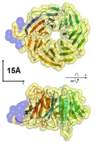 Fig. 1 Two orthogonal views of the homology model of human RACK1.1
The key structural characteristics of RACK1:
- Typical seven-leaf β-screw structure
- WD40 repeat sequences form multiple protein binding interfaces
- The central hydrophobic cavity maintains the overall conformational stability
- Multiple surface sites simultaneously interact with various signaling molecules such as PKC
Functions of RACK1
RACK1, as a multifunctional scaffold protein, plays a central regulatory role in the cell signal transduction network. It participates in the regulation of key physiological processes such as gene expression, protein synthesis, and cell growth by dynamically binding to various signaling molecules.
| Function | Description |
| Signal Integration Platform | Integrates various signaling molecules such as protein kinase C (PKC), coordinating cross-dialogues among different pathways. |
| Ribosome-related Regulation | As a component of the 40S subunit of the ribosome, participates in precise regulation of translation initiation and protein synthesis. |
| Cell Migration Regulation | Through the integration of focal adhesion kinase (FAK) and Src kinase signals, affects cytoskeleton reorganization and migration. |
| Stress Response Mediation | Mediates protective response reactions in conditions such as oxidative stress and viral infection. |
| Tumor Progression Regulation | Regulates the activity of transcription factors such as NF-κB, influencing tumor cell proliferation, invasion, and drug resistance. |
RACK1 is different from traditional enzyme proteins. It functions through protein-protein interactions. This "molecular hub" characteristic enables it to simultaneously participate in multiple signaling pathways, playing a crucial role in maintaining cellular homeostasis and in the occurrence and development of diseases.
Applications of RACK1 and RACK1 Antibody in Literature
1. Gonçalves, Kaliandra A., et al. "Solution structure of the human signaling protein RACK1." BMC structural biology 10.1 (2010): 15. https://doi.org/10.1186/1472-6807-10-15
The article indicates that RACK1 exists mainly as a monomer in solution, in a spherical shape, and its low-resolution structure is highly similar to the homologous crystal structure of Arabidopsis thaliana. RACK1 binds to Ki-1/57 (122-413) in a 1:1 stoichiometric ratio, with a binding constant of approximately (1.5 ± 0.2) × 10⁶ M⁻¹.
2. Pi, Yanan, et al. "Loss of SMURF2 expression enhances RACK1 stability and promotes ovarian cancer progression." Cell Death & Differentiation 30.11 (2023): 2382-2392. https://doi.org/10.1038/s41418-023-01226-w
The article indicates that SMURF2 acts as the E3 ligase of RACK1, mediating its ubiquitination and degradation. In ovarian cancer, SMURF2 is expressed at a low level, which leads to an increase in the stability of RACK1, promoting tumor progression and being associated with poor prognosis for patients. The SMURF2-RACK1 axis may serve as a therapeutic target.
3. Dan, Hongxia, et al. "RACK1 promotes cancer progression by increasing the M2/M1 macrophage ratio via the NF‐κB pathway in oral squamous cell carcinoma." Molecular oncology 14.4 (2020): 795-807. https://doi.org/10.1002/1878-0261.12644
The article indicates that high expression of RACK1 promotes M2-type macrophage polarization and infiltration in oral squamous cell carcinoma by regulating NF-κB. The expression level and the M2/M1 ratio of RACK1 can predict the poor prognosis of patients, and it may become a target for anti-tumor immunotherapy.
4. Wang, Qiuhao, et al. "Prognostic and clinicopathological role of RACK1 for cancer patients: a systematic review and meta-analysis." PeerJ 11 (2023): e15873. https://doi.org/10.7717/peerj.15873
The article indicates that the meta-analysis shows that high expression of RACK1 is negatively correlated with the overall survival and disease-free survival of patients with various cancers, and is significantly associated with lymph node infiltration. RACK1 may serve as a global predictor of poor prognosis for cancer.
5. Shen, Xin, et al. "RACK1 and NEK7 mediate GSDMD-dependent macrophage pyroptosis upon Streptococcus suis infection." Veterinary Research 55.1 (2024): 120. https://doi.org/10.1186/s13567-024-01376-w
The article indicates that Streptococcus suis type 2 activates P65 phosphorylation through the NEK7-RACK1-NLRP3 complex axis, promoting NLRP3 transcription, and subsequently inducing pyroptosis in macrophages. RACK1 plays a key regulatory role in this process.
Creative Biolabs: RACK1 Antibodies for Research
Creative Biolabs specializes in the production of high-quality RACK1 antibodies for research and industrial applications. Our portfolio includes monoclonal and polyclonal antibodies tailored for ELISA, Flow Cytometry, Western blot, immunohistochemistry, and other diagnostic methodologies.
- Custom RACK1 Antibody Development: Tailor-made solutions to meet specific research requirements.
- Bulk Production: Large-scale antibody manufacturing for industry partners.
- Technical Support: Expert consultation for protocol optimization and troubleshooting.
- Aliquoting Services: Conveniently sized aliquots for long-term storage and consistent experimental outcomes.
For more details on our RACK1 antibodies, custom preparations, or technical support, contact us at email.
Reference
- Gonçalves, Kaliandra A., et al. "Solution structure of the human signaling protein RACK1." BMC structural biology 10.1 (2010): 15. Distributed under Open Access license CC BY 2.0, and cropped from the original figure. https://doi.org/10.1186/1472-6807-10-15
Anti-RACK1 antibodies
 Loading...


Hot products 
-
Mouse Anti-CASP8 Recombinant Antibody (CBYY-C0987) (CBMAB-C2424-YY)

-
Mouse Anti-CEMIP Recombinant Antibody (3C12) (CBMAB-K0296-LY)

-
Rat Anti-CD63 Recombinant Antibody (7G4.2E8) (CBMAB-C8725-LY)

-
Mouse Anti-FLI1 Recombinant Antibody (CBXF-0733) (CBMAB-F0435-CQ)

-
Mouse Anti-AHCYL1 Recombinant Antibody (V2-180270) (CBMAB-A1703-YC)

-
Rabbit Anti-ATF4 Recombinant Antibody (D4B8) (CBMAB-A3872-YC)

-
Mouse Anti-BSN Recombinant Antibody (219E1) (CBMAB-1228-CN)

-
Mouse Anti-GIPC2 Recombinant Antibody (10) (CBMAB-G0476-LY)

-
Mouse Anti-ENO1 Recombinant Antibody (8G8) (CBMAB-E1329-FY)

-
Rabbit Anti-Acetyl-Histone H3 (Lys36) Recombinant Antibody (V2-623395) (CBMAB-CP0994-LY)

-
Mouse Anti-ENO2 Recombinant Antibody (85F11) (CBMAB-0276CQ)

-
Mouse Anti-AGO2 Recombinant Antibody (V2-634169) (CBMAB-AP203LY)

-
Mouse Anti-BLK Recombinant Antibody (CBYY-0618) (CBMAB-0621-YY)

-
Mouse Anti-BIRC5 Recombinant Antibody (6E4) (CBMAB-CP2646-LY)

-
Mouse Anti-ACKR3 Recombinant Antibody (V2-261265) (CBMAB-C1023-LY)

-
Mouse Anti-Acetyl SMC3 (K105/K106) Recombinant Antibody (V2-634053) (CBMAB-AP052LY)

-
Mouse Anti-CCS Recombinant Antibody (CBFYC-1093) (CBMAB-C1150-FY)

-
Mouse Anti-ACTG1 Recombinant Antibody (V2-179597) (CBMAB-A0916-YC)

-
Mouse Anti-BZLF1 Recombinant Antibody (BZ.1) (CBMAB-AP705LY)

-
Mouse Anti-AMACR Recombinant Antibody (CB34A) (CBMAB-CA034LY)

- AActivation
- AGAgonist
- APApoptosis
- BBlocking
- BABioassay
- BIBioimaging
- CImmunohistochemistry-Frozen Sections
- CIChromatin Immunoprecipitation
- CTCytotoxicity
- CSCostimulation
- DDepletion
- DBDot Blot
- EELISA
- ECELISA(Cap)
- EDELISA(Det)
- ESELISpot
- EMElectron Microscopy
- FFlow Cytometry
- FNFunction Assay
- GSGel Supershift
- IInhibition
- IAEnzyme Immunoassay
- ICImmunocytochemistry
- IDImmunodiffusion
- IEImmunoelectrophoresis
- IFImmunofluorescence
- IGImmunochromatography
- IHImmunohistochemistry
- IMImmunomicroscopy
- IOImmunoassay
- IPImmunoprecipitation
- ISIntracellular Staining for Flow Cytometry
- LALuminex Assay
- LFLateral Flow Immunoassay
- MMicroarray
- MCMass Cytometry/CyTOF
- MDMeDIP
- MSElectrophoretic Mobility Shift Assay
- NNeutralization
- PImmunohistologyp-Paraffin Sections
- PAPeptide Array
- PEPeptide ELISA
- PLProximity Ligation Assay
- RRadioimmunoassay
- SStimulation
- SESandwich ELISA
- SHIn situ hybridization
- TCTissue Culture
- WBWestern Blot
