RPS3 Antibodies
Background
RPS3 is a highly conserved small subunit component of ribosomes, mainly existing in the cytoplasm of eukaryotes. This protein not only participates in the translation process of mRNA, but also plays a significant role in non-classical functions such as DNA damage repair and apoptosis regulation. Studies have shown that RPS3 can specifically recognize and bind to DNA sites with oxidative damage, maintaining genomic stability through the base excision repair pathway. This gene was first identified in yeast in the 1980s. Its three-dimensional structure analysis revealed a unique bifunctional domain feature: the N-terminal is responsible for ribosome assembly, and the C-terminal has endonuclease activity. As a typical representative of ribosomal protein multi-functionalization, the research on RPS3 provides an important model for understanding the functional evolution of proteins and the interaction between nucleic acids and proteins, and shows potential application value in the study of cancer and neurodegenerative diseases.
Structure of RPS3
RPS3 is a multifunctional ribosomal protein with a molecular weight of approximately 26.8 kDa. There are minor differences in its molecular weight among different species, mainly due to conserved variations in amino acid sequences.
| Species | Human | Mouse | Yeast | Drosophila |
| Molecular Weight (kDa) | 26.8 | 26.7 | 26.5 | 26.6 |
| Primary Structural Differences | Highly conservative, including fixed-position signals | Highly homologous to humans | The core functional domains are similar | Adapt to the demand for rapid translation |
RPS3 is composed of 243 amino acids and has a typical ribosomal protein folding structure. Its N-terminal domain is involved in the assembly of the 40S subunit of the ribosome, while the C-terminal domain contains endonuclease active sites and can play a role in DNA damage repair. The secondary structure of this protein contains multiple α -helices and β -folds, forming a stable RNA binding interface, among which the KH domain is crucial for mRNA recognition. In addition, the Lys residues of RPS3 play a key role in translation regulation, while its Cys residues are involved in oxidative stress responses.
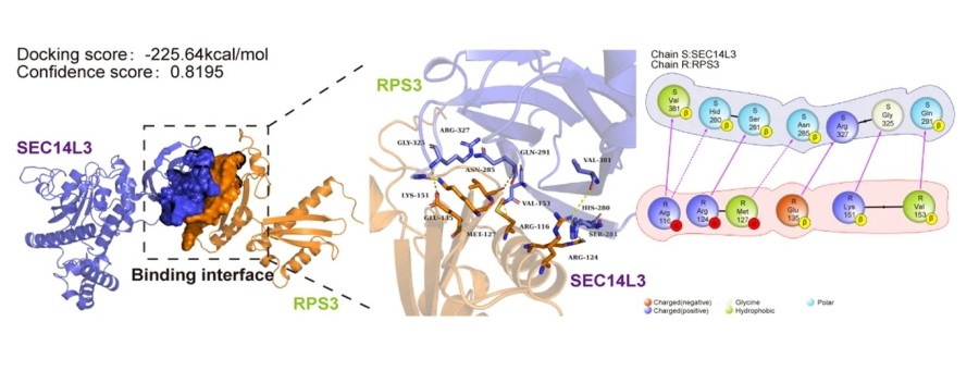 Fig. 1 PyMOL software illustrates the interaction between SEC14L3 and RPS3 within their respective 3D protein structures.1
Key structural properties of RPS3:
- Typical folding structure of ribosomal protein
- Dual-function domain
- KH domain
- Key amino acid sites
Functions of RPS3
The main function of the RPS3 gene is to participate in protein translation as a component of the 40S subunit of the ribosome, and it also has multiple non-classical functions, playing a key role in cellular stress response and maintaining genomic stability.
| Function | Description |
| Ribosome assembly | As a 40S small subunit structural protein, it participates in the initiation and extension processes of mRNA translation. |
| DNA damage repair | The genomic stability is maintained by identifying and removing DNA bases with oxidative damage through endonuclease activity. |
| Regulation of apoptosis | It interacts with signaling molecules such as NF-κB to regulate cell fate determination under stress conditions. |
| Oxidative stress response | Sensing reactive oxygen species levels through Cys residues activates antioxidant defense pathways. |
| Virus defense | Interfere with the translation efficiency of viral mRNA and participate in the innate immune response. |
Unlike typical ribosomal proteins, RPS3 exhibits unique dual-functional characteristics: its translation activity is expressed in a compositional manner, while its DNA repair function is specifically activated under oxidative stress conditions. This functional switch is achieved through phosphorylation modifications (such as at the Tyr42 site), reflecting the dual role of ribosomal proteins in basic life activities and stress responses.
Applications of RPS3 and RPS3 Antibody in Literature
1. Wang, Tuo, et al. "UBE2J1 inhibits colorectal cancer progression by promoting ubiquitination and degradation of RPS3." Oncogene 42.9 (2023): 651-664. https://doi.org/10.1038/s41388-022-02581-7
Research has found that UBE2J1 is lowly expressed in colorectal cancer through DNA methylation and is associated with a poor prognosis. The UBE2J1-TRIM25 complex mediates the ubiquitination and degradation of the K214 site of RPS3, inhibits the NF-κB signaling pathway, and thereby hinders tumor progression. The RPS3 degradation mechanism provides a new therapeutic target for colorectal cancer.
2. Jiang, Ziming, et al. "SEC14L3 knockdown inhibited clear cell renal cell carcinoma proliferation, metastasis and sunitinib resistance through an SEC14L3/RPS3/NFκB positive feedback loop." Journal of Experimental & Clinical Cancer Research 43.1 (2024): 288. https://doi.org/10.1186/s13046-024-03206-5
Research has found that SEC14L3 is highly expressed in clear cell renal cell carcinoma (ccRCC), and its down-regulation hinders tumor progression by promoting the ubiquitination and degradation of RPS3 and inhibiting the NF-κB signaling pathway. The SEC14L3/RPS3/NF-κB positive feedback loop provides a new therapeutic target for ccRCC.
3. Rui, et al. "RPS3 promotes the metastasis and cisplatin resistance of adenoid cystic carcinoma." Frontiers in Oncology 12 (2022): 804439. https://doi.org/10.3389/fonc.2022.804439
Research has found that RPS3 is highly expressed in adenoid cystic carcinoma (ACC) and is associated with a poor prognosis for patients. RPS3 activates the NF-κB signaling pathway by binding to STAT1, promoting the metastasis, invasion and cisplatin resistance of ACC, suggesting its potential value as a therapeutic target.
4. Piloto, Ana Margarida, et al. "Plastic antibodies tailored on quantum dots for an optical detection of myoglobin down to the femtomolar range." Scientific reports 8.1 (2018): 4944. https://doi.org/10.18632/aging.204211
Research has found that E3 ubiquitin ligase SIAH1 is lowly expressed in ovarian cancer. It inhibits the NF-κB signaling pathway by promoting the ubiquitination and degradation of RPS3, thereby enhancing cisplatin sensitivity. The SIAH1-RPS3-NF-κB axis provides a new target for overcoming drug resistance in ovarian cancer.
5. Wu, Miaomiao, Samir El Qaidi, and Philip R. Hardwidge. "SseL deubiquitinates RPS3 to inhibit its nuclear translocation." Pathogens 7.4 (2018): 86. https://doi.org/10.3390/pathogens7040086
Research has found that the Salmonella effector protein SseL inhibits the host's innate immune response by specifically removing the K63 ubiquitination modification of RPS3 and blocking its nuclear translocation. This mechanism, together with Escherichia coli NleH1/NleC, jointly reveals the multiple escape strategies of pathogenic bacteria targeting the RPS3-NF-κB pathway.
Creative Biolabs: RPS3 Antibodies for Research
Creative Biolabs specializes in the production of high-quality RPS3 antibodies for research and industrial applications. Our portfolio includes monoclonal antibodies tailored for ELISA, Flow Cytometry, Western blot, immunohistochemistry, and other diagnostic methodologies.
- Custom RPS3 Antibody Development: Tailor-made solutions to meet specific research requirements.
- Bulk Production: Large-scale antibody manufacturing for industry partners.
- Technical Support: Expert consultation for protocol optimization and troubleshooting.
- Aliquoting Services: Conveniently sized aliquots for long-term storage and consistent experimental outcomes.
For more details on our RPS3 antibodies, custom preparations, or technical support, contact us at email.
Reference
- Jiang, Ziming, et al. "SEC14L3 knockdown inhibited clear cell renal cell carcinoma proliferation, metastasis and sunitinib resistance through an SEC14L3/RPS3/NFκB positive feedback loop." Journal of Experimental & Clinical Cancer Research 43.1 (2024): 288. https://doi.org/10.1186/s13046-024-03206-5
Anti-RPS3 antibodies
 Loading...


Hot products 
-
Mouse Anti-ACTG1 Recombinant Antibody (V2-179597) (CBMAB-A0916-YC)

-
Mouse Anti-APC Recombinant Antibody (CBYC-A661) (CBMAB-A3036-YC)

-
Rabbit Anti-DLK1 Recombinant Antibody (9D8) (CBMAB-D1061-YC)

-
Mouse Anti-APP Recombinant Antibody (5C2A1) (CBMAB-A3314-YC)

-
Mouse Anti-CD59 Recombinant Antibody (CBXC-2097) (CBMAB-C4421-CQ)

-
Mouse Anti-ACTN4 Recombinant Antibody (V2-6075) (CBMAB-0020CQ)

-
Mouse Anti-CD24 Recombinant Antibody (2Q1282) (CBMAB-C1624-CN)

-
Mouse Anti-G6PD Recombinant Antibody (13B331) (CBMAB-G1553-LY)

-
Mouse Anti-Acetyl-α-Tubulin (Lys40) Recombinant Antibody (V2-623485) (CBMAB-CP2897-LY)

-
Mouse Anti-CFL1 Recombinant Antibody (CBFYC-1771) (CBMAB-C1833-FY)

-
Mouse Anti-dsRNA Recombinant Antibody (2) (CBMAB-D1807-YC)

-
Mouse Anti-ABCA3 Recombinant Antibody (V2-178911) (CBMAB-A0145-YC)

-
Rat Anti-EPO Recombinant Antibody (16) (CBMAB-E1578-FY)

-
Rat Anti-CD34 Recombinant Antibody (MEC 14.7) (CBMAB-C10196-LY)

-
Mouse Anti-GGT1 Recombinant Antibody (1F9) (CBMAB-G3273-LY)

-
Mouse Anti-BHMT Recombinant Antibody (CBYY-0547) (CBMAB-0550-YY)

-
Mouse Anti-CCS Recombinant Antibody (CBFYC-1093) (CBMAB-C1150-FY)

-
Mouse Anti-ANXA7 Recombinant Antibody (A-1) (CBMAB-A2941-YC)

-
Mouse Anti-APP Recombinant Antibody (DE2B4) (CBMAB-1122-CN)

-
Mouse Anti-AKR1B1 Antibody (V2-2449) (CBMAB-1001CQ)

- AActivation
- AGAgonist
- APApoptosis
- BBlocking
- BABioassay
- BIBioimaging
- CImmunohistochemistry-Frozen Sections
- CIChromatin Immunoprecipitation
- CTCytotoxicity
- CSCostimulation
- DDepletion
- DBDot Blot
- EELISA
- ECELISA(Cap)
- EDELISA(Det)
- ESELISpot
- EMElectron Microscopy
- FFlow Cytometry
- FNFunction Assay
- GSGel Supershift
- IInhibition
- IAEnzyme Immunoassay
- ICImmunocytochemistry
- IDImmunodiffusion
- IEImmunoelectrophoresis
- IFImmunofluorescence
- IGImmunochromatography
- IHImmunohistochemistry
- IMImmunomicroscopy
- IOImmunoassay
- IPImmunoprecipitation
- ISIntracellular Staining for Flow Cytometry
- LALuminex Assay
- LFLateral Flow Immunoassay
- MMicroarray
- MCMass Cytometry/CyTOF
- MDMeDIP
- MSElectrophoretic Mobility Shift Assay
- NNeutralization
- PImmunohistologyp-Paraffin Sections
- PAPeptide Array
- PEPeptide ELISA
- PLProximity Ligation Assay
- RRadioimmunoassay
- SStimulation
- SESandwich ELISA
- SHIn situ hybridization
- TCTissue Culture
- WBWestern Blot
