RRAS Antibodies
Background
The RRAS gene encodes a small GTP-binding protein belonging to the RAS superfamily, which is mainly present in various cell types of vertebrates. This protein participates in regulating intracellular signal transduction pathways by converting between its active GTP-binding form and its inactive GDP-binding form, thereby influencing key life processes such as cell proliferation, differentiation and survival. The RRAS gene plays a significant role in vascular development and maintaining the structure of the cytoskeleton. Abnormalities in its function are associated with certain tumors and developmental disorders. This gene was first identified as a member of the RAS proto-oncogene family in the 1980s. Its highly conserved structure and function provide an important model for studying G protein-mediated signaling networks, deepening people's understanding of cell growth regulation and carcinogenic mechanisms.
Structure of RRAS
The RRAS gene encodes a small GTP-binding protein with a molecular weight of approximately 21.5 kDa. There are slight differences in this molecular weight among different species, mainly due to the substitution of individual amino acids in the RAS domain.
| Species | Human | Mouse | Rat | Zebrafish | African clawed toad |
| Molecular Weight (kDa) | 21.5 | 21.4 | 21.5 | 21.6 | 21.3 |
| Primary Structural Differences | As the typical structure of RAS domain | Very high homology with humans | Highly conserved sequence | There are subtle variations in the effector junction area | Has the G1 and G5 nucleotide combining with key motif |
This protein is composed of 189 amino acids and presents a classic GTPase folding structure. Its three-dimensional structure contains a RAS domain composed of six β -folded sheets and five α -helices, which forms a highly conserved nucleotide binding pocket. The GTP hydrolytic activity of this protein depends on the glycine residue in its P-ring (G1 motif) and the glutamine residue in the G3 motif. The threonine residue located in the switch I region (G2 motif) directly participates in the hydrolysis process of GTP, while the switch II region (G4 motif) is responsible for recognizing downstream effector molecules, thereby transmitting the activation signal of GTP binding to pathways such as MAPK.
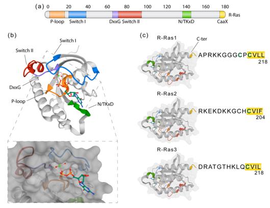 Fig. 1 R-Ras molecular structure and domains.1
Key structural properties of RRAS:
- Classical GTPase folded conformation
- Conserved nucleotide-binding pockets
- Switch I/II conformational changes determine the functional state
- The effector domain mediates downstream signal conduction
Functions of RRAS
The core function of the RRAS gene is to act as a molecular switch to regulate intracellular signal transduction. However, it is also involved in a variety of cellular physiological processes, including cytoskeletal recombination and regulation of cell adhesion.
| Function | Description |
| Signal transduction switch | Switch between the active GTP bound state and the inactive GDP bound state, precisely controlling the signal duration like a timer. |
| Regulation of cell proliferation | By activating the RAF-MAPK pathway to promote the cell cycle process, its abnormal activation can directly drive tumorigenesis. |
| Cytoskeletal recombination | The integration of integrin signals regulates the dynamics of actin cytoskeleton, influencing cell morphology and motor capacity. |
| Regulation of cell adhesion | Enriched at local adhesion spots and coordinating the interaction between cells and extracellular matrix, this function is particularly crucial in angiogenesis. |
| Resistance to apoptosis | Survival signals are transmitted through the PI3K-AKT pathway to inhibit programmed cell death and promote cell survival. |
Compared with the classic HRAS and KRAS, RRAS has a higher sensitivity to GAP proteins, which enables a faster GTP hydrolysis rate. This characteristic makes it particularly suitable for functioning in physiological processes that require rapid signal termination, such as local adhesion spot turnover during cell migration.
Applications of RRAS and RRAS Antibody in Literature
1. Miller, John P., et al. "A genome-scale RNA–Interference screen identifies RRAS signaling as a pathologic feature of Huntington's disease." PLoS genetics 8.11 (2012): e1003042. https://doi.org/10.1371/journal.pgen.1003042
The article indicates that a genomic screening found that the pathogenic protein of Huntington's disease abnormally activates the RRAS signaling pathway. In cell and animal models, reducing the function of RRAS or its pathway components can effectively alleviate toxicity, suggesting that this pathway is a potential new target for drug treatment.
2. Perrot, Carole Y., Junko Sawada, and Masanobu Komatsu. "Prolonged activation of cAMP signaling leads to endothelial barrier disruption via transcriptional repression of RRAS." The FASEB Journal 32.11 (2018): 5793. https://doi.org/10.1096/fj.201700818RRR
The article indicates that long-term elevated cAMP signaling can inhibit the expression of the RRAS gene through the transcription factor CREB3. The reduction of RRAS disrupts endothelial cell junctions, ultimately leading to impaired vascular barrier function and abnormally increased permeability.
3. Alcover‐Sanchez, Berta, et al. "R‐Ras1 and R‐Ras2 regulate mature oligodendrocyte subpopulations." Glia 73.4 (2025): 701-719. https://doi.org/10.1002/glia.24643
Studies have shown that R-Ras1 and R-Ras2 are the key factors for balancing different subsets of mature oligodendrocytes (MOL). When the function of RRAS is reduced or absent, it will disrupt the normal proportion of subgroups, leading to an abnormal increase in the MOL1 subgroup and a decrease in the MOL2 and MOL5/6 subgroups, thereby affecting myelin formation.
4. Iida, Takaya, et al. "Functional analysis of RRAS2 pathogenic variants with a Noonan-like phenotype." Frontiers in Genetics 15 (2024): 1383176. https://doi.org/10.3389/fgene.2024.1383176
Research has found that novel pathogenic mutations in the RRAS2 gene (such as P. Gly23val) can abnormally enhance its function, thereby overactivating the RAS signaling pathway. This functional gain causes developmental malformations and high mortality rates in fruit fly and zebrafish models, leading to a Nounan syndrome-like phenotype in humans.
5. Alcover-Sanchez, Berta, et al. "R-Ras GTPases signaling role in myelin neurodegenerative diseases." International Journal of Molecular Sciences 21.16 (2020): 5911. https://doi.org/10.3390/ijms21165911
The review indicates that myelin formation is regulated by signaling pathways such as PI3K, ERK and Wnt. Ras GTPases (including R-Ras) are key participants in these pathways and may act as cross-dialogue nodes among them to coordinate the myelination process, thus being regarded as potential therapeutic research targets.
Creative Biolabs: RRAS Antibodies for Research
Creative Biolabs specializes in the production of high-quality RRAS antibodies for research and industrial applications. Our portfolio includes monoclonal antibodies tailored for ELISA, Flow Cytometry, Western blot, immunohistochemistry, and other diagnostic methodologies.
- Custom RRAS Antibody Development: Tailor-made solutions to meet specific research requirements.
- Bulk Production: Large-scale antibody manufacturing for industry partners.
- Technical Support: Expert consultation for protocol optimization and troubleshooting.
- Aliquoting Services: Conveniently sized aliquots for long-term storage and consistent experimental outcomes.
For more details on our RRAS antibodies, custom preparations, or technical support, contact us at email.
Reference
- Alcover-Sanchez, Berta, et al. "R-Ras GTPases signaling role in myelin neurodegenerative diseases." International Journal of Molecular Sciences 21.16 (2020): 5911. https://doi.org/10.3390/ijms21165911
Anti-RRAS antibodies
 Loading...


Hot products 
-
Rat Anti-CD300A Recombinant Antibody (172224) (CBMAB-C0423-LY)

-
Mouse Anti-CFL1 (Phospho-Ser3) Recombinant Antibody (CBFYC-1770) (CBMAB-C1832-FY)

-
Mouse Anti-EMP3 Recombinant Antibody (CBFYE-0100) (CBMAB-E0207-FY)

-
Mouse Anti-FN1 Monoclonal Antibody (71) (CBMAB-1241CQ)

-
Mouse Anti-CCL18 Recombinant Antibody (64507) (CBMAB-C7910-LY)

-
Mouse Anti-FTH1 Recombinant Antibody (CBXF-1896) (CBMAB-F3426-CQ)

-
Mouse Anti-BCL6 Recombinant Antibody (CBYY-0442) (CBMAB-0445-YY)

-
Mouse Anti-ARSA Recombinant Antibody (CBYC-A799) (CBMAB-A3679-YC)

-
Mouse Anti-COL1A2 Recombinant Antibody (CF108) (V2LY-1206-LY626)

-
Mouse Anti-ACTG1 Recombinant Antibody (V2-179597) (CBMAB-A0916-YC)

-
Mouse Anti-CASP7 Recombinant Antibody (10-01-62) (CBMAB-C2005-LY)

-
Mouse Anti-ENPP1 Recombinant Antibody (CBFYE-0159) (CBMAB-E0375-FY)

-
Mouse Anti-ADGRE5 Recombinant Antibody (V2-360335) (CBMAB-C2088-CQ)

-
Mouse Anti-CD59 Recombinant Antibody (CBXC-2097) (CBMAB-C4421-CQ)

-
Mouse Anti-CD2AP Recombinant Antibody (BR083) (CBMAB-BR083LY)

-
Mouse Anti-ADRB2 Recombinant Antibody (V2-180026) (CBMAB-A1420-YC)

-
Mouse Anti-CTNND1 Recombinant Antibody (CBFYC-2414) (CBMAB-C2487-FY)

-
Rabbit Anti-Acetyl-Histone H3 (Lys36) Recombinant Antibody (V2-623395) (CBMAB-CP0994-LY)

-
Mouse Anti-CORO1A Recombinant Antibody (4G10) (V2LY-1206-LY806)

-
Human Anti-SARS-CoV-2 Spike Recombinant Antibody (CBC05) (CBMAB-CR005LY)

- AActivation
- AGAgonist
- APApoptosis
- BBlocking
- BABioassay
- BIBioimaging
- CImmunohistochemistry-Frozen Sections
- CIChromatin Immunoprecipitation
- CTCytotoxicity
- CSCostimulation
- DDepletion
- DBDot Blot
- EELISA
- ECELISA(Cap)
- EDELISA(Det)
- ESELISpot
- EMElectron Microscopy
- FFlow Cytometry
- FNFunction Assay
- GSGel Supershift
- IInhibition
- IAEnzyme Immunoassay
- ICImmunocytochemistry
- IDImmunodiffusion
- IEImmunoelectrophoresis
- IFImmunofluorescence
- IGImmunochromatography
- IHImmunohistochemistry
- IMImmunomicroscopy
- IOImmunoassay
- IPImmunoprecipitation
- ISIntracellular Staining for Flow Cytometry
- LALuminex Assay
- LFLateral Flow Immunoassay
- MMicroarray
- MCMass Cytometry/CyTOF
- MDMeDIP
- MSElectrophoretic Mobility Shift Assay
- NNeutralization
- PImmunohistologyp-Paraffin Sections
- PAPeptide Array
- PEPeptide ELISA
- PLProximity Ligation Assay
- RRadioimmunoassay
- SStimulation
- SESandwich ELISA
- SHIn situ hybridization
- TCTissue Culture
- WBWestern Blot
