ASS1 Antibodies
Background
ASS1, full name Arginine Aminosuccinate Synthetase 1, is one of the key enzymes in the urea cycle. It is mainly expressed in liver and kidney cells. This enzyme catalyzes the reaction of citrulline and aspartate to form argininosuccinate, which is the rate-limiting step in the synthesis of arginine in the body. Studies have found that in many tumor cells, ASS1 is often downregulated or absent, forcing the cells to rely on exogenous arginine for survival. This metabolic weakness provides a theoretical basis for arginine deprivation therapy. Currently, a targeted drug ADI-PEG20 has been developed, which selectively kills tumor cells by consuming the arginine in the blood. In addition, ASS1 also regulates the flow of aspartate metabolism within the cell, and its expression changes can affect nucleotide synthesis and the cell cycle progression. The research on ASS1 not only reveals the important role of metabolic reprogramming in tumors but also opens up new directions for targeted therapy.
Structure of ASS1
The protein encoded by the ASS1 gene has a molecular weight of approximately 46.7 kDa and is composed of 412 amino acids. It shows high conservation across different species.
| Species | Human | Mouse | Rat | Cow | Pig |
| Molecular Weight (kDa) | 46.7 | 46.9 | 46.8 | 46.6 | 46.7 |
| Primary Structural Differences | Classical structure, containing C-terminal domain | Homology reaches 92% | Active site is completely conserved | Some amino acids are replaced | Highly homologous to humans |
ASS1 exists in a homotrimeric form, with each subunit containing three domains: the N-terminal nucleotide-binding domain, the central hinge domain, and the C-terminal domain. The active center is located at the interface of the subunits and is composed of multiple conserved residues. The enzyme activity depends on the participation of Mg2+ ions and catalyzes the formation of arginine succinate from citrulline and aspartic acid. This enzyme plays a rate-limiting role in the urea cycle and its expression is regulated by transcriptional levels and post-translational modifications.
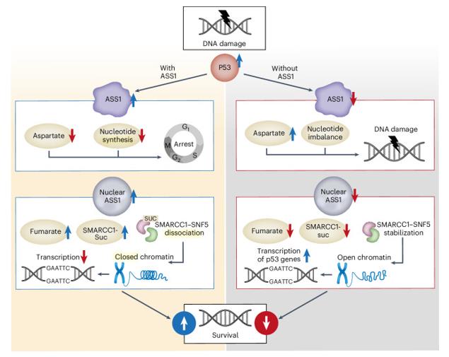 Fig. 1 Suggested model for the requirement of ASS1 in the p53-response to DNA damage.1
The key structural features of ASS1:
- Homodimeric structure, with each subunit consisting of three domains
- The active center is located at the interface of the subunits and contains multiple conserved catalytic residues
- Mg²⁺ ions participate in the catalytic reaction and stabilize the transition state conformation
- The C-terminal domain mediates the assembly of the dimer and maintains the stability of the enzyme activity
Functions of ASS1
The core function of ASS1 is to catalyze the formation of argininosuccinate from citrulline and aspartic acid, which is a rate-limiting enzyme in the urea cycle. However, it also plays a role in various physiological and pathological processes, including arginine synthesis, nitric oxide regulation, and tumor metabolic remodeling.
| Function | Description |
| Urea Cycle | Catalyzes the condensation of citrulline and aspartic acid in the liver, which is a crucial step for ammonia detoxification and urea production. |
| Arginine Synthesis | Provides endogenous arginine to the body, maintaining cell proliferation, protein synthesis and signal transduction. |
| Aspartate Metabolism Regulation | By consuming aspartate and influencing its flow, it regulates nucleotide synthesis and cell cycle progression. |
| Regulation of Nitric Oxide Production | As a source of arginine, it indirectly affects the activity of NOS enzymes and participates in vasodilation and oxidative stress responses. |
| Tumor Metabolic Remodeling | It is expressed at a lower level in various cancer cells, causing the cells to rely on exogenous arginine and creating a metabolic weakness. |
In tumor cells, ASS1 is often silenced due to promoter methylation. Its absence leads to the redirection of aspartic acid to pyrimidine synthesis, promoting the proliferation of cancer cells. This metabolic reprogramming provides a theoretical basis for arginine deprivation therapy.
Applications of ASS1 and ASS1 Antibody in Literature
1. Lim, Lisha Qiu Jin, et al. "ASS1 metabolically contributes to the nuclear and cytosolic p53-mediated DNA damage response." Nature metabolism 6.7 (2024): 1294-1309. https://doi.org/10.1038/s42255-024-01060-5
The research has found that ASS1 is part of the p53 network. After DNA damage, it inhibits nucleotide synthesis and regulates chromatin remodeling to arrest the cell cycle for DNA repair. The absence of ASS1 leads to DNA damage and accelerated cell cycle, promoting tumor mutations and adaptation.
2. Luo, Wensong, et al. "ASS1 inhibits triple-negative breast cancer by regulating PHGDH stability and de novo serine synthesis." Cell death & disease 15.5 (2024): 319. https://doi.org/10.1038/s41419-024-06672-z
The research has found that ASS1 can bind to PHGDH and promote its degradation, thereby inhibiting serine synthesis and exerting an anti-cancer effect. Knocking out PHGDH can reverse the anti-cancer effect of ASS1. The absence of ASS1 will enhance the proliferation ability of cells in an environment lacking serine.
3. Panda, Prashanta Kumar, et al. "BCL-XL Protects ASS1-Deficient Cancers from Arginine Starvation–Induced Apoptosis." Clinical Cancer Research 31.7 (2025): 1333-1345. https://doi.org/10.1158/1078-0432.CCR-24-2548
The research has found that cancer cells lacking ASS1 depend on arginine for survival. ADI-PEG20 can inhibit the cell cycle, but its efficacy is limited as BCL-XL combines with BAX/BAK to block apoptosis. Combining with BCL-XL inhibitors can remove the blockade and synergistically induce apoptosis, providing a basis for clinical combination therapy.
4. Qian, Xiangjun, et al. "ZFPL1 Promotes Colorectal Cancer Progression by Stabilizing ASS1 to Drive the Urea Cycle and M2 Macrophage‐Mediated Metastatic Colonization." Advanced Science 12.46 (2025): e05291. https://doi.org/10.1002/advs.202505291
The study found that ZFPL1 is highly expressed in colorectal cancer. It activates the urea cycle by binding to ASS1 and inhibiting its degradation, thereby promoting tumor progression. The absence of ZFPL1 can reshape the immune microenvironment. Compound Sal B can block the binding of ZFPL1 and ASS1, and synergistically enhance the efficacy of immunotherapy.
5. Khare, Sanika, et al. "ASS1 and ASL suppress growth in clear cell renal cell carcinoma via altered nitrogen metabolism." Cancer & Metabolism 9.1 (2021): 40. https://doi.org/10.1186/s40170-021-00271-8
The study found that in renal clear cell carcinoma, the expression of urea cycle enzymes ASS1 and ASL was downregulated. Their absence could enable aspartate to be redirected to pyrimidine synthesis, thereby promoting cancer cell proliferation. Restoring the expression of both enzymes could inhibit tumor growth by depleting aspartate and regulating nitric oxide synthesis.
Creative Biolabs: ASS1 Antibodies for Research
Creative Biolabs specializes in the production of high-quality ASS1 antibodies for research and industrial applications. Our portfolio includes monoclonal and polyclonal antibodies tailored for ELISA, Flow Cytometry, Western blot, immunohistochemistry, and other diagnostic methodologies.
- Custom ASS1 Antibody Development: Tailor-made solutions to meet specific research requirements.
- Bulk Production: Large-scale antibody manufacturing for industry partners.
- Technical Support: Expert consultation for protocol optimization and troubleshooting.
- Aliquoting Services: Conveniently sized aliquots for long-term storage and consistent experimental outcomes.
For more details on our ASS1 antibodies, custom preparations, or technical support, contact us at email.
Reference
- Lim, Lisha Qiu Jin, et al. "ASS1 metabolically contributes to the nuclear and cytosolic p53-mediated DNA damage response." Nature metabolism 6.7 (2024): 1294-1309. Distributed under Open Access license CC BY 4.0, without modification. https://doi.org/10.1038/s42255-024-01060-5
Anti-ASS1 antibodies
 Loading...


Hot products 
-
Mouse Anti-ENO1 Recombinant Antibody (CBYC-A950) (CBMAB-A4388-YC)

-
Rabbit Anti-BAD (Phospho-Ser136) Recombinant Antibody (CAP219) (CBMAB-AP536LY)

-
Mouse Anti-AAV-5 Recombinant Antibody (V2-503417) (CBMAB-V208-1369-FY)

-
Mouse Anti-B2M Recombinant Antibody (CBYY-0050) (CBMAB-0050-YY)

-
Mouse Anti-DLL4 Recombinant Antibody (D1090) (CBMAB-D1090-YC)

-
Mouse Anti-CDKL5 Recombinant Antibody (CBFYC-1629) (CBMAB-C1689-FY)

-
Rabbit Anti-ADRA1A Recombinant Antibody (V2-12532) (CBMAB-1022-CN)

-
Mouse Anti-ACO2 Recombinant Antibody (V2-179329) (CBMAB-A0627-YC)

-
Mouse Anti-GGT1 Recombinant Antibody (1F9) (CBMAB-G3273-LY)

-
Mouse Anti-ADAM29 Recombinant Antibody (V2-179787) (CBMAB-A1149-YC)

-
Mouse Anti-Acetyl SMC3 (K105/K106) Recombinant Antibody (V2-634053) (CBMAB-AP052LY)

-
Mouse Anti-AGK Recombinant Antibody (V2-258056) (CBMAB-M0989-FY)

-
Mouse Anti-COL1A2 Recombinant Antibody (CF108) (V2LY-1206-LY626)

-
Mouse Anti-BZLF1 Recombinant Antibody (BZ.1) (CBMAB-AP705LY)

-
Mouse Anti-FTH1 Recombinant Antibody (CBXF-1896) (CBMAB-F3426-CQ)

-
Mouse Anti-ALPL Antibody (B4-78) (CBMAB-1009CQ)

-
Mouse Anti-FPR2 Recombinant Antibody (1D6) (CBMAB-F2628-CQ)

-
Rabbit Anti-B2M Recombinant Antibody (CBYY-0059) (CBMAB-0059-YY)

-
Mouse Anti-ASTN1 Recombinant Antibody (H-9) (CBMAB-1154-CN)

-
Mouse Anti-GFP Recombinant Antibody (28) (CBMAB-G3038-LY)

- AActivation
- AGAgonist
- APApoptosis
- BBlocking
- BABioassay
- BIBioimaging
- CImmunohistochemistry-Frozen Sections
- CIChromatin Immunoprecipitation
- CTCytotoxicity
- CSCostimulation
- DDepletion
- DBDot Blot
- EELISA
- ECELISA(Cap)
- EDELISA(Det)
- ESELISpot
- EMElectron Microscopy
- FFlow Cytometry
- FNFunction Assay
- GSGel Supershift
- IInhibition
- IAEnzyme Immunoassay
- ICImmunocytochemistry
- IDImmunodiffusion
- IEImmunoelectrophoresis
- IFImmunofluorescence
- IGImmunochromatography
- IHImmunohistochemistry
- IMImmunomicroscopy
- IOImmunoassay
- IPImmunoprecipitation
- ISIntracellular Staining for Flow Cytometry
- LALuminex Assay
- LFLateral Flow Immunoassay
- MMicroarray
- MCMass Cytometry/CyTOF
- MDMeDIP
- MSElectrophoretic Mobility Shift Assay
- NNeutralization
- PImmunohistologyp-Paraffin Sections
- PAPeptide Array
- PEPeptide ELISA
- PLProximity Ligation Assay
- RRadioimmunoassay
- SStimulation
- SESandwich ELISA
- SHIn situ hybridization
- TCTissue Culture
- WBWestern Blot
