BATF3 Antibodies
Background
BATF3, as a transcription factor belonging to the basic leucine zipper family, plays a crucial role in the development and differentiation of lymphocytes. This protein binds to specific DNA sequences through homodimerization or heterodimerization, precisely regulating the expression of target genes, especially participating in the lineage specialization and functional maintenance of CD8α+ dendritic cells. The immune pathway mediated by BATF3 is essential for initiating antiviral responses and anti-tumor immune surveillance in the body. Abnormal function of BATF3 will directly weaken the activation efficiency of cytotoxic T cells. Initially identified as a member of the AP-1 family in the early 2000s, BATF3 was later confirmed to be an indispensable regulatory node in the development of classical type I dendritic cells. The breakthrough work has provided new targets for tumor immunotherapy and vaccine development. The simple and precise regulatory network of this molecule is continuously revealing the deep logic of the immune system in maintaining homeostasis and pathological states.
Structure of BATF3
BATF3 is an alkaline leucine zipper transcription factor with a molecular weight of approximately 16.8 kDa. There is a slight variation in its sequence among different species.
| Species | Human | Mouse | Rat | Bovine |
| Molecular Weight (kDa) | 16.8 | 17.1 | 17.0 | 16.9 |
| Primary Structural Differences | Leucine zipper and DNA-binding domain are highly conserved | About 85% homology with human | Similar to mice | Adaptive variation |
BATF3 contains approximately 142 amino acids. Its protein structure is centered around an acidic domain and a leucine zipper. The former is responsible for recognizing AP-1-like sites on DNA, while the latter mediates the formation of a dimer with Jun family proteins. This dimer regulates the transcription of target genes by inserting into specific DNA sequences. BATF3 does not contain an endogenous activation domain and typically functions by interacting with other transcription factors. It plays a decisive role in the differentiation of CD8α+ dendritic cells and in antiviral immunity.
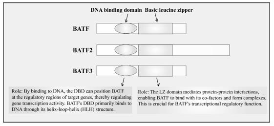 Fig. 1 Members of the BATF family possess both a bZIP and a DBD.1
Key structural properties of BATF3:
- The basic domain is responsible for DNA recognition and binding
- The leucine zipper mediates dimerization with Jun family proteins
- Lack of classical transcriptional activation domain structure, rely on each other for protein function
Functions of BATF3
The main function of BATF3 is to regulate the differentiation of immune cells and the anti-infection response. Additionally, it also plays a role in various physiological processes, including metabolic adaptation and tumor immune surveillance.
| Function | Description |
| Dendritic Cell Differentiation | BATF3 is a crucial transcription factor necessary for the development of CD8α+ classical dendritic cells. |
| Antiviral Immunity | By regulating the expression of target genes, it participates in initiating the cytotoxic T cell response against intracellular pathogens. |
| Tumor immune surveillance | Maintaining the antigen cross-presentation ability of dendritic cells is conducive to the recognition and elimination of tumor cells by effector T cells. |
| Metabolic Adaptation | Regulates mitochondrial function and energy metabolism in specific immune subsets, influencing cell survival and effector functions. |
| T Cell Function Support | Although it does not directly activate transcription, it cooperatively regulates T cell differentiation and cytokine expression through dimerization with Jun protein. |
BATF3 itself lacks the typical trans-activating domain. Its function is highly dependent on the recruitment of dimerization partners and auxiliary factors, and it acts as a hub node rather than a terminal executor in the immune transcriptional regulatory network.
Applications of BATF3 and BATF3 Antibody in Literature
1. Wang, Xiaomeng, et al. "The role of BATF in immune cell differentiation and autoimmune diseases." Biomarker Research 13.1 (2025): 22. https://doi.org/10.1186/s40364-025-00733-x
The article indicates that BATF, as a member of the AP-1 family, regulates the differentiation and function of immune cells, and participates in the progression of autoimmune diseases, allergies, and tumors, providing new directions for related treatment strategies.
2. Hamade, Hussein, et al. "BATF3 protects against metabolic syndrome and maintains intestinal epithelial homeostasis." Frontiers in Immunology 13 (2022): 841065. https://doi.org/10.3389/fimmu.2022.841065
The article indicates that the absence of BATF3 disrupts the intestinal barrier and leads to dysbiosis (such as a decrease in Akkermansia), thereby triggering metabolic syndrome; inhibiting glycolysis or using antibiotics can alleviate these symptoms, suggesting that it plays a protective role in maintaining intestinal homeostasis.
3. Reschke, Robin, and Daniel J. Olson. "Leveraging STING, batf3 dendritic cells, CXCR3 ligands, and other components related to innate immunity to induce A "Hot" Tumor microenvironment that is responsive to immunotherapy." Cancers 14.10 (2022): 2458. https://doi.org/10.3390/cancers14102458
The article indicates that the tumor immune response relies on Batf3-expressing CD103+ DCs for the activation and recruitment of T cells. For the absence of this mechanism in "cold tumors", strategies such as delivering STING agonists or activating DCs can enhance the efficacy of immunotherapy.
4. Benckendorff, Julian, et al. "Usefulness of BATF3 immunohistochemistry in diagnosing classical Hodgkin lymphoma." Diagnostics 11.6 (2021): 1123. https://doi.org/10.3390/diagnostics11061123
The article indicates that BATF3 is stably and highly expressed in classical Hodgkin lymphoma and anaplastic large cell lymphoma, and can serve as a sensitive and specific immunohistochemical diagnostic marker, which is helpful for differentiating it from other lymphoma subtypes.
5. Lee, Woo Ho, et al. "BATF3 is sufficient for the induction of Il9 expression and can compensate for BATF during Th9 cell differentiation." Experimental & molecular medicine 51.11 (2019): 1-12. https://doi.org/10.1038/s12276-019-0348-6
The article indicates that in the differentiation of Th9 cells, the OX40 signal induces the expression of BATF3. BATF3, by binding to IRF4, activates IL-9, which can replace the function of BATF and thereby drive the inflammatory responses associated with allergies and asthma.
Creative Biolabs: BATF3 Antibodies for Research
Creative Biolabs specializes in the production of high-quality BATF3 antibodies for research and industrial applications. Our portfolio includes monoclonal and polyclonal antibodies tailored for ELISA, Flow Cytometry, Western blot, immunohistochemistry, and other diagnostic methodologies.
- Custom BATF3 Antibody Development: Tailor-made solutions to meet specific research requirements.
- Bulk Production: Large-scale antibody manufacturing for industry partners.
- Technical Support: Expert consultation for protocol optimization and troubleshooting.
- Aliquoting Services: Conveniently sized aliquots for long-term storage and consistent experimental outcomes.
For more details on our BATF3 antibodies, custom preparations, or technical support, contact us at info@creative-biolabs.com.
Reference
- Wang, Xiaomeng, et al. "The role of BATF in immune cell differentiation and autoimmune diseases." Biomarker Research 13.1 (2025): 22. Distributed under Open Access license CC BY 4.0, without modification. https://doi.org/10.1186/s40364-025-00733-x
Anti-BATF3 antibodies
 Loading...


Hot products 
-
Mouse Anti-ARID3A Antibody (A4) (CBMAB-0128-YC)

-
Mouse Anti-NSUN6 Recombinant Antibody (D-5) (CBMAB-N3674-WJ)

-
Mouse Anti-AFM Recombinant Antibody (V2-634159) (CBMAB-AP185LY)

-
Mouse Anti-AZGP1 Recombinant Antibody (CBWJZ-007) (CBMAB-Z0012-WJ)

-
Mouse Anti-DISP2 Monoclonal Antibody (F66A4B1) (CBMAB-1112CQ)

-
Rabbit Anti-ADRA1A Recombinant Antibody (V2-12532) (CBMAB-1022-CN)

-
Rat Anti-FABP3 Recombinant Antibody (CBXF-2299) (CBMAB-F1612-CQ)

-
Mouse Anti-APC Recombinant Antibody (CBYC-A661) (CBMAB-A3036-YC)

-
Mouse Anti-G6PD Recombinant Antibody (13B331) (CBMAB-G1553-LY)

-
Mouse Anti-dsRNA Recombinant Antibody (2) (CBMAB-D1807-YC)

-
Mouse Anti-B2M Recombinant Antibody (CBYY-0050) (CBMAB-0050-YY)

-
Mouse Anti-DLC1 Recombinant Antibody (D1009) (CBMAB-D1009-YC)

-
Mouse Anti-ALX1 Recombinant Antibody (96k) (CBMAB-C0616-FY)

-
Mouse Anti-CD247 Recombinant Antibody (6B10.2) (CBMAB-C1583-YY)

-
Mouse Anti-AMIGO2 Recombinant Antibody (CBYY-C0756) (CBMAB-C2192-YY)

-
Mouse Anti-CCT6A/B Recombinant Antibody (CBXC-0168) (CBMAB-C5570-CQ)

-
Mouse Anti-ENO2 Recombinant Antibody (H14) (CBMAB-E1341-FY)

-
Mouse Anti-Acetyl SMC3 (K105/K106) Recombinant Antibody (V2-634053) (CBMAB-AP052LY)

-
Mouse Anti-CASQ1 Recombinant Antibody (CBFYC-0863) (CBMAB-C0918-FY)

-
Mouse Anti-CD24 Recombinant Antibody (HIS50) (CBMAB-C10123-LY)

- AActivation
- AGAgonist
- APApoptosis
- BBlocking
- BABioassay
- BIBioimaging
- CImmunohistochemistry-Frozen Sections
- CIChromatin Immunoprecipitation
- CTCytotoxicity
- CSCostimulation
- DDepletion
- DBDot Blot
- EELISA
- ECELISA(Cap)
- EDELISA(Det)
- ESELISpot
- EMElectron Microscopy
- FFlow Cytometry
- FNFunction Assay
- GSGel Supershift
- IInhibition
- IAEnzyme Immunoassay
- ICImmunocytochemistry
- IDImmunodiffusion
- IEImmunoelectrophoresis
- IFImmunofluorescence
- IGImmunochromatography
- IHImmunohistochemistry
- IMImmunomicroscopy
- IOImmunoassay
- IPImmunoprecipitation
- ISIntracellular Staining for Flow Cytometry
- LALuminex Assay
- LFLateral Flow Immunoassay
- MMicroarray
- MCMass Cytometry/CyTOF
- MDMeDIP
- MSElectrophoretic Mobility Shift Assay
- NNeutralization
- PImmunohistologyp-Paraffin Sections
- PAPeptide Array
- PEPeptide ELISA
- PLProximity Ligation Assay
- RRadioimmunoassay
- SStimulation
- SESandwich ELISA
- SHIn situ hybridization
- TCTissue Culture
- WBWestern Blot
