
ETFA Antibodies
Background
The ETFA gene encodes the α subunit of the electron transfer flavoprotein. This subunit constitutes a crucial flavin adenine dinucleotide (FAD) binding protein in the mitochondrial matrix. This protein acts as an electron carrier and participates in the electron transfer during fatty acid β-oxidation and the decomposition metabolism of various amino acids, which is crucial for maintaining the homeostasis of cellular energy metabolism. If the ETFA gene mutates, it can lead to various acyl-CoA dehydrogenase deficiency (GA type II), causing metabolic disorders. This gene was first identified in the study of mitochondrial enzyme systems in the 1970s. The structural and functional analysis of its protein has deepened our understanding of the molecular mechanism of metabolic diseases and provided important targets for the diagnosis and treatment of genetic metabolic abnormalities.
Structure of ETFA
The α subunit of the electron transfer flavoprotein encoded by the ETFA gene is a protein with a molecular weight of approximately 35 kDa. This molecular weight remains relatively stable across different species, reflecting its core conserved function in energy metabolism.
| Species | Human | Mouse | Bovine | Rat |
| Molecular Weight (kDa) | 35.0 | 34.8 | 35.1 | 34.9 |
| Primary Structural Differences | Form a heterodimer with ETFB and bind to the FAD cofactor | Highly homologous, participating in the same metabolic pathway | Structurally highly conservative | Often used in the research of metabolic disease models |
This protein is composed of approximately 300 amino acid residues and its tertiary structure forms a typical α/β sandwich fold. The core region is responsible for binding the flavin adenine dinucleotide (FAD) cofactor. This FAD binding domain is crucial for the electron transfer function, capable of reversibly accepting and releasing electrons. The protein surface contains specific charged residues and hydrophobic patches, which are essential for the correct assembly with the other subunit ETFB and the specific interaction with the downstream electron acceptor ETF-CoQ reductase (ETF-QO), thereby ensuring the efficient transfer of electrons in the metabolic pathway.
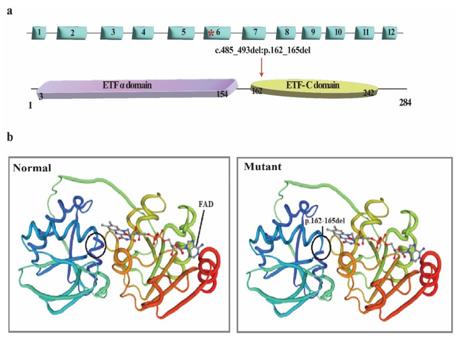 Fig. 1 ETFA Gene/Protein Structure and Mutational Bioinformatic Analysis.1
Key structural properties of ETFA:
- Typical α/β sandwich folding structure
- Conservative FAD binding pockets, surrounded by hydrophobic residues
- Flavin adenine dinucleotide (FAD) cofactors serve as electron carriers
Functions of ETFA
The core function of the protein encoded by the ETFA gene is to act as an electron transfer carrier in the mitochondrial matrix. However, it also plays a role in various physiological and pathological processes, including the maintenance of cellular redox homeostasis and the regulation of metabolic diseases.
| Function | Description |
| Electron Transfer | As an electron intermediate, it receives electrons from various acyl-CoA dehydrogenases and transfers them to the downstream ETF-QO, which is a crucial step in fatty acid β-oxidation and amino acid catabolism. |
| Energy Metabolism Support | Through efficient electron transfer, ensure that the mitochondrial respiratory chain obtains reducing equivalents, thereby supporting the continuous production of ATP. |
| Metabolic Disease Association | The defect in ETFA function directly leads to various acyl-CoA dehydrogenase deficiencies (GAII type), causing metabolic crises and energy supply disorders. |
| Redox Regulation | It participates in maintaining the redox balance between the cytoplasm and the mitochondrial matrix, and affects the level of reactive oxygen species (ROS). |
| Cell Protection | Under metabolic stress, it helps alleviate mitochondrial oxidative damage caused by electron leakage by maintaining the stability of the electron flow. |
This protein acts as a soluble electron carrier and its mode of action is a linear single-electron transfer, which is completely different from the S-shaped curve of cooperative oxygen binding of hemoglobin. This reflects its characteristic of being an efficient and specific "transit station" in the metabolic network. Its dysfunction will immediately interrupt multiple metabolic pathways, highlighting its pivotal role in basic metabolism.
Applications of ETFA and ETFA Antibody in Literature
1. Chen, Wankun, et al. "RETRACTED: NBPF4 mitigates progression in colorectal cancer through the regulation of EZH2‐associated ETFA." Journal of cellular and molecular medicine 25.18 (2021): 9038-9050. https://doi.org/10.1111/jcmm.16867
The article indicates that NBPF4 is lowly expressed in colorectal cancer. It avoids being inhibited by miR-17-3p by binding to ZFP36, thereby down-regulating EZH2 and inhibiting the expression of ETFA, thus slowing down tumor development.
2. Chautard, Robin, et al. "A case report of a mild form of multiple acyl-CoA dehydrogenase deficiency due to compound heterozygous mutations in the ETFA gene." BMC medical genomics 13.1 (2020): 12. https://doi.org/10.1186/s12920-020-0665-6
This study reports for the first time two novel heterozygous mutations (c.354C>A and c.652G>A) in the ETFA gene, which lead to mild MADD with onset in childhood. After avoiding fasting and undergoing treatment with levocarnitine and riboflavin, the patient's development and academic performance returned to normal.
3. Guo, Jingjing, et al. "Hepatocyte-derived Igκ promotes HCC progression by stabilizing electron transfer flavoprotein subunit α to facilitate fatty acid β-oxidation." Journal of Experimental & Clinical Cancer Research 43.1 (2024): 280. https://doi.org/10.1186/s13046-024-03203-8
Research has found that the Vκ4-1/Jκ 3-type immunoglobulin κ chain (Igκ), which is highly expressed in liver cancer cells, can bind to ETFA protein, stabilize its expression, thereby interfering with fatty acid β -oxidation and promoting tumor development. Targeting this pathway may become a new strategy for the treatment of liver cancer.
4. Chen, Fuying, et al. "S1P defects cause a new entity of cataract, alopecia, oral mucosal disorder, and psoriasis‐like syndrome." EMBO Molecular Medicine 14.5 (2022): e14904. https://doi.org/10.15252/emmm.202114904
This study has for the first time discovered that mutations in the MBTPS1 gene lead to abnormal function of the S1P protease encoded by it, which cannot stabilize the ETFA/ETFB complex, thereby causing mitochondrial dysfunction and CAOP syndrome. Supplementation of riboflavin can restore the stability of ETFA/ETFB and improve symptoms.
5. Parvini, Farshid, Mobarakeh Ajam-Hosseini, and Marziyeh Shadpour. "First report of neonatal-onset glutaric aciduria type II in the Iranian population caused by a novel deleterious ETFA variant." Orphanet Journal of Rare Diseases 20.1 (2025): 590. https://doi.org/10.1186/s13023-025-04107-2
This study reports GA2 caused by a novel heterozygous deletion mutation (c.485_493del) of the ETFA gene for the first time in Iran. This discovery clarifies the importance of genetic diagnosis in the prognosis judgment and family counseling of rare metabolic diseases.
Creative Biolabs: ETFA Antibodies for Research
Creative Biolabs specializes in the production of high-quality ETFA antibodies for research and industrial applications. Our portfolio includes monoclonal antibodies tailored for ELISA, Flow Cytometry, Western blot, immunohistochemistry, and other diagnostic methodologies.
- Custom ETFA Antibody Development: Tailor-made solutions to meet specific research requirements.
- Bulk Production: Large-scale antibody manufacturing for industry partners.
- Technical Support: Expert consultation for protocol optimization and troubleshooting.
- Aliquoting Services: Conveniently sized aliquots for long-term storage and consistent experimental outcomes.
For more details on our ETFA antibodies, custom preparations, or technical support, contact us at email.
Reference
- Parvini, Farshid, Mobarakeh Ajam-Hosseini, and Marziyeh Shadpour. "First report of neonatal-onset glutaric aciduria type II in the Iranian population caused by a novel deleterious ETFA variant." Orphanet Journal of Rare Diseases 20.1 (2025): 590. https://doi.org/10.1186/s13023-025-04107-2
Anti-ETFA antibodies
 Loading...
 Compare




Hot products 
- AActivation
- AGAgonist
- APApoptosis
- BBlocking
- BABioassay
- BIBioimaging
- CImmunohistochemistry-Frozen Sections
- CIChromatin Immunoprecipitation
- CTCytotoxicity
- CSCostimulation
- DDepletion
- DBDot Blot
- EELISA
- ECELISA(Cap)
- EDELISA(Det)
- ESELISpot
- EMElectron Microscopy
- FFlow Cytometry
- FNFunction Assay
- GSGel Supershift
- IInhibition
- IAEnzyme Immunoassay
- ICImmunocytochemistry
- IDImmunodiffusion
- IEImmunoelectrophoresis
- IFImmunofluorescence
- IGImmunochromatography
- IHImmunohistochemistry
- IMImmunomicroscopy
- IOImmunoassay
- IPImmunoprecipitation
- ISIntracellular Staining for Flow Cytometry
- LALuminex Assay
- LFLateral Flow Immunoassay
- MMicroarray
- MCMass Cytometry/CyTOF
- MDMeDIP
- MSElectrophoretic Mobility Shift Assay
- NNeutralization
- PImmunohistologyp-Paraffin Sections
- PAPeptide Array
- PEPeptide ELISA
- PLProximity Ligation Assay
- RRadioimmunoassay
- SStimulation
- SESandwich ELISA
- SHIn situ hybridization
- TCTissue Culture
- WBWestern Blot
