GCLC Antibodies
Background
The GCLC gene encodes a key enzyme involved in the synthesis of glutathione, which is mainly found in tissues such as the liver, kidneys, and brain of vertebrates. The protein expressed by this gene catalyzes the first step of the rate-limiting reaction in glutathione synthesis. It maintains the redox balance by regulating the levels of antioxidant substances within the cells and supports the cellular defense mechanism. It plays a core role in responding to oxidative stress, detoxification metabolism, and immune regulation. Since it was systematically characterized in the 1990s, the regulatory mechanism of GCLC and its association with various diseases (such as neurodegenerative diseases, metabolic syndrome) have been continuously explored. This gene and its product have become an important model for exploring cellular antioxidant pathways, developing drug targets, and analyzing pathological mechanisms, promoting the systematic understanding of the molecular network of the life form's response to oxidative stress.
Structure of GCLC
The protein molecule encoded by the GCLC gene has a molecular weight of approximately 73 kDa. Its size varies slightly among different species due to differences in amino acid sequences.
| Species | Human | Mouse | Rat | Bovine | Zebrafish |
|---|---|---|---|---|---|
| Molecular Weight (kDa) | ~73 | ~72.8 | ~72.5 | ~72.9 | ~73.2 |
| Primary Structural Differences | Highly conservative catalytic core domain | There are subtle differences in the N-terminal regulatory regions | Extremely similar to humans in origin | Catalytically active centers are highly similar | There were specific functional domain variants |
This protein is composed of approximately 637 amino acids and presents an overall conformation combining a spherical functional domain with an extended domain. Its core domain adopts a typical α/β folding pattern, forming a catalytic pocket that precisely accommodates the substrate glutamate and cysteine. The key function relies on a cysteine residue located at the active center (such as human Cys-269), which directly participates in peptide bond formation. The protein's secondary structure consists of multiple parallel β-sheet segments, surrounded by α-helices, jointly constructing a stable active site environment. Adjacent glutamate residues (such as Glu-267) precisely stabilize the reaction intermediate through a hydrogen bond network, ensuring the efficiency and specificity of the synthesis reaction.
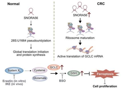 Fig. 1 SNORA56 Promotes Tumor Growth by Enhancing GCLC Translation to Inhibit Ferroptosis.1
Key structural properties of GCLC:
- Typical α/β fold domain
- Hydrophobic active center
- Key cysteine residue
- Synergistic regulation of arginine and glutamate
Functions of GCLC
The core function of the GCLC gene is to catalyze the rate-limiting step in the biosynthesis of glutathione. Additionally, it is involved in a variety of physiological and pathological processes such as redox signal transduction and maintenance of cellular homeostasis.
| Function | Description |
|---|---|
| Glutathione Synthesis | Catalyzes the ATP-dependent condensation reaction between glutamate and cysteine to produce γ-glutamylcysteine, which is the first crucial step in the synthesis of the intracellular endogenous antioxidant glutathione. |
| Oxidative Stress Defense | By regulating the level of glutathione, it directly eliminates reactive oxygen free radicals and protects cells from oxidative damage. This is particularly crucial in high-metabolism tissues such as the liver, kidneys, and nervous system. |
| Detoxification Metabolism Support | The provided glutathione acts as a co-substrate for the second-phase detoxification reaction, binding to toxins to increase their water solubility and facilitating their excretion through bile or urine. |
| Immune Regulation | It affects the function and proliferation of lymphocytes, and regulates inflammatory responses and immune responses by maintaining the level of reduced glutathione within immune cells. |
| Cell Apoptosis Regulation | The level of glutathione directly affects mitochondrial function and the apoptotic signaling pathway. Insufficient GCLC activity can lead to apoptosis induced by oxidative stress. |
The kinetic characteristics of this enzyme are characterized by a high affinity for the substrates glutamate and cysteine. However, its activity is subject to feedback inhibition by the final product glutathione and precise regulation by the transcription factor Nrf2. This ensures that cells can rapidly upregulate their antioxidant defense capabilities under stress conditions.
Applications of GCLC and GCLC Antibody in Literature
1. Niu, Kaifeng, et al. "NSUN2 lactylation drives cancer cell resistance to ferroptosis through enhancing GCLC-dependent glutathione synthesis." Redox biology 79 (2025): 103479. https://doi.org/10.1016/j.redox.2024.103479
This study reveals that lactate-mediated lactylation of the NSUN2 protein can promote the m5C modification and stability of GCLC mRNA, thereby increasing the glutathione level in gastric cancer cells and enhancing their resistance to ferroptosis. This mechanism is mediated by NAA10, providing a potential target for improving cancer prognosis.
2. Deng, Jiao, et al. "Histone lactylation enhances GCLC expression and thus promotes chemoresistance of colorectal cancer stem cells through inhibiting ferroptosis." Cell Death & Disease 16.1 (2025): 1-15. https://doi.org/10.1038/s41419-025-07498-z
The study found that the lactylation modification of histone H4K12 is regulated by p300/HDAC1, upregulates the expression of GCLC and inhibits ferroptosis, thereby enhancing the chemotherapy resistance of colorectal cancer stem cells. Targeting this pathway may become a new strategy for reversing drug resistance.
3. Chen, Zixiang, et al. "GCLC desuccinylation regulated by oxidative stress protects human cancer cells from ferroptosis." Cell Death & Differentiation (2025): 1-12. https://doi.org/10.1038/s41418-025-01505-8
The research has found that SIRT2 can remove the succinylated modifications of the GCLC protein (at the K38/126/326 sites), thereby activating its function of synthesizing glutathione, helping tumor cells resist oxidative stress and ferroptosis, and maintaining redox homeostasis.
4. Xu, Chang, et al. "SNORA56-mediated pseudouridylation of 28 S rRNA inhibits ferroptosis and promotes colorectal cancer proliferation by enhancing GCLC translation." Journal of Experimental & Clinical Cancer Research 42.1 (2023): 331. https://doi.org/10.1186/s13046-023-02906-8
The study found that SNORA56 mediates the modification of pseudouridine on 28S rRNA, promoting the translation of GCLC protein, enhancing glutathione synthesis to inhibit lipid peroxidation and ferroptosis, thereby driving the progression of colorectal cancer. This pathway has diagnostic and therapeutic potential.
5. Luo, Lianxiang, et al. "Ferroptosis-related gene GCLC is a novel prognostic molecular and correlates with immune infiltrates in lung adenocarcinoma." Cells 11.21 (2022): 3371. https://doi.org/https://doi.org/10.3390/cells11213371
The study found that high expression of GCLC is associated with poor prognosis of lung adenocarcinoma. This gene inhibits ferroptosis by enhancing glutathione synthesis, thereby promoting tumor proliferation and invasion. It can serve as a potential prognostic marker and therapeutic target.
Creative Biolabs: GCLC Antibodies for Research
Creative Biolabs specializes in the production of high-quality GCLC antibodies for research and industrial applications. Our portfolio includes monoclonal antibodies tailored for ELISA, Flow Cytometry, Western blot, immunohistochemistry, and other diagnostic methodologies.
- Custom GCLC Antibody Development: Tailor-made solutions to meet specific research requirements.
- Bulk Production: Large-scale antibody manufacturing for industry partners.
- Technical Support: Expert consultation for protocol optimization and troubleshooting.
- Aliquoting Services: Conveniently sized aliquots for long-term storage and consistent experimental outcomes.
For more details on our GCLC antibodies, custom preparations, or technical support, contact us at email.
Reference
- Xu, Chang, et al. "SNORA56-mediated pseudouridylation of 28 S rRNA inhibits ferroptosis and promotes colorectal cancer proliferation by enhancing GCLC translation." Journal of Experimental & Clinical Cancer Research 42.1 (2023): 331. https://doi.org/10.1186/s13046-023-02906-8
Anti-GCLC antibodies
 Loading...


Hot products 
-
Mouse Anti-ABL2 Recombinant Antibody (V2-179121) (CBMAB-A0364-YC)

-
Rabbit Anti-CBL Recombinant Antibody (D4E10) (CBMAB-CP0149-LY)

-
Mouse Anti-C5AR1 Recombinant Antibody (R63) (CBMAB-C9553-LY)

-
Mouse Anti-ENPP1 Recombinant Antibody (CBFYE-0159) (CBMAB-E0375-FY)

-
Mouse Anti-AGK Recombinant Antibody (V2-258056) (CBMAB-M0989-FY)

-
Mouse Anti-CAT Recombinant Antibody (724810) (CBMAB-C8431-LY)

-
Mouse Anti-BSN Recombinant Antibody (219E1) (CBMAB-1228-CN)

-
Mouse Anti-ADAM29 Recombinant Antibody (V2-179787) (CBMAB-A1149-YC)

-
Mouse Anti-BAD (Phospho-Ser136) Recombinant Antibody (CBYY-0138) (CBMAB-0139-YY)

-
Mouse Anti-FPR2 Recombinant Antibody (1D6) (CBMAB-F2628-CQ)

-
Mouse Anti-FeLV g27 Recombinant Antibody (1) (CBMAB-V208-1714-FY)

-
Mouse Anti-ASH1L Monoclonal Antibody (ASH5H03) (CBMAB-1372-YC)

-
Mouse Anti-AAV-5 Recombinant Antibody (V2-503417) (CBMAB-V208-1369-FY)

-
Mouse Anti-ESR1 Recombinant Antibody (Y31) (CBMAB-1208-YC)

-
Mouse Anti-2C TCR Recombinant Antibody (V2-1556) (CBMAB-0951-LY)

-
Mouse Anti-AGO2 Recombinant Antibody (V2-634169) (CBMAB-AP203LY)

-
Mouse Anti-BRCA2 Recombinant Antibody (CBYY-1728) (CBMAB-2077-YY)

-
Mouse Anti-DDC Recombinant Antibody (8E8) (CBMAB-0992-YC)

-
Mouse Anti-AHCYL1 Recombinant Antibody (V2-180270) (CBMAB-A1703-YC)

-
Rat Anti-AChR Recombinant Antibody (V2-12500) (CBMAB-0990-CN)

- AActivation
- AGAgonist
- APApoptosis
- BBlocking
- BABioassay
- BIBioimaging
- CImmunohistochemistry-Frozen Sections
- CIChromatin Immunoprecipitation
- CTCytotoxicity
- CSCostimulation
- DDepletion
- DBDot Blot
- EELISA
- ECELISA(Cap)
- EDELISA(Det)
- ESELISpot
- EMElectron Microscopy
- FFlow Cytometry
- FNFunction Assay
- GSGel Supershift
- IInhibition
- IAEnzyme Immunoassay
- ICImmunocytochemistry
- IDImmunodiffusion
- IEImmunoelectrophoresis
- IFImmunofluorescence
- IGImmunochromatography
- IHImmunohistochemistry
- IMImmunomicroscopy
- IOImmunoassay
- IPImmunoprecipitation
- ISIntracellular Staining for Flow Cytometry
- LALuminex Assay
- LFLateral Flow Immunoassay
- MMicroarray
- MCMass Cytometry/CyTOF
- MDMeDIP
- MSElectrophoretic Mobility Shift Assay
- NNeutralization
- PImmunohistologyp-Paraffin Sections
- PAPeptide Array
- PEPeptide ELISA
- PLProximity Ligation Assay
- RRadioimmunoassay
- SStimulation
- SESandwich ELISA
- SHIn situ hybridization
- TCTissue Culture
- WBWestern Blot
