
IGFBP7 Antibodies
Background
IGFBP7 is a secreted glycoprotein belonging to the insulin-like growth factor binding protein family. This protein binds to insulin-like growth factors with high affinity, regulating their biological activity and independently participating in processes such as cell proliferation, aging, and angiogenesis. Its encoding gene is located on human chromosome 4q12. It was initially identified in 1991 and has now been confirmed to be widely expressed in various tissues, especially playing a crucial role in vascular endothelial and epithelial cells. Studies have shown that IGFBP7 inhibits tumor growth through p53-dependent and -independent pathways, thus being regarded as an important cancer suppressor. The research on the structure and function of this protein has deepened our understanding of the growth factor signaling network and the regulatory mechanism of the cellular microenvironment, providing potential targets for the diagnosis and treatment of related diseases.
Structure of IGFBP7
IGFBP7 is a secreted glycoprotein with a molecular weight of approximately 29 kDa. The molecular weight varies slightly among different species due to differences in the level of glycosylation modification.
| Species | Human | Mouse | Rat |
| Molecular Weight (kDa) | ~29 | ~28 | ~28.5 |
| Primary Structural Differences | Mature peptide containing 282 amino acids, containing the IB structure domain and immune globulin structure domain | Highly conserved amino acid sequences, tiny differences glycosylation sites | High homology with human IGFBP7 and high functional domain identity |
This protein is composed of approximately 250 amino acids, and its spatial structure depends on a rigid core formed by conserved cysteine residues. The structural core contains an insulin-like growth factor binding domain, which, through specific β-sheets and α-helices, forms a hydrophobic pocket for ligand binding. A crucial "cell adhesion module" is located at the C-terminal end, mediating interactions with extracellular matrix components, which is the structural basis for its non-IGF-dependent functions.
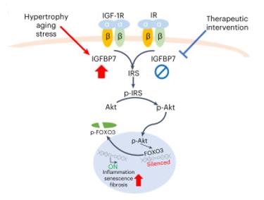 Fig. 1 Diagram shows proposed IGFBP7 action in the stress myocardium.1
Key structural properties of IGFBP7:
- Secretory glycoprotein, containing insulin-like growth factor binding domain (IB domain)
- The core structure is stabilized by a network of disulfide bonds formed by conserved cysteine residues
- The unique "Kazal-like domain" mediates its interaction with the extracellular matrix and cell surfaces
- The C-terminal region contains a functional "cell adhesion motif" responsible for IGF-independent signaling regulation
Functions of IGFBP7
The main function of the IGFBP7 gene is to regulate the biological activity of insulin-like growth factors (IGFs) by binding to them, and to participate in the regulation of cell proliferation and aging. In addition, it also plays a key role independently of the IGF pathway in tumor suppression, angiogenesis, and cell adhesion.
| Function | Description |
| IGF Signal Regulation | By binding to IGF-1 and IGF-2 with high affinity, it restricts their interaction with receptors, thereby negatively regulating the growth and survival signaling pathways. |
| Tumor Suppression | By activating p53-dependent and -independent pathways (such as inducing cellular senescence and apoptosis), it inhibits the growth of various cancer cells. |
| Vascularization Regulation | Expressed in vascular endothelial cells, it can exert dual regulatory effects of promoting or inhibiting angiogenesis depending on the signals from the microenvironment. |
| Cell Adhesion and Migration | By directly interacting with extracellular matrix proteins through its C-terminal cell adhesion module, it influences the adhesion, spreading, and migration behaviors of cells. |
| Aging induction | It can induce premature senescence in various cell types (including tumor cells), which is one of its important tumor suppression mechanisms. |
Unlike typical members of the IGFBP family, IGFBP7 has a relatively lower affinity for IGFs, but its functions are more pleiotropic. It plays a complex and crucial role in the regulation of the tumor microenvironment and tissue homeostasis.
Applications of IGFBP7 and IGFBP7 Antibody in Literature
1. Zhang, Liyong, et al. "Insulin-like growth factor-binding protein-7 (IGFBP7) links senescence to heart failure." Nature Cardiovascular Research 1.12 (2022): 1195-1214. https://doi.org/10.1038/s44161-022-00181-y
The article indicates that in the research on heart failure, the increased expression of IGFBP7 promotes myocardial aging. Mechanistically, IGFBP7 inhibits FOXO3a, thereby weakening DNA repair and antioxidant capacity, and exacerbating cardiac damage. Targeted inhibition of IGFBP7 can delay the progression of heart failure and has therapeutic potential.
2. Yi, Xianyanling, et al. "IGFBP7 and the tumor immune landscape: a novel target for immunotherapy in bladder cancer." Frontiers in Immunology 13 (2022): 898493. https://doi.org/10.3389/fimmu.2022.898493
The article indicates that in bladder cancer, high expression of IGFBP7 is associated with adverse clinical features. It promotes lymphocyte infiltration but inhibits T-cell recognition and killing functions, resulting in a reduced immune treatment response. Studies have shown that targeting IGFBP7 has the potential to become a new strategy for combined immunotherapy in bladder cancer.
3. Chen, Zhe, et al. "Exercise protects proliferative muscle satellite cells against exhaustion via the Igfbp7-Akt-mTOR axis." Theranostics 10.14 (2020): 6448. https://doi.org/10.7150/thno.43577
The research reveals that exercise inhibits the Akt-mTOR pathway by upregulating IGFBP7, restricts mitochondrial metabolism in muscle satellite cells, maintains their proliferative potential, prevents exhaustion, and thereby promotes muscle hypertrophy and regeneration, providing potential therapeutic targets for muscle diseases.
4. He, Rui, et al. "IGFBP7 promotes endothelial cell repair in the recovery phase of acute lung injury." Clinical Science 138.13 (2024): 797-815. https://doi.org/10.1042/CS20240179
The study found that during the recovery period of acute lung injury, endothelial cells' IGFBP7 promotes endothelial proliferation through the FOS/YAP1 signaling pathway, which is crucial for tissue repair. This suggests that the targeted IGFBP7 treatment should take into account the specificity of the disease stage.
5. Li, Dandan, et al. "Cancer-associated fibroblast-secreted IGFBP7 promotes gastric cancer by enhancing tumor associated macrophage infiltration via FGF2/FGFR1/PI3K/AKT axis." Cell Death Discovery 9.1 (2023): 17. https://doi.org/10.1038/s41420-023-01336-x
The study found that in gastric cancer, IGFBP7 drives the polarization of tumor-associated macrophages towards the M2 type through the FGF2/FGFR1/PI3K/AKT axis, promoting tumor progression and being closely related to poor prognosis and remodeling of the tumor microenvironment.
Creative Biolabs: IGFBP7 Antibodies for Research
Creative Biolabs specializes in the production of high-quality IGFBP7 antibodies for research and industrial applications. Our portfolio includes monoclonal antibodies tailored for ELISA, Flow Cytometry, Western blot, immunohistochemistry, and other diagnostic methodologies.
- Custom IGFBP7 Antibody Development: Tailor-made solutions to meet specific research requirements.
- Bulk Production: Large-scale antibody manufacturing for industry partners.
- Technical Support: Expert consultation for protocol optimization and troubleshooting.
- Aliquoting Services: Conveniently sized aliquots for long-term storage and consistent experimental outcomes.
For more details on our IGFBP7 antibodies, custom preparations, or technical support, contact us at email.
Reference
- Zhang, Liyong, et al. "Insulin-like growth factor-binding protein-7 (IGFBP7) links senescence to heart failure." Nature Cardiovascular Research 1.12 (2022): 1195-1214. https://doi.org/10.1038/s44161-022-00181-y
Anti-IGFBP7 antibodies
 Loading...
 Compare




Hot products 
-
Mouse Anti-ATP1A2 Recombinant Antibody (M7-PB-E9) (CBMAB-A4013-YC)

-
Mouse Anti-CD8 Recombinant Antibody (C1083) (CBMAB-C1083-LY)

-
Mouse Anti-BAX Recombinant Antibody (CBYY-0216) (CBMAB-0217-YY)

-
Mouse Anti-FLI1 Recombinant Antibody (CBXF-0733) (CBMAB-F0435-CQ)

-
Mouse Anti-ACVR1C Recombinant Antibody (V2-179685) (CBMAB-A1041-YC)

-
Mouse Anti-BZLF1 Recombinant Antibody (BZ.1) (CBMAB-AP705LY)

-
Mouse Anti-A2M Recombinant Antibody (V2-178822) (CBMAB-A0036-YC)

-
Rabbit Anti-AKT3 Recombinant Antibody (V2-12567) (CBMAB-1057-CN)

-
Rat Anti-(1-5)-α-L-Arabinan Recombinant Antibody (V2-501861) (CBMAB-XB0003-YC)

-
Mouse Anti-FOXA3 Recombinant Antibody (2A9) (CBMAB-0377-YC)

-
Mouse Anti-FTH1 Recombinant Antibody (CBXF-1896) (CBMAB-F3426-CQ)

-
Mouse Anti-14-3-3 Pan Recombinant Antibody (V2-9272) (CBMAB-1181-LY)

-
Mouse Anti-HTLV-1 gp46 Recombinant Antibody (CBMW-H1006) (CBMAB-V208-1154-FY)

-
Mouse Anti-ADV Recombinant Antibody (V2-503423) (CBMAB-V208-1364-FY)

-
Mouse Anti-8-oxoguanine Recombinant Antibody (V2-7697) (CBMAB-1869CQ)

-
Mouse Anti-G6PD Recombinant Antibody (13B331) (CBMAB-G1553-LY)

-
Human Anti-SARS-CoV-2 S1 Monoclonal Antibody (CBFYR-0120) (CBMAB-R0120-FY)

-
Mouse Anti-BCL2L1 Recombinant Antibody (H5) (CBMAB-1025CQ)

-
Mouse Anti-CHRNA9 Recombinant Antibody (8E4) (CBMAB-C9161-LY)

-
Mouse Anti-FOXL1 Recombinant Antibody (CBXF-0845) (CBMAB-F0462-CQ)

- AActivation
- AGAgonist
- APApoptosis
- BBlocking
- BABioassay
- BIBioimaging
- CImmunohistochemistry-Frozen Sections
- CIChromatin Immunoprecipitation
- CTCytotoxicity
- CSCostimulation
- DDepletion
- DBDot Blot
- EELISA
- ECELISA(Cap)
- EDELISA(Det)
- ESELISpot
- EMElectron Microscopy
- FFlow Cytometry
- FNFunction Assay
- GSGel Supershift
- IInhibition
- IAEnzyme Immunoassay
- ICImmunocytochemistry
- IDImmunodiffusion
- IEImmunoelectrophoresis
- IFImmunofluorescence
- IGImmunochromatography
- IHImmunohistochemistry
- IMImmunomicroscopy
- IOImmunoassay
- IPImmunoprecipitation
- ISIntracellular Staining for Flow Cytometry
- LALuminex Assay
- LFLateral Flow Immunoassay
- MMicroarray
- MCMass Cytometry/CyTOF
- MDMeDIP
- MSElectrophoretic Mobility Shift Assay
- NNeutralization
- PImmunohistologyp-Paraffin Sections
- PAPeptide Array
- PEPeptide ELISA
- PLProximity Ligation Assay
- RRadioimmunoassay
- SStimulation
- SESandwich ELISA
- SHIn situ hybridization
- TCTissue Culture
- WBWestern Blot
