MPP3 Antibodies
Background
MPP3 is a scaffold protein located on the cell membrane, mainly present in specific regions of epithelial cells and neurons in vertebrates. This protein binds to various transmembrane proteins and signaling molecules through its PDZ domain, thereby participating in the establishment of cell polarity, the assembly of intercellular connections, and the temporal regulation of signaling pathways. Its function in the auditory system is particularly crucial. MPP3 maintains the structural integrity of the basal hair cells in the inner ear by interacting with transmembrane proteins such as cadherins and ion channels, which is essential for the mechanical electro-transduction of auditory signals. Since its identification in the early 21st century, MPP3 has become an important model for studying cell polarity complexes and the function of sensory epithelia. The elucidation of its mechanism has deepened our understanding of cell organization, neural development, and the mechanisms of related diseases.
Structure of MPP3
MPP3 is a scaffold protein belonging to the membrane-associated guanylate kinase (MAGUK) family. Its molecular weight is roughly similar among different vertebrate species, approximately ranging from 70 to 75 kDa. The molecular weight difference mainly results from minor variations in its protein domains (such as PDZ, SH3, and GUK domains), which adapt to the different functional requirements for mediating cell polarity and signal transduction in specific cell types (such as inner ear hair cells, epithelial cells).
| Species | Human | Mouse | Rat | Zebrafish |
| Molecular Weight (kDa) | ~75 | ~74 | ~75 | ~72 |
| Primary Structural Differences | Auditory function, epithelial polarity | Inner ear development, neural synapses | Cell junctions, polarity establishment | Development of lateral organs |
MPP3 is composed of approximately 550-650 amino acids. Its core feature lies in the tandem PDZ domains, which can specifically recognize and bind to the C-terminal sequences of target proteins (such as cadherins, receptors), thereby assembling multi-protein complexes at specific regions of the cell membrane (such as tight junctions, basal regions of stereocilia). This interaction is crucial for maintaining the integrity of the epithelial barrier and the stability of the mechanical sensing structure of inner ear hair cells. Dysfunction of this function is closely related to diseases such as hearing impairment.
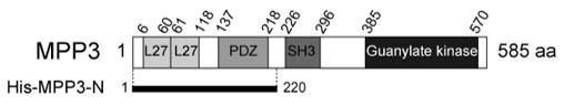 Fig. 1 Schematic representation of the structures of MPP3.1
Key structural properties of MPP3:
- Has a more conservative structure domain (e.g., PDZ, SH3, GUK), forming a modular framework
- Specifically recognize and bind to the C-terminal sequence of the target protein through the PDZ domain
- As a member of the membrane-associated guanylate kinase (MAGUK) family
- Protein conformation is flexible and tunable to precisely adapt the connectivity and polarity requirements in different cellular environments
Functions of MPP3
The main function of the MPP3 gene is to act as a scaffold protein, participating in the establishment and maintenance of cell polarity, especially in epithelial tissues and sensory hair cells. However, it is also widely involved in various cellular physiological processes, including the assembly of intercellular connections and the regulation of local signaling pathways.
| Function | Description |
| Establishment of cell polarity | Through its PDZ and other domains, it organizes and anchors protein complexes at specific regions of the cell membrane (such as the apical membrane or basolateral membrane), serving as the structural basis for the formation of cell asymmetry. |
| Cellular Junction Assembly | As a molecular scaffold at sites such as tight junctions and adherens junctions, it recruits and stabilizes transmembrane proteins (such as cadherin) and cytoplasmic signaling molecules, maintaining the integrity of the tissue barrier. |
| Mechanical Sensory Transduction | A complex is formed at the base of the basal cilia of inner ear hair cells, which is crucial for converting mechanical vibrations into electrical signals and is the core process of auditory function. |
| Signal Pathway Regulation | As a signaling platform, it coordinates and precisely regulates the spatiotemporal activity of various developmental and homeostasis-related signaling pathways (such as the Hippo and Wnt pathways). |
| Tissue Development and Homeostasis | It plays an indispensable role in epithelial tissue morphogenesis, formation of neuronal synapses, and inner ear development, and its dysfunction can lead to diseases such as hearing loss. |
The function of MPP3 relies on its modular domains for specific protein-protein interactions. Its mode of action is highly localized and context-dependent, which is consistent with its core role in precisely integrating structure and signaling in different cell types.
Applications of MPP3 and MPP3 Antibody in Literature
1. Wang, Xinyue, et al. "A novel rabbit anti-myoglobin monoclonal antibody's potential application in rhabdomyolysis associated acute kidney injury."International Journal of Molecular Sciences 24.9 (2023): 7822. https://doi.org/10.1371/journal.pone.0082894
Through the cell spreading experiment in this study, it was first discovered that the allosteric interaction of CADM1 activates the PI3K signaling pathway through the MPP3/Dlg complex, and regulates the cytoskeleton reorganization and epithelial structure formation by relying on Akt and Rac1.
2. Kang, Yoon-A., et al. "Secretory MPP3 reinforce myeloid differentiation trajectory and amplify myeloid cell production." Journal of Experimental Medicine 220.8 (2023): e20230088. https://doi.org/10.1084/jem.20230088
The study found that within the hematopoietic multipotent progenitor cell MPP3, there exists a FcγR-positive subgroup with secretory function, and its endoplasmic reticulum is well-developed. This subgroup not only serves as a reserve pool for granulocyte/macrophage progenitor cells, but also can enhance itself by secreting cytokines during inflammation, rapidly amplifying myeloid generation in the bone marrow microenvironment.
3. Dudok, Jacobus J., et al. "MPP3 is required for maintenance of the apical junctional complex, neuronal migration, and stratification in the developing cortex." Journal of Neuroscience 33.19 (2013): 8518-8527. https://doi.org/10.1523/JNEUROSCI.5627-12.2013
The research has found that during the development of the mammalian cortex, the membrane protein MPP3 is crucial for maintaining the top protein complex and adhesion junctions. The absence of MPP3 leads to disordered spindle orientation, delayed cell migration, and subsequently results in defects in neuronal layering and abnormal localization.
4. Lenaerts, Aurelie, et al. "EBF1 primes B-lymphoid enhancers and limits the myeloid bias in murine multipotent progenitors." Journal of Experimental Medicine 219.11 (2022): e20212437. https://doi.org/10.1084/jem.20212437
The study found that the transcription factor EBF1 regulates the lineage preference of multipotent progenitor cell MPP3 by directly inhibiting C/EBPα, limiting its excessive myeloid differentiation and simultaneously initiating the B lymphocyte fate program.
5. Sakurai-Yageta, Mika, et al. "Dynamic regulation of a cell adhesion protein complex including CADM1 by combinatorial analysis of FRAP with exponential curve-fitting." PLoS One 10.3 (2015): e0116637. https://doi.org/10.1371/journal.pone.0116637
The research found that through long-term FRAP analysis, it was discovered that CADM1 is the core that stabilizes the 4.1B-MPP3 complex. 4.1B and MPP3 exist in two states: free and bound. The ratios are approximately 3:2 and 3:1 respectively, revealing the dynamic regulation mechanism of the cell adhesion complex.
Creative Biolabs: MPP3 Antibodies for Research
Creative Biolabs specializes in the production of high-quality MPP3 antibodies for research and industrial applications. Our portfolio includes monoclonal antibodies tailored for ELISA, Flow Cytometry, Western blot, immunohistochemistry, and other diagnostic methodologies.
- Custom MPP3 Antibody Development: Tailor-made solutions to meet specific research requirements.
- Bulk Production: Large-scale antibody manufacturing for industry partners.
- Technical Support: Expert consultation for protocol optimization and troubleshooting.
- Aliquoting Services: Conveniently sized aliquots for long-term storage and consistent experimental outcomes.
For more details on our MPP3 antibodies, custom preparations, or technical support, contact us at email.
Reference
- Wang, Xinyue, et al. "A novel rabbit anti-myoglobin monoclonal antibody's potential application in rhabdomyolysis associated acute kidney injury."International Journal of Molecular Sciences 24.9 (2023): 7822. Distributed under Open Access license CC BY 4.0, without modification. https://doi.org/10.1371/journal.pone.0082894
Anti-MPP3 antibodies
 Loading...


Hot products 
-
Rabbit Anti-BAD (Phospho-Ser136) Recombinant Antibody (CAP219) (CBMAB-AP536LY)

-
Mouse Anti-CCND2 Recombinant Antibody (DCS-3) (CBMAB-G1318-LY)

-
Rabbit Anti-ALDOA Recombinant Antibody (D73H4) (CBMAB-A2314-YC)

-
Rabbit Anti-ADRA1A Recombinant Antibody (V2-12532) (CBMAB-1022-CN)

-
Rat Anti-C5AR1 Recombinant Antibody (8D6) (CBMAB-C9139-LY)

-
Rabbit Anti-ABL1 (Phosphorylated Y245) Recombinant Antibody (V2-505716) (PTM-CBMAB-0465LY)

-
Rabbit Anti-Acetyl-Histone H3 (Lys36) Recombinant Antibody (V2-623395) (CBMAB-CP0994-LY)

-
Mouse Anti-AOC3 Recombinant Antibody (CBYY-0014) (CBMAB-0014-YY)

-
Mouse Anti-ENO2 Recombinant Antibody (85F11) (CBMAB-0276CQ)

-
Mouse Anti-CHRNA9 Recombinant Antibody (8E4) (CBMAB-C9161-LY)

-
Rabbit Anti-AKT3 Recombinant Antibody (V2-12567) (CBMAB-1057-CN)

-
Mouse Anti-F11R Recombinant Antibody (402) (CBMAB-0026-WJ)

-
Mouse Anti-CD59 Recombinant Antibody (CBXC-2097) (CBMAB-C4421-CQ)

-
Mouse Anti-ALB Recombinant Antibody (V2-180650) (CBMAB-A2186-YC)

-
Mouse Anti-NSUN6 Recombinant Antibody (D-5) (CBMAB-N3674-WJ)

-
Mouse Anti-ELAVL4 Recombinant Antibody (6B9) (CBMAB-1132-YC)

-
Mouse Anti-BRCA2 Recombinant Antibody (CBYY-1728) (CBMAB-2077-YY)

-
Mouse Anti-BIRC3 Recombinant Antibody (315304) (CBMAB-1214-CN)

-
Mouse Anti-ASTN1 Recombinant Antibody (H-9) (CBMAB-1154-CN)

-
Mouse Anti-BBS2 Recombinant Antibody (CBYY-0253) (CBMAB-0254-YY)

- AActivation
- AGAgonist
- APApoptosis
- BBlocking
- BABioassay
- BIBioimaging
- CImmunohistochemistry-Frozen Sections
- CIChromatin Immunoprecipitation
- CTCytotoxicity
- CSCostimulation
- DDepletion
- DBDot Blot
- EELISA
- ECELISA(Cap)
- EDELISA(Det)
- ESELISpot
- EMElectron Microscopy
- FFlow Cytometry
- FNFunction Assay
- GSGel Supershift
- IInhibition
- IAEnzyme Immunoassay
- ICImmunocytochemistry
- IDImmunodiffusion
- IEImmunoelectrophoresis
- IFImmunofluorescence
- IGImmunochromatography
- IHImmunohistochemistry
- IMImmunomicroscopy
- IOImmunoassay
- IPImmunoprecipitation
- ISIntracellular Staining for Flow Cytometry
- LALuminex Assay
- LFLateral Flow Immunoassay
- MMicroarray
- MCMass Cytometry/CyTOF
- MDMeDIP
- MSElectrophoretic Mobility Shift Assay
- NNeutralization
- PImmunohistologyp-Paraffin Sections
- PAPeptide Array
- PEPeptide ELISA
- PLProximity Ligation Assay
- RRadioimmunoassay
- SStimulation
- SESandwich ELISA
- SHIn situ hybridization
- TCTissue Culture
- WBWestern Blot
