
MSR1 Antibodies
Background
The MSR1 gene encodes a transmembrane protein, which is mainly expressed on the surface of macrophages. This protein acts as a scavenger receptor and can recognize and internalize various ligands, including modified lipoproteins, pathogen-associated molecular patterns, and apoptotic cell debris. By mediating the clearance of these substances, MSR1 plays a crucial role in innate immune defense, atherosclerotic plaque formation, and maintenance of tissue homeostasis. Studies have found that mutations in this gene are associated with susceptibility to various diseases, such as when the function of MSR1 is impaired, the ability of macrophages to clear oxidized low-density lipoproteins decreases, which may promote foam cell formation and the development of atherosclerosis. Additionally, this gene is involved in the process of pathogen recognition, and its polymorphism may affect the immune response of the body to intracellular pathogens such as Mycobacterium tuberculosis.
Structure of MSR1
The protein encoded by the MSR1 gene has a molecular weight of approximately 75-80 kDa and is expressed by macrophages. This protein contains 451 amino acids and its secondary structure is mainly composed of α-helices and collagen-like domains, forming a characteristic trimeric helical structure. The protein structure of MSR1 contains a cysteine-rich domain, which can recognize various ligands through the coiled-coil structure, including acetylated low-density lipoprotein and pathogen surface molecules. The ligand-binding domain of this protein contains multiple positively charged amino acid residues, which can form electrostatic interactions with negatively charged ligands. The recombinant protein of its specific domain has been widely applied in the study of immune regulatory mechanisms.
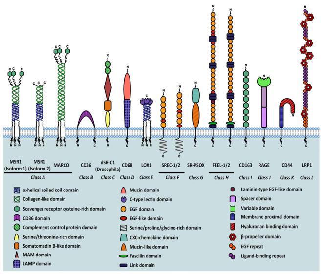 Fig. 1 Classification of scavenger receptors and MSR1 isoforms.1
Key structural properties of MSR1:
- Contains cysteine domain, collagen-like domain and helical coiled-coil domain
- The transmembrane region anchors to the macrophage membrane
- The trimeric structure forms the ligand binding site
- Positive charge amino acids mediate the binding with negatively charged ligands
Functions of MSR1
The scavenger receptor encoded by the MSR1 gene mainly functions to recognize and remove harmful substances in the body, and is involved in immune regulation and lipid metabolism.
| Function | Description |
| Clearing modified lipoproteins | Mediating the internalization of oxidized low-density lipoprotein by macrophages and promoting the accumulation of cholesterol within the cells. |
| Pathogen recognition and clearance | Recognizing pathogen-associated molecules such as lipopolysaccharides on bacterial surfaces and participating in innate immune defense. |
| Clearance of apoptotic cells | Binding to phosphatidylserine exposed on the surface of apoptotic cells to promote their phagocytosis and clearance. |
| Cell adhesion regulation | Mediating the interaction between macrophages and other cells, and participating in cell recruitment during inflammatory responses. |
| Signal transduction regulation | Activating intracellular signaling pathways upon ligand binding and regulating the expression of inflammatory factors . |
The broad-spectrum recognition ability of MSR1 for various ligands enables it to play a dual role in atherosclerosis and infectious diseases, and its expression level is strictly regulated by the inflammatory microenvironment.
Applications of MSR1 and MSR1 Antibody in Literature
1. Gudgeon, Jack, José Luis Marín-Rubio, and Matthias Trost. "The role of macrophage scavenger receptor 1 (MSR1) in inflammatory disorders and cancer." Frontiers in immunology 13 (2022): 1012002. https://doi.org/10.3389/fimmu.2022.1012002
The research has found that the macrophage scavenger receptor 1 (MSR1/CD204) plays a dual role in atherosclerosis, immunity and cancer. This article reviews its expression regulation mechanism, the impact on macrophage function, the progress in signal pathway research, and the therapeutic potential in tumor-associated macrophages.
2. Zhao, Shu-Jie, et al. "Macrophage MSR1 promotes BMSC osteogenic differentiation and M2-like polarization by activating PI3K/AKT/GSK3β/β-catenin pathway." Theranostics 10.1 (2020): 17. https://doi.org/10.7150/thno.36930
The study found that macrophages with MSR1 regulate PGC1α through the PI3K/AKT/GSK3β/β-catenin pathway, promoting M2 polarization and mitochondrial oxidative phosphorylation, thereby inducing osteogenic differentiation of BMSCs and accelerating fracture healing. MSR1 may be a new target for fracture repair.
3. Jia, Jinchao, et al. "Ferritin triggers neutrophil extracellular trap-mediated cytokine storm through Msr1 contributing to adult-onset Still’s disease pathogenesis." Nature Communications 13.1 (2022): 6804. https://doi.org/10.1038/s41467-022-34560-7
The study found that ferritin induces the formation of NETs by upregulating and binding to neutrophil Msr1, thereby driving the inflammation of hyperferritinemia syndrome. Removing neutrophils or Msr1 can alleviate the damage. This mechanism has been confirmed in adult Still's disease, suggesting that Msr1 is a potential therapeutic target.
4. Nance, Sierra A., et al. "MSR1 is not required for obesity-associated inflammation and insulin resistance in mice." Scientific Reports 13.1 (2023): 2651. https://doi.org/10.1038/s41598-023-29736-0
The study found that MSR1 is highly expressed in the adipose tissue of obese humans and is associated with diabetes, and can serve as a biomarker. However, in the mouse model, regardless of the gene dosage, the absence of Msr1 did not improve obesity, insulin resistance, and macrophage accumulation, suggesting that it is not a key pathogenic gene in mice.
5. Sheng, Wei, Guang Ji, and Li Zhang. "Role of macrophage scavenger receptor MSR1 in the progression of non-alcoholic steatohepatitis." Frontiers in Immunology 13 (2022): 1050984. https://doi.org/10.3389/fimmu.2022.1050984
The research has found that in non-alcoholic steatohepatitis (NASH), lipid metabolism disorders lead to the generation of modified lipoproteins through oxidative stress. Macrophage scavenger receptor 1 (MSR1) takes in these modified lipoproteins to form foam cells, thereby promoting the progression of NASH. This article reviews the regulation of MSR1 expression and its role in the pathogenesis of NASH.
Creative Biolabs: MSR1 Antibodies for Research
Creative Biolabs specializes in the production of high-quality MSR1 antibodies for research and industrial applications. Our portfolio includes monoclonal and polyclonal antibodies tailored for ELISA, Flow Cytometry, Western blot, immunohistochemistry, and other diagnostic methodologies.
- Custom MSR1 Antibody Development: Tailor-made solutions to meet specific research requirements.
- Bulk Production: Large-scale antibody manufacturing for industry partners.
- Technical Support: Expert consultation for protocol optimization and troubleshooting.
- Aliquoting Services: Conveniently sized aliquots for long-term storage and consistent experimental outcomes.
For more details on our MSR1 antibodies, custom preparations, or technical support, contact us at email.
Reference
- Gudgeon, Jack, José Luis Marín-Rubio, and Matthias Trost. "The role of macrophage scavenger receptor 1 (MSR1) in inflammatory disorders and cancer." Frontiers in immunology 13 (2022): 1012002. Distributed under Open Access license CC BY 4.0, without modification. https://doi.org/10.3389/fimmu.2022.1012002
Anti-MSR1 antibodies
 Loading...
 Compare




Hot products 
-
Mouse Anti-CD63 Recombinant Antibody (CBXC-1200) (CBMAB-C1467-CQ)

-
Mouse Anti-ACLY Recombinant Antibody (V2-179314) (CBMAB-A0610-YC)

-
Mouse Anti-FOXA3 Recombinant Antibody (2A9) (CBMAB-0377-YC)

-
Mouse Anti-CGAS Recombinant Antibody (CBFYM-0995) (CBMAB-M1146-FY)

-
Mouse Anti-CAT Recombinant Antibody (724810) (CBMAB-C8431-LY)

-
Mouse Anti-4-Hydroxynonenal Recombinant Antibody (V2-502280) (CBMAB-C1055-CN)

-
Mouse Anti-GIPC2 Recombinant Antibody (10) (CBMAB-G0476-LY)

-
Mouse Anti-APP Recombinant Antibody (DE2B4) (CBMAB-1122-CN)

-
Mouse Anti-AAV8 Recombinant Antibody (V2-634028) (CBMAB-AP022LY)

-
Mouse Anti-CFL1 (Phospho-Ser3) Recombinant Antibody (CBFYC-1770) (CBMAB-C1832-FY)

-
Mouse Anti-GNA12 Recombinant Antibody (CBFYH-0402) (CBMAB-H1272-FY)

-
Rat Anti-EMCN Recombinant Antibody (28) (CBMAB-E0280-FY)

-
Mouse Anti-CASP7 Recombinant Antibody (10-01-62) (CBMAB-C2005-LY)

-
Mouse Anti-ADGRE5 Recombinant Antibody (V2-360335) (CBMAB-C2088-CQ)

-
Mouse Anti-CSPG4 Recombinant Antibody (CBFYM-1050) (CBMAB-M1203-FY)

-
Rabbit Anti-ATF4 Recombinant Antibody (D4B8) (CBMAB-A3872-YC)

-
Mouse Anti-DMD Recombinant Antibody (D1190) (CBMAB-D1190-YC)

-
Mouse Anti-AAV-5 Recombinant Antibody (V2-503417) (CBMAB-V208-1369-FY)

-
Mouse Anti-CD46 Recombinant Antibody (CBFYC-0076) (CBMAB-C0085-FY)

-
Mouse Anti-CD59 Recombinant Antibody (CBXC-2097) (CBMAB-C4421-CQ)

- AActivation
- AGAgonist
- APApoptosis
- BBlocking
- BABioassay
- BIBioimaging
- CImmunohistochemistry-Frozen Sections
- CIChromatin Immunoprecipitation
- CTCytotoxicity
- CSCostimulation
- DDepletion
- DBDot Blot
- EELISA
- ECELISA(Cap)
- EDELISA(Det)
- ESELISpot
- EMElectron Microscopy
- FFlow Cytometry
- FNFunction Assay
- GSGel Supershift
- IInhibition
- IAEnzyme Immunoassay
- ICImmunocytochemistry
- IDImmunodiffusion
- IEImmunoelectrophoresis
- IFImmunofluorescence
- IGImmunochromatography
- IHImmunohistochemistry
- IMImmunomicroscopy
- IOImmunoassay
- IPImmunoprecipitation
- ISIntracellular Staining for Flow Cytometry
- LALuminex Assay
- LFLateral Flow Immunoassay
- MMicroarray
- MCMass Cytometry/CyTOF
- MDMeDIP
- MSElectrophoretic Mobility Shift Assay
- NNeutralization
- PImmunohistologyp-Paraffin Sections
- PAPeptide Array
- PEPeptide ELISA
- PLProximity Ligation Assay
- RRadioimmunoassay
- SStimulation
- SESandwich ELISA
- SHIn situ hybridization
- TCTissue Culture
- WBWestern Blot
