PTP1B Antibodies
Background
PTP1B is a non-receptor type tyrosine phosphatase that mainly exists on the surface of the endoplasmic reticulum. This enzyme catalyzes the dephosphorylation of specific protein tyrosine residues, negatively regulating the insulin and leptin signaling pathways, thereby influencing glucose homeostasis and energy metabolism. Due to its crucial role in insulin resistance and the pathogenesis of type 2 diabetes, PTP1B has rapidly become a key research focus in the field of metabolic disease treatment since it was cloned and identified in 1990. In 2003, the high-resolution crystal structure of its inhibitor complex was resolved, laying a structural biological foundation for the design of specific drugs. The regulatory mechanism of the protein's activity and its pivotal position in the cellular signaling network continue to provide crucial theoretical basis for the treatment strategies and drug development of metabolic diseases.
Structure of PTP1B
PTP1B is a crucial non-receptor type protein tyrosine phosphatase. Its molecular weight is highly conserved in mammals, approximately 50 kDa.
| Species | Human | Mouse | Rat |
| Molecular Weight (kDa) | 50.0 | 49.8 | 49.9 |
| Primary Structural Differences | Composed of 435 amino acids, the C-terminal hydrophobic sequence is anchored in the endoplasmic reticulum. | The sequence homology is extremely high, and the catalytic domain is completely conserved. | The structural and functional domains are highly similar to those of humans |
This protein is composed of approximately 435 amino acids. Its primary structure includes a catalytic domain at the N-terminus and a hydrophobic tail at the C-terminus. Its secondary and tertiary structures form a classic phosphatase fold, in which the catalytic active site contains a crucial "P-ring" (sequence motif: HCXXGXXRS/T), and the cysteine residue at position 215 (Cys215) is the core for performing nucleophilic attack and hydrolyzing the phosphotyrosine bond. An adjacent "WPD ring" (dominated by ASP181) undergoes conformational changes after substrate binding, transitioning from "open" to "closed" state, precisely positioning the substrate at the active site. Its quaternary structure usually functions as a monomer, but it can be regulated in terms of its activity and localization through interactions with other proteins. The precise three-dimensional structure of PTP1B (especially its deep and positively charged active pocket) has provided a solid structural basis for designing highly selective inhibitors for treating type 2 diabetes and obesity.
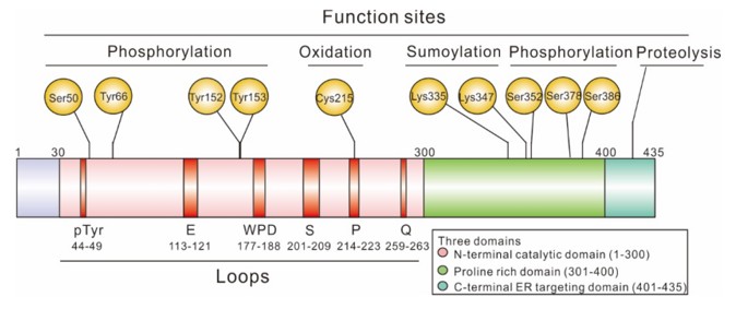 Fig. 1 Schematic representation of the domain structures of PTP1B full length.1
Key structural properties of PTP1B:
- Classical phosphatases fold domains to form deep and positively charged active site pockets
- Conserved "P-loop" structure with catalytic core (Cys215 key residue)
- "WPD ring" (including Asp181) closed conformation in the substrate when combined
- Carbon hydrophobic sequence formation endoplasmic reticulum anchor structure domain
Functions of PTP1B
The main function of PTP1B is to act as a key negative regulatory factor in the insulin and leptin signaling pathways, mediating metabolic regulation by catalyzing dephosphorylation reactions. Additionally, it is involved in various cellular processes, including cell growth, adhesion, and endoplasmic reticulum stress responses.
| Function | Description |
| Inhibition of Insulin Signaling | By dephosphorylating the tyrosine residues of insulin receptors (IR) and their substrates (IRS), it directly interrupts the downstream signaling pathway, which is one of the core mechanisms leading to insulin resistance. |
| Leptin Signal Inhibition | It acts on hypothalamic neurons, dephosphorylating the JAK2 downstream of the leptin receptor, blocking the appetite suppression and energy consumption signals of leptin, and promoting obesity development. |
| Cell Growth and Adhesion Regulation | By dephosphorylating key proteins such as focal adhesion kinase (FAK) and SRC family kinases, the proliferation, migration and adhesion of cells are regulated. |
| Endoplasmic Reticulum Stress Response | Under endoplasmic reticulum stress conditions, its expression and activity can be regulated, interacting with the Unfolded Protein Response (UPR), which affects cell survival and metabolic adaptation. |
| Cancer-related regulation | It plays a complex role in different types of cancers. It can exert tumor-suppressing effects by negatively regulating growth-promoting signaling pathways, and may also exhibit oncogenic activity by promoting cell migration. |
Unlike multi-subunit enzymes with synergistic effects, PTP1B acts as a monomeric enzyme, and its enzymatic reaction kinetics typically exhibit the classic Michaelis-Menten curve characteristics. This reflects its direct and highly efficient hydrolysis ability on individual phosphorylated tyrosine sites, enabling it to play a precise "molecular switch" role in complex signaling networks.
Applications of PTP1B and PTP1B Antibody in Literature
1. Liu, Rongxing, et al. "Human protein tyrosine phosphatase 1B (PTP1B): from structure to clinical inhibitor perspectives." International Journal of Molecular Sciences 23.13 (2022): 7027. https://doi.org/10.3390/ijms23137027
The article indicates that phosphorylation is a key process for regulating biological functions, which is jointly regulated by protein kinases and phosphatases. PTP1B, as a core protein tyrosine phosphatase, is associated with diseases such as diabetes and obesity. This article reviews three types of highly specific inhibitors for it: catalytic site targeting, allosteric site targeting, and mRNA targeting strategies, providing a new direction for the development of efficient small molecule drugs.
2. Kołodziej-Sobczak, Dominika, Łukasz Sobczak, and Krzysztof Z. Łączkowski. "Protein tyrosine phosphatase 1B (PTP1B): A comprehensive review of its role in pathogenesis of human diseases." International Journal of Molecular Sciences 25.13 (2024): 7033. https://doi.org/10.3390/ijms25137033
The article indicates that overexpression of PTP1B is associated with various diseases. Although the development of its inhibitors has been slow in clinical trials, this target has great potential. This article reviews the latest progress and aims to clarify the intrinsic connections of PTP1B in multiple diseases, advocating the adoption of a holistic treatment strategy to promote the development of new inhibitors.
3. Danquah, Bright D., et al. "Mass Spectrometric analysis of antibody—Epitope peptide complex dissociation: Theoretical concept and practical procedure of binding strength characterization." Molecules 25.20 (2020): 4776. https://doi.org/10.1097/MD.0000000000030826
This article utilizes mass spectrometry to characterize the binding strength and dissociation dynamics of myoglobin and its antibody-epitope complexes, providing a methodological framework for quantifying protein-ligand interactions and advancing epitope mapping in antibody research.
4. Zhang, Xiao, et al. "PTP1B Modulates Carotid Plaque Vulnerability in Atherosclerosis Through Rab5‐PDGFRβ‐Mediated Endocytosis Disruption and Apoptosis." CNS Neuroscience & Therapeutics 30.11 (2024): e70071. https://doi.org/10.1111/cns.70071
This study reveals that PTP1B regulates the Rab5-mediated PDGFRβ endocytosis, affecting the balance of the intracellular endocytic system in vascular smooth muscle cells, inducing cell apoptosis, and thereby increasing the instability of carotid artery plaques. Inhibiting PTP1B can alleviate atherosclerosis, suggesting it as a potential therapeutic target.
5. Oliver, Helk, et al. "Myeloid PTP1B deficiency protects against atherosclerosis by improving cholesterol homeostasis through an AMPK-dependent mechanism." Journal of translational medicine 21.1 (2023): 715. https://doi.org/10.1186/s12967-023-04598-2
Studies have confirmed that inhibiting PTP1B in myeloid cells can enhance the uptake of ApoB-containing lipoproteins by Kupffer cells, reduce the formation of foam cells, and promote reverse cholesterol transport, thereby exerting an anti-atherosclerotic effect. This provides a new basis for targeted PTP1B therapy.
Creative Biolabs: PTP1B Antibodies for Research
Creative Biolabs specializes in the production of high-quality PTP1B antibodies for research and industrial applications. Our portfolio includes monoclonal antibodies tailored for ELISA, Flow Cytometry, Western blot, immunohistochemistry, and other diagnostic methodologies.
- Custom PTP1B Antibody Development: Tailor-made solutions to meet specific research requirements.
- Bulk Production: Large-scale antibody manufacturing for industry partners.
- Technical Support: Expert consultation for protocol optimization and troubleshooting.
- Aliquoting Services: Conveniently sized aliquots for long-term storage and consistent experimental outcomes.
For more details on our PTP1B antibodies, custom preparations, or technical support, contact us at email.
Reference
- Liu, Rongxing, et al. "Human protein tyrosine phosphatase 1B (PTP1B): from structure to clinical inhibitor perspectives." International Journal of Molecular Sciences 23.13 (2022): 7027. Distributed under Open Access license CC BY 4.0, without modification. https://doi.org/10.3390/ijms23137027
Anti-PTP1B antibodies
 Loading...


Hot products 
-
Mouse Anti-AMIGO2 Recombinant Antibody (CBYY-C0756) (CBMAB-C2192-YY)

-
Mouse Anti-BIRC3 Recombinant Antibody (315304) (CBMAB-1214-CN)

-
Mouse Anti-CD83 Recombinant Antibody (HB15) (CBMAB-C1765-CQ)

-
Mouse Anti-CSPG4 Recombinant Antibody (CBFYM-1050) (CBMAB-M1203-FY)

-
Mouse Anti-BSN Recombinant Antibody (219E1) (CBMAB-1228-CN)

-
Mouse Anti-FLI1 Recombinant Antibody (CBXF-0733) (CBMAB-F0435-CQ)

-
Mouse Anti-DMD Recombinant Antibody (D1190) (CBMAB-D1190-YC)

-
Mouse Anti-APP Recombinant Antibody (5C2A1) (CBMAB-A3314-YC)

-
Mouse Anti-CASP7 Recombinant Antibody (10-01-62) (CBMAB-C2005-LY)

-
Mouse Anti-AFDN Recombinant Antibody (V2-58751) (CBMAB-L0408-YJ)

-
Mouse Anti-BIRC3 Recombinant Antibody (16E63) (CBMAB-C3367-LY)

-
Mouse Anti-CA9 Recombinant Antibody (CBXC-2079) (CBMAB-C0131-CQ)

-
Mouse Anti-ENO1 Recombinant Antibody (CBYC-A950) (CBMAB-A4388-YC)

-
Mouse Anti-BMI1 Recombinant Antibody (CBYC-P026) (CBMAB-P0108-YC)

-
Mouse Anti-AKT1 (Phosphorylated S473) Recombinant Antibody (V2-505430) (PTM-CBMAB-0067LY)

-
Mouse Anti-DES Monoclonal Antibody (440) (CBMAB-AP1857LY)

-
Mouse Anti-BHMT Recombinant Antibody (CBYY-0547) (CBMAB-0550-YY)

-
Mouse Anti-AGO2 Recombinant Antibody (V2-634169) (CBMAB-AP203LY)

-
Mouse Anti-APOE Recombinant Antibody (A1) (CBMAB-0078CQ)

-
Mouse Anti-dsRNA Recombinant Antibody (2) (CBMAB-D1807-YC)

- AActivation
- AGAgonist
- APApoptosis
- BBlocking
- BABioassay
- BIBioimaging
- CImmunohistochemistry-Frozen Sections
- CIChromatin Immunoprecipitation
- CTCytotoxicity
- CSCostimulation
- DDepletion
- DBDot Blot
- EELISA
- ECELISA(Cap)
- EDELISA(Det)
- ESELISpot
- EMElectron Microscopy
- FFlow Cytometry
- FNFunction Assay
- GSGel Supershift
- IInhibition
- IAEnzyme Immunoassay
- ICImmunocytochemistry
- IDImmunodiffusion
- IEImmunoelectrophoresis
- IFImmunofluorescence
- IGImmunochromatography
- IHImmunohistochemistry
- IMImmunomicroscopy
- IOImmunoassay
- IPImmunoprecipitation
- ISIntracellular Staining for Flow Cytometry
- LALuminex Assay
- LFLateral Flow Immunoassay
- MMicroarray
- MCMass Cytometry/CyTOF
- MDMeDIP
- MSElectrophoretic Mobility Shift Assay
- NNeutralization
- PImmunohistologyp-Paraffin Sections
- PAPeptide Array
- PEPeptide ELISA
- PLProximity Ligation Assay
- RRadioimmunoassay
- SStimulation
- SESandwich ELISA
- SHIn situ hybridization
- TCTissue Culture
- WBWestern Blot
