WNT3A Antibodies
Background
The WNT3A gene encodes a secreted glycoprotein and serves as the core ligand of the Wnt/β-catenin signaling pathway, activating downstream signaling by binding to the capretin receptor. This gene regulates cell proliferation, differentiation and polarity establishment during embryonic development, especially playing a key role in neural tube formation and limb development. Since its discovery in a mouse breast cancer model in 1982, WNT3A has become an important model molecule in developmental biology and cancer research. Its abnormal expression is closely related to the occurrence of various tumors and tissue aplastic disorders. In-depth research on this signaling pathway not only reveals the molecular mechanism of embryonic morphogenesis but also provides a new theoretical basis for targeted therapy.
Structure of WNT3A
WNT3A is a secretory glycoprotein with a molecular weight of approximately 39-44 kDa, and its precise molecular weight varies depending on the degree of glycosylation modification.
| Species | Human | Mouse | Rat | Chicken | Zebrafish |
| Molecular Weight (kDa) | 42.5 | 41.8 | 42.1 | 40.9 | 39.7 |
| Primary Structural Differences | Conservative structure domain containing 24 cysteine | N-terminal signal peptide sequence differences | Variation of the glycosylation site | The receptor binding region is partially conserved | The core domains have a relatively high similarity |
This protein is composed of 343 amino acids, and its three-dimensional structure contains highly conserved palmitoylation sites, a modification that is crucial for its secretion and function. Protein folding forms a typical "cysteine junction" framework, with multiple α -helices constituting the receptor binding domain. The lipid modification sites in the active center are directly involved in the recognition and binding of Frizzled receptors, while the C-terminal domain is responsible for the specificity of signal transduction. This unique structural feature enables WNT3A to effectively activate the Wnt/β-catenin signaling pathway and play a key role in embryonic development.
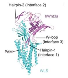 Fig. 1 Overall structure of WLS-Wnt3a complex.1
Key structural properties of WNT3A:
- Highly conserved cysteine nodular skeleton structure
- N-terminal palmitoylation modification mediated membrane-bound domains
- Receptor-binding pockets formed in the hydrophobic core region
Functions of WNT3A
The core function of the WNT3A protein is to activate the Wnt signaling pathway and regulate cell fate determination. In addition, it is also involved in various physiological and pathological processes, including the maintenance of tissue homeostasis and tumorigenesis.
| Function | Description |
| Activation of signal pathways | As a key ligand of the Wnt/β-catenin pathway, it initiates downstream signal transduction after binding to the Frizzled receptor. |
| Regulation of cell fate | Guide cells to a particular lineage differentiation in embryonic development, especially in the neural tube and body in the process of the formation. |
| Maintenance of tissue homeostasis | In the adult tissues through regulate stem cell self-renewal and differentiation in tissue repair. |
| Tumorigenesis promotion | Abnormal activation leads to dysregulation of signaling pathways and is closely related to the occurrence and development of various tumors, including colorectal cancer. |
| Establishment of cell polarity | Through the classic pathways involved in regulating cell polarity and motility. |
Unlike the widely expressed WNT1, the expression of WNT3A has stronger spatiotemporal specificity. Its signal output strictly depends on the local receptor expression level and the concentration of inhibitory factors in the microenvironment. This characteristic enables it to achieve precise morphogenetic regulation in the developmental program.
Applications of WNT3A and WNT3A Antibody in Literature
1. Zhong, Qing, et al. "Cryo-EM structure of human Wntless in complex with Wnt3a." Nature communications 12.1 (2021): 4541. https://doi.org/10.1038/s41467-021-24731-3
This study reveals the cryo-electron microscopy structure of the human WNT3A-Wntless complex, providing insights into the molecular mechanism of WNT3A secretion and its interaction with Wntless, which could inform therapeutic strategies targeting WNT signaling.
2. Willert, Kim, et al. "Wnt proteins are lipid-modified, soluble factors that can initiate pathway transduction." Development 130.20 (2003): 4877-4886. https://doi.org/10.1242/dev.00686
This paper demonstrates that Wnt proteins, including WNT3A, are lipid-modified and secreted as soluble factors, and their palmitoylation is essential for their function in initiating pathway transduction in developmental processes.
3. Liu, Jia, et al. "Wnt3a is required for endothelial differentiation from embryonic stem cells." Stem cells 27.7 (2009): 1569-1578. https://doi.org/10.1002/stem.95
Research shows that WNT3A is essential for endothelial differentiation from embryonic stem cells, highlighting its role in vascular development and potential applications in regenerative medicine.
4. Bafico, Andrea, et al. "A mechanism by which the Wnt signaling pathway regulates GSK3 activity." Journal of Biological Chemistry 279.22 (2004): 23890-23896. https://doi.org/10.1074/jbc.M313266200
This study elucidates how the WNT3A signaling pathway regulates GSK3 activity, providing mechanistic insights into the canonical Wnt/β-catenin pathway and its role in cellular processes.
5. Sato, Atsushi, et al. "Wnt3a-induced growth cone collapse requires the Dvl1-CK1ε-mediated microtubule-remodeling pathway." Development 142.14 (2015): 2464-2474. https://doi.org/10.1242/dev.121550
This research demonstrates that WNT3A induces growth cone collapse through a Dvl1-CK1ε-mediated microtubule-remodeling pathway, revealing its role in neural development and axon guidance.
Creative Biolabs: WNT3A Antibodies for Research
Creative Biolabs specializes in the production of high-quality WNT3A antibodies for research and industrial applications. Our portfolio includes monoclonal antibodies tailored for ELISA, Flow Cytometry, Western blot, immunohistochemistry, and other diagnostic methodologies.
- Custom WNT3A Antibody Development: Tailor-made solutions to meet specific research requirements.
- Bulk Production: Large-scale antibody manufacturing for industry partners.
- Technical Support: Expert consultation for protocol optimization and troubleshooting.
- Aliquoting Services: Conveniently sized aliquots for long-term storage and consistent experimental outcomes.
For more details on our WNT3A antibodies, custom preparations, or technical support, contact us at email.
Reference
- Zhong, Qing, et al. "Cryo-EM structure of human Wntless in complex with Wnt3a." Nature communications 12.1 (2021): 4541. https://doi.org/10.1038/s41467-021-24731-3
Anti-WNT3A antibodies
 Loading...


Hot products 
-
Mouse Anti-4-Hydroxynonenal Recombinant Antibody (V2-502280) (CBMAB-C1055-CN)

-
Mouse Anti-ELAVL4 Recombinant Antibody (6B9) (CBMAB-1132-YC)

-
Mouse Anti-ALDOA Recombinant Antibody (A2) (CBMAB-A2316-YC)

-
Human Anti-SARS-CoV-2 S1 Monoclonal Antibody (CBFYR-0120) (CBMAB-R0120-FY)

-
Rabbit Anti-B2M Recombinant Antibody (CBYY-0059) (CBMAB-0059-YY)

-
Mouse Anti-GFP Recombinant Antibody (28) (CBMAB-G3038-LY)

-
Mouse Anti-ARID1B Recombinant Antibody (KMN1) (CBMAB-A3546-YC)

-
Mouse Anti-ASB9 Recombinant Antibody (1D8) (CBMAB-A0529-LY)

-
Mouse Anti-GDF5 Recombinant Antibody (1F4) (CBMAB-G2740-LY)

-
Mouse Anti-EMP3 Recombinant Antibody (CBFYE-0100) (CBMAB-E0207-FY)

-
Mouse Anti-BCL6 Recombinant Antibody (CBYY-0442) (CBMAB-0445-YY)

-
Mouse Anti-FAS2 Monoclonal Antibody (1D4) (CBMAB-0071-CN)

-
Mouse Anti-COL12A1 Recombinant Antibody (CBYY-C3117) (CBMAB-C4560-YY)

-
Rabbit Anti-ALOX5AP Recombinant Antibody (CBXF-1219) (CBMAB-F0750-CQ)

-
Mouse Anti-EGR1 Recombinant Antibody (CBWJZ-100) (CBMAB-Z0289-WJ)

-
Mouse Anti-BLNK Recombinant Antibody (CBYY-0623) (CBMAB-0626-YY)

-
Mouse Anti-DHFR Recombinant Antibody (D0821) (CBMAB-D0821-YC)

-
Mouse Anti-ACTN4 Recombinant Antibody (V2-6075) (CBMAB-0020CQ)

-
Mouse Anti-C5AR1 Recombinant Antibody (R63) (CBMAB-C9553-LY)

-
Human Anti-SARS-CoV-2 Spike Recombinant Antibody (CR3022) (CBMAB-CR014LY)

- AActivation
- AGAgonist
- APApoptosis
- BBlocking
- BABioassay
- BIBioimaging
- CImmunohistochemistry-Frozen Sections
- CIChromatin Immunoprecipitation
- CTCytotoxicity
- CSCostimulation
- DDepletion
- DBDot Blot
- EELISA
- ECELISA(Cap)
- EDELISA(Det)
- ESELISpot
- EMElectron Microscopy
- FFlow Cytometry
- FNFunction Assay
- GSGel Supershift
- IInhibition
- IAEnzyme Immunoassay
- ICImmunocytochemistry
- IDImmunodiffusion
- IEImmunoelectrophoresis
- IFImmunofluorescence
- IGImmunochromatography
- IHImmunohistochemistry
- IMImmunomicroscopy
- IOImmunoassay
- IPImmunoprecipitation
- ISIntracellular Staining for Flow Cytometry
- LALuminex Assay
- LFLateral Flow Immunoassay
- MMicroarray
- MCMass Cytometry/CyTOF
- MDMeDIP
- MSElectrophoretic Mobility Shift Assay
- NNeutralization
- PImmunohistologyp-Paraffin Sections
- PAPeptide Array
- PEPeptide ELISA
- PLProximity Ligation Assay
- RRadioimmunoassay
- SStimulation
- SESandwich ELISA
- SHIn situ hybridization
- TCTissue Culture
- WBWestern Blot
