ATP7A Antibodies
Background
ATP7A is a copper-translocating ATPase mainly located in the Golgi network. It is responsible for transporting copper ions to copper-dependent enzymes in the secretion pathway and maintaining the copper homeostasis throughout the body. This gene mutation is the main cause of Menkes disease. Patients with this disease experience progressive neurological degeneration and abnormal connective tissues due to copper absorption disorders. The functional study of this gene began in the 1990s. Scientists first isolated this gene through the localization cloning technique. Subsequent studies gradually revealed its multiple roles in intracellular copper transport, polar cell directional transport, and immune defense, laying the foundation for understanding the molecular mechanisms of copper metabolism-related diseases.
Structure of ATP7A
ATP7A is a transmembrane copper transporter with a molecular weight of approximately 178 kDa. Its size varies slightly among different species, mainly due to minor changes in amino acid sequences and domain compositions.
| Species | Humans | Mice | Rats | Zebrafish | Fruit flies |
| Molecular Weight (kDa) | 178 | 179 | 179 | 176 | 175 |
| Primary Structural Differences | Contains 8 copper-binding sites | Highly homologous to humans | Copper-binding domains slightly different | Structure is simpler | Only 6 copper-binding sites |
This protein is composed of approximately 1500 amino acids and includes 8 N-terminal copper-binding domains, 8 transmembrane segments, and an ATP-binding domain and other core functional regions. ATP7A uses the energy generated from ATP hydrolysis to transport copper ions from the cytoplasm to the Golgi apparatus, providing copper for copper-dependent enzymes. When the intracellular copper concentration increases, this protein relocates from the Golgi apparatus to the cell membrane or vesicles, participating in copper excretion. Its molecular structure contains multiple highly conserved motifs, such as phosphorylation domains and nucleotide-binding domains, which together ensure the directional transport of copper ions and the regulation of copper homeostasis in the cell.
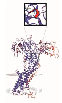 Fig. 1 Three-dimensional structure and key positions of ATP7A protein.1
Key structural characteristics of ATP7A protein:
- Contains 8 N-terminal copper-binding domains, each with a conserved GMXCXXC copper-binding motif
- Has 8 transmembrane segments, forming a copper ion transmembrane transport channel
- Contains a highly conserved P-type ATPase phosphorylation domain (DKTGT sequence)
- The C-terminal nucleotide-binding domain (N-domain) provides the core for ATP hydrolysis catalysis
- The transmembrane region CPC motif is involved in the selective binding and release of copper ions
- The conformational change of the phosphorylated intermediate E1-E2 drives the directional transport of copper
Functions of ATP7A
The core function of ATP7A is to maintain intracellular copper homeostasis by transporting copper ions to the secretion pathway or out of the cell, thereby regulating the activity of various copper-dependent enzymes.
| Function | Description |
| Copper ion transmembrane transport | Using ATP hydrolysis energy, it pumps copper ions from the cytoplasm into the Golgi apparatus to supply copper-dependent enzymes for synthesis. |
| Copper excretion and detoxification | When the intracellular copper concentration is too high, it repositions to the plasma membrane and excretes excess copper ions out of the cell to prevent copper toxicity. |
| Copper-dependent enzyme activation | Provides copper cofactors for tyrosinase, lysyl oxidase, etc., participating in melanin synthesis, connective tissue cross-linking, etc. |
| Polarity cell directional transport | In neurons and other cells, it is responsible for transporting copper to the distal axons and dendrites, maintaining the normal function of the nervous system. |
| Whole-body copper homeostasis regulation | It mediates copper absorption in intestinal epithelial cells and participates in the redistribution and excretion of copper in other tissues. |
ATP7A has high specificity in copper transport. Its transport activity is precisely regulated by copper concentration and kinase phosphorylation modification to ensure that the intracellular copper level is maintained at the optimal physiological range required.
Applications of ATP7A and ATP7A Antibody in Literature
1. Li, Hao, et al. "Genetic diversity, tissue-specific expression, and functional analysis of the ATP7A gene in sheep." Frontiers in Genetics 14 (2023): 1239979. https://doi.org/10.3389/fgene.2023.1239979
The study revealed that the ATP7A gene is highly expressed in the horns of sheep, and the expression level in large horn individuals is significantly higher than that in small horn individuals. Through genome-wide analysis, multiple SNPs and potential functional sites with significant differences in frequency between the horned and hornless populations were screened out. Among them, three mutations exhibit allele-specific expression.
2. Horn, Nina, and Pernilla Wittung-Stafshede. "ATP7A-regulated enzyme metalation and trafficking in the menkes disease puzzle." Biomedicines 9.4 (2021): 391. https://doi.org/10.3390/biomedicines9040391
The research reveals that copper ions are crucial for maintaining cellular functions, and the transporter protein ATP7A is the core for regulating the copper homeostasis throughout the body. Defects in this gene can lead to Menkes disease. This article reviews how the mechanism of ATP7A-mediated copper enzyme activation explains the diverse symptoms of this disease, and emphasizes that clarifying its mechanism is helpful for early diagnosis.
3. Gao, Wei, et al. "Elesclomol induces copper‐dependent ferroptosis in colorectal cancer cells via degradation of ATP7A." Molecular Oncology 15.12 (2021): 3527-3544. https://doi.org/10.1002/1878-0261.13079
Studies have shown that the copper transporter ATP7A is a key target of the anti-cancer drug Elsclomol. This drug can induce the degradation of ATP7A, and when combined with copper, it leads to the accumulation of copper in mitochondria, triggering oxidative stress and ferroptosis, thereby inhibiting colorectal cancer.
4. Zhou, Yixuan, and Leiliang Zhang. "The interplay between copper metabolism and microbes: in perspective of host copper-dependent ATPases ATP7A/B." Frontiers in cellular and infection microbiology 13 (2023): 1267931. https://doi.org/10.3389/fcimb.2023.1267931
Studies have shown that the copper transporter ATP7A/B maintains the copper homeostasis of the organism through intracellular transport. Certain microorganisms (such as Salmonella and influenza viruses) can interfere with its regulatory mechanism to facilitate their own survival. This article reviews the transport characteristics of ATP7A/B and discusses the interaction between microorganisms and host copper metabolism.
5. Writzl, Karin, et al. "Novel ATP7A Splice-Site Variant Causing Distal Motor Neuropathy and Occipital Horn Syndrome: Two Siblings and Literature Review." Genes 16.9 (2025): 1077. https://doi.org/10.3390/genes16091077
The study identified a novel splicing variant of the ATP7A gene that can cause a rare neurological disorder combined with an overlapping phenotype of occipital bone angle syndrome. The patients initially presented with chronic diarrhea and later developed distal motor neuropathy. This finding expands the mutation spectrum of ATP7A-related diseases.
Creative Biolabs: ATP7A Antibodies for Research
Creative Biolabs specializes in the production of high-quality ATP7A antibodies for research and industrial applications. Our portfolio includes monoclonal and polyclonal antibodies tailored for ELISA, Flow Cytometry, Western blot, immunohistochemistry, and other diagnostic methodologies.
- Custom ATP7A Antibody Development: Tailor-made solutions to meet specific research requirements.
- Bulk Production: Large-scale antibody manufacturing for industry partners.
- Technical Support: Expert consultation for protocol optimization and troubleshooting.
- Aliquoting Services: Conveniently sized aliquots for long-term storage and consistent experimental outcomes.
For more details on our ATP7A antibodies, custom preparations, or technical support, contact us at email.
Reference
- Li, Hao, et al. "Genetic diversity, tissue-specific expression, and functional analysis of the ATP7A gene in sheep." Frontiers in Genetics 14 (2023): 1239979. Distributed under Open Access license CC BY 4.0, and cropped from the original figure. https://doi.org/10.3389/fgene.2023.1239979
Anti-ATP7A antibodies
 Loading...


Hot products 
-
Rat Anti-FABP3 Recombinant Antibody (CBXF-2299) (CBMAB-F1612-CQ)

-
Mouse Anti-ADIPOR1 Recombinant Antibody (V2-179982) (CBMAB-A1368-YC)

-
Mouse Anti-8-oxoguanine Recombinant Antibody (V2-7719) (CBMAB-1898CQ)

-
Mouse Anti-ABCA3 Recombinant Antibody (V2-178911) (CBMAB-A0145-YC)

-
Mouse Anti-ACTG1 Recombinant Antibody (V2-179597) (CBMAB-A0916-YC)

-
Mouse Anti-AKR1B1 Antibody (V2-2449) (CBMAB-1001CQ)

-
Mouse Anti-GFAP Recombinant Antibody (20) (CBMAB-G2914-LY)

-
Mouse Anti-dsRNA Recombinant Antibody (2) (CBMAB-D1807-YC)

-
Mouse Anti-AAV8 Recombinant Antibody (V2-634028) (CBMAB-AP022LY)

-
Rabbit Anti-AKT2 (Phosphorylated S474) Recombinant Antibody (V2-556130) (PTM-CBMAB-0605LY)

-
Rat Anti-C5AR1 Recombinant Antibody (8D6) (CBMAB-C9139-LY)

-
Mouse Anti-BRD3 Recombinant Antibody (CBYY-0801) (CBMAB-0804-YY)

-
Mouse Anti-ELAVL4 Recombinant Antibody (6B9) (CBMAB-1132-YC)

-
Mouse Anti-GLP1R Recombinant Antibody (4F3) (CBMAB-G0521-LY)

-
Mouse Anti-ALDOA Recombinant Antibody (A2) (CBMAB-A2316-YC)

-
Mouse Anti-EMP3 Recombinant Antibody (CBFYE-0100) (CBMAB-E0207-FY)

-
Mouse Anti-CAT Recombinant Antibody (724810) (CBMAB-C8431-LY)

-
Mouse Anti-ACE2 Recombinant Antibody (V2-179293) (CBMAB-A0566-YC)

-
Mouse Anti-AKT1/AKT2/AKT3 (Phosphorylated T308, T309, T305) Recombinant Antibody (V2-443454) (PTM-CBMAB-0030YC)

-
Mouse Anti-CD24 Recombinant Antibody (HIS50) (CBMAB-C10123-LY)

- AActivation
- AGAgonist
- APApoptosis
- BBlocking
- BABioassay
- BIBioimaging
- CImmunohistochemistry-Frozen Sections
- CIChromatin Immunoprecipitation
- CTCytotoxicity
- CSCostimulation
- DDepletion
- DBDot Blot
- EELISA
- ECELISA(Cap)
- EDELISA(Det)
- ESELISpot
- EMElectron Microscopy
- FFlow Cytometry
- FNFunction Assay
- GSGel Supershift
- IInhibition
- IAEnzyme Immunoassay
- ICImmunocytochemistry
- IDImmunodiffusion
- IEImmunoelectrophoresis
- IFImmunofluorescence
- IGImmunochromatography
- IHImmunohistochemistry
- IMImmunomicroscopy
- IOImmunoassay
- IPImmunoprecipitation
- ISIntracellular Staining for Flow Cytometry
- LALuminex Assay
- LFLateral Flow Immunoassay
- MMicroarray
- MCMass Cytometry/CyTOF
- MDMeDIP
- MSElectrophoretic Mobility Shift Assay
- NNeutralization
- PImmunohistologyp-Paraffin Sections
- PAPeptide Array
- PEPeptide ELISA
- PLProximity Ligation Assay
- RRadioimmunoassay
- SStimulation
- SESandwich ELISA
- SHIn situ hybridization
- TCTissue Culture
- WBWestern Blot
