
BCOR Antibodies
Background
BCOR gene encoding a core transcriptional total inhibitory factor, mainly as a comb more inhibition compounds 1 (PRC1.1) scaffold proteins play a role. This protein participates in key biological processes such as embryonic development, cell differentiation and tumor suppression by regulating histone modification and chromatin remodeling. In diseases such as acute myeloid leukemia and retinoblastoma, mutations in the BCOR gene can disrupt its normal function, leading to abnormal regulation of gene expression. The structural analysis of its complex reveals the specific interaction mechanism with DNA-binding proteins, providing a structural biology basis for the development of targeted therapeutic strategies and having significant scientific value for understanding the epigenetic regulatory network.
Structure of BCOR
BCOR is a relatively large nucleoprotein with a molecular weight of approximately 175 kDa. Its molecular weight is highly conserved across different species, mainly due to the well-preserved key functional domains in chromatin regulation. This protein is composed of 1,674 amino acids and adopts a multi-domain organizational form, including the BCOR motif at the N-terminal, the ANK repeat sequence in the middle, and the PUFD domain at the C-terminal. BCOR interacts with proteins such as PCGF1 through ANK repeat sequences to form the core framework of the PRC1.1 complex, and its PUFD domain directly participates in the deubiquitination regulation of histone H2A. These structural features enable BCOR to act as a scaffold protein to coordinate the assembly and function of epigenetic modification complexes, which is crucial for maintaining gene silencing and cell differentiation.
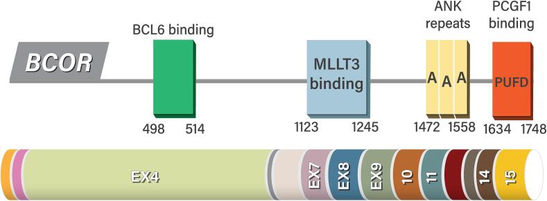 Fig. 1 Structure and functional domains of BCOR.1
Key structural properties of BCOR:
- Multi-domain organizational form, including BCOR motif, ANK repeat sequence and PUFD functional domain
- ANK repeat regions mediate specific protein interactions
- PUFD domains specifically recognize histones and participate in epigenetic regulation
Functions of BCOR
The core function of the BCOR gene is to regulate epigenetic modifications as a transcriptional co-suppressor. In addition, it is also involved in a variety of key cellular processes, including cell differentiation, developmental regulation and tumor suppression.
| Function | Description |
| Transcriptional inhibition | BCOR, as a scaffold protein of the PRC1.1 complex, inhibits the transcription of target genes by promoting the deubiquitination of histone H2A. |
| Regulation of cell differentiation | Regulating the pluripotency and directed differentiation of stem cells, especially playing a key role in hematopoietic system and bone development. |
| Embryonic development | Participate in the regulation of gene expression and the formation of tissues and organs during the early embryonic development process. |
| Tumor suppression | As a tumor suppressor gene, its dysfunctional inactivation mutations are associated with the occurrence of cancers such as acute myeloid leukemia and retinoblastoma. |
| Chromatin remodeling | By recruiting histone modification enzymes, chromatin structure and accessibility are regulated to influence gene expression patterns. |
BCOR specifically recognizes histone modifications through its C-terminal PUFD domain and forms complexes with multiple epigenetic regulatory factors to precisely regulate gene expression in a synergistic manner. This mechanism is significantly different from the traditional sequence-specific transcription factor action mode.
Applications of BCOR and BCOR Antibody in Literature
1. Fayoumi, Abdulkareem. "BCOR abnormalities in endometrial stromal sarcoma." Gynecologic Oncology Reports (2025): 101672. https://doi.org/10.1016/j.gore.2024.101672
The article indicates that BCOR gene abnormalities (such as fusion or mutation) are key features of high-grade endometrial stromal sarcoma, affecting diagnosis and prognosis. Molecular testing can assist in identification. Surgery combined with radiotherapy and chemotherapy is the main therapy, and treatment strategies targeting BCOR and related pathways are under exploration.
2. Tauziède-Espariat, Arnault, et al. "CNS tumor with EP300:: BCOR fusion: discussing its prevalence in adult population." Acta neuropathologica communications 11.1 (2023): 26. https://doi.org/10.1186/s40478-023-01523-y
The article indicates that EP300::BCOR fusion in CNS tumors is a newly discovered type, more common in adults. Its clinical, imaging and pathological features are different from the common BCOR internal duplication (ITD) in children, and it may become a new subtype in the future WHO classification.
3. Ebrahimi, Azadeh, et al. "BCOR: CREBBP fusion in malignant neuroepithelial tumor of CNS expands the spectrum of methylation class CNS tumor with BCOR/BCOR (L1)-fusion." Acta Neuropathologica Communications 12.1 (2024): 60. https://doi.org/10.1186/s40478-024-01780-5
The article indicates that BCOR/BCORL1 fusion type CNS tumors are a new subtype defined recently through methylation analysis, and their diagnosis still needs to combine molecular and histological features. This article reports for the first time a case of adult BCOR::CREBBP fusion and, based on the literature, presents the typical characteristics of this type of tumor, emphasizing the importance of complementary molecular testing for diagnosis.
4. Kelly, Madison J., et al. "Bcor loss perturbs myeloid differentiation and promotes leukaemogenesis." Nature communications 10.1 (2019): 1347. https://doi.org/10.1038/s41467-019-09250-6
The article indicates that BCOR gene mutations are associated with the occurrence of acute myeloid leukemia. Mouse model studies have shown that the absence of BCOR can lead to abnormal differentiation of hematopoietic stem cells, expansion of myeloid progenitor cells, and synergistic induction of aggressive leukemia with carcinogenic KrasG12D. The mechanism is related to the dysregulation of PRC1 target genes (such as Hoxa7/9).
5. Rompietti, Chiara, et al. "Bcor loss promotes Richter transformation of chronic lymphocytic leukemia associated with Notch1 activation in mice." Leukemia (2025): 1-12. https://doi.org/10.1038/s41375-025-02557-y
The article indicates that BCOR deletion can work in synergy with NOTCH1 activation to drive the transformation of chronic lymphocytic leukemia (CLL) into aggressive Richter (RT). In mouse models, Bcor absence accelerates lymphoid proliferation and alters the tumor microenvironment, while NOTCH1 inhibition reduces tumor infiltration and prolongs survival, suggesting targeting potential.
Creative Biolabs: BCOR Antibodies for Research
Creative Biolabs specializes in the production of high-quality BCOR antibodies for research and industrial applications. Our portfolio includes monoclonal antibodies tailored for ELISA, Flow Cytometry, Western blot, immunohistochemistry, and other diagnostic methodologies.
- Custom BCOR Antibody Development: Tailor-made solutions to meet specific research requirements.
- Bulk Production: Large-scale antibody manufacturing for industry partners.
- Technical Support: Expert consultation for protocol optimization and troubleshooting.
- Aliquoting Services: Conveniently sized aliquots for long-term storage and consistent experimental outcomes.
For more details on our BCOR antibodies, custom preparations, or technical support, contact us at email.
Reference
- Fayoumi, Abdulkareem. "BCOR abnormalities in endometrial stromal sarcoma." Gynecologic Oncology Reports (2025): 101672. https://doi.org/10.1016/j.gore.2024.101672
Anti-BCOR antibodies
 Loading...
 Compare




Hot products 
-
Mouse Anti-CD59 Recombinant Antibody (CBXC-2097) (CBMAB-C4421-CQ)

-
Rabbit Anti-BAD (Phospho-Ser136) Recombinant Antibody (CAP219) (CBMAB-AP536LY)

-
Mouse Anti-BIRC3 Recombinant Antibody (315304) (CBMAB-1214-CN)

-
Mouse Anti-CALR Recombinant Antibody (CBFYC-0763) (CBMAB-C0818-FY)

-
Mouse Anti-CD1C Recombinant Antibody (L161) (CBMAB-C2173-CQ)

-
Mouse Anti-CD2AP Recombinant Antibody (BR083) (CBMAB-BR083LY)

-
Mouse Anti-14-3-3 Pan Recombinant Antibody (V2-9272) (CBMAB-1181-LY)

-
Mouse Anti-ARSA Recombinant Antibody (CBYC-A799) (CBMAB-A3679-YC)

-
Mouse Anti-BZLF1 Recombinant Antibody (BZ.1) (CBMAB-AP705LY)

-
Mouse Anti-GFAP Recombinant Antibody (24) (CBMAB-G2927-LY)

-
Mouse Anti-DES Monoclonal Antibody (440) (CBMAB-AP1857LY)

-
Mouse Anti-AHCYL1 Recombinant Antibody (V2-180270) (CBMAB-A1703-YC)

-
Mouse Anti-Acetyl SMC3 (K105/K106) Recombinant Antibody (V2-634053) (CBMAB-AP052LY)

-
Rabbit Anti-B2M Recombinant Antibody (CBYY-0059) (CBMAB-0059-YY)

-
Mouse Anti-8-oxoguanine Recombinant Antibody (V2-7719) (CBMAB-1898CQ)

-
Mouse Anti-DMD Recombinant Antibody (D1190) (CBMAB-D1190-YC)

-
Mouse Anti-BCL2L1 Recombinant Antibody (H5) (CBMAB-1025CQ)

-
Mouse Anti-AKR1C3 Recombinant Antibody (V2-12560) (CBMAB-1050-CN)

-
Human Anti-SARS-CoV-2 Spike Recombinant Antibody (CBC05) (CBMAB-CR005LY)

-
Mouse Anti-ALOX5 Recombinant Antibody (33) (CBMAB-1890CQ)

- AActivation
- AGAgonist
- APApoptosis
- BBlocking
- BABioassay
- BIBioimaging
- CImmunohistochemistry-Frozen Sections
- CIChromatin Immunoprecipitation
- CTCytotoxicity
- CSCostimulation
- DDepletion
- DBDot Blot
- EELISA
- ECELISA(Cap)
- EDELISA(Det)
- ESELISpot
- EMElectron Microscopy
- FFlow Cytometry
- FNFunction Assay
- GSGel Supershift
- IInhibition
- IAEnzyme Immunoassay
- ICImmunocytochemistry
- IDImmunodiffusion
- IEImmunoelectrophoresis
- IFImmunofluorescence
- IGImmunochromatography
- IHImmunohistochemistry
- IMImmunomicroscopy
- IOImmunoassay
- IPImmunoprecipitation
- ISIntracellular Staining for Flow Cytometry
- LALuminex Assay
- LFLateral Flow Immunoassay
- MMicroarray
- MCMass Cytometry/CyTOF
- MDMeDIP
- MSElectrophoretic Mobility Shift Assay
- NNeutralization
- PImmunohistologyp-Paraffin Sections
- PAPeptide Array
- PEPeptide ELISA
- PLProximity Ligation Assay
- RRadioimmunoassay
- SStimulation
- SESandwich ELISA
- SHIn situ hybridization
- TCTissue Culture
- WBWestern Blot
