GPC3 Antibodies
Background
GPC3 is a glycoprotein anchored to the cell membrane and is mainly present in embryonic tissues and various malignant tumors, such as hepatocellular carcinoma. This protein participates in the tumorigenesis and development process by regulating the signaling pathways related to cell proliferation and apoptosis, and its interaction with pathways such as Wnt and Hippo is particularly crucial. As it is hardly expressed in normal adult tissues, GPC3 has become an important biomarker for the diagnosis of hepatocellular carcinoma. In recent years, significant progress has been made in the development of immunotherapy and antibody-drug conjugations based on its targeting characteristics. Since its first report by the Keichi Kadomatsu research team in 1996, the structural analysis of GPC3 has revealed its characteristic of interacting with the cell membrane through the anchoring domain of glycoside phosphatidylinositol. This special structural feature provides a molecular basis for the development of targeted therapeutic strategies against GPC3 and promotes the development of the field of precision oncology.
Structure of GPC3
GPC3 is a cell membrane protein with a relatively large molecular weight, and the molecular weight of its core protein is approximately 70 kDa. Due to the extensive glycosylation modification of this protein after translation, the actual molecular weight of its glycosylation form shows significant heterogeneity within the range of 100-150 kDa. This difference is mainly attributed to the different glycosylation patterns among different cell types or species.
| Species | Human | Mouse | Rat |
| Molecular weight (core protein) | ~70 kDa | ~69 kDa | ~69 kDa |
| Molecular weight after glycosylation | 100-150 kDa | 90-140 kDa | 95-145 kDa |
| Main structural differences | With a conservative sugar-based phosphatidyl inositol anchor domain | With the human GPC3 high homology, core protein sequence similarity | The sugar chain structure may have subtle differences from that of other mammals |
The GPC3 protein is composed of approximately 580 amino acids. Its structural feature is that there is a unique heparan sulfate chain attachment region at the N-terminal, and the C-terminal is anchored to the cell membrane by glycosylphosphatidylinositol. The tertiary structure of this protein forms a specific proteoglycan conformation, enabling it to act as a co-receptor and directly interact with cell signaling molecules (such as Wnt and Hedgehog) through its core protein. At the same time, it regulates the concentration and activity of growth factors through its sugar chain, thereby playing a core role in the regulation of cell proliferation.
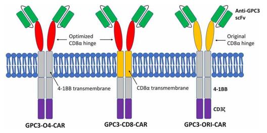 Fig. 1 Diagrams of different GPC3-CAR structures.1
Key structural properties of GPC3:
- Located in the approximately 1MB genomic region of chromosome Xq26.1
- Contains eight highly conserved exon structures
- Protein core sequence encoding 580 amino acids
- As the typical sugar-based phosphatidyl inositol anchor signal sequence
Functions of GPC3
The main function of GPC3 is to act as a co-receptor to regulate the cell growth signaling pathway. However, it is also widely involved in various pathophysiological processes such as embryonic development, tissue regeneration and tumorigenesis.
| Function | Description |
| Regulation of embryonic development | High expression in embryonic liver, lung, kidney development, morphogenesis, by adjusting the gradient guidance organ formation. |
| Cell proliferation promotion | As a co-receptor of growth signaling pathways such as Wnt and Hedgehog, it enhances downstream signal transduction and drives cell cycle progression. |
| Tumorigenesis driver | In hepatocellular carcinoma (HCC) abnormally high expression in malignant tumors, such as by inhibiting tumor cell apoptosis and promote angiogenesis to accelerate progress. |
| Regulation of cell migration | By interacting with adhesion molecules such as integrins, it affects the adhesion of cells to the matrix and regulates the motility of cells. |
| Immune regulatory effect | Its extracellular domain can be cut and release to the environment, regulating local immune cell activity in paracrine manner. |
The interaction between GPC3 and its ligands exhibits a multivalent binding characteristic, which conceptually shares similarities with the synergistic effect of hemoglobin. However, its molecular mechanism involves simultaneously binding multiple signaling molecules (such as Wnt and its co-receptor) to form a signaling complex, thereby significantly amplifying growth-promoting signals. This feature enables it to play a core role in the rapid growth of tumors.
Applications of GPC3 and GPC3 Antibody in Literature
1. Akkermans, Onno, et al. "GPC3-Unc5 receptor complex structure and role in cell migration." Cell 185.21 (2022): 3931-3949. https://doi.org/10.1016/j.cell.2022.09.025
This article reveals the octamer complex structure formed by the central sugar-sugar interaction between GPC3 and Unc5D, which regulates the migration of pyramidal neurons and neuroblastoma cells in the cerebral cortex. Studies have shown that precisely balanced Unc5-GPC3 interactions play a significant guiding role in cancer development and spread.
2. Li, Dinuo, et al. "Targeting GPC3high cancer-associated fibroblasts sensitizing the PD-1 blockage therapy in gastric cancer." Annals of Medicine 55.1 (2023): 2189295. https://doi.org/10.1080/07853890.2023.2189295
Studies have found that Glypican-3 (GPC3) is highly expressed in cancer-related fibroblasts (CAFs) associated with advanced gastric cancer and is associated with poor prognosis and resistance to PD-1 immunotherapy. Targeting CAFs with high GPC3 can effectively sensitize the efficacy of PD-1 inhibitors, indicating that GPC3 is a key prognostic biomarker and potential therapeutic target.
3. Ning, Jing, et al. "GPC3 affects the prognosis of lung adenocarcinoma and lung squamous cell carcinoma." BMC pulmonary medicine 21.1 (2021): 199. https://doi.org/10.1186/s12890-021-01549-9
This study confirmed that GPC3 is significantly highly expressed in non-small cell lung cancer (NSCLC), especially in lung squamous cell carcinoma (LUSC) tissues. The risk scoring model constructed based on GPC3 and its interacting genes can effectively predict the prognosis of lung adenocarcinoma and lung squamous cell carcinoma, providing a new tool for improving the prognosis assessment of patients.
4. Muralidharan, Sujatha, et al. "Glypican-3 (GPC3) is associated with MCPyV-negative status and impaired outcome in Merkel cell carcinoma." Oncotarget 13 (2022): 960. https://doi.org/10.18632/oncotarget.28260
This study found that the expression rate of GPC3 in Merkel cell carcinoma reached 67.7%, and it was particularly common in McPyv-negative cases. GPC3 positivity is an independent risk factor for disease-specific mortality, and its high expression is significantly associated with poor prognosis, suggesting that it can serve as a potential immunotherapy target.
5. Qin, Lijuan, et al. "GPC3 and PEG10 peptides associated with placental gp96 elicit specific T cell immunity against hepatocellular carcinoma." Cancer Immunology, Immunotherapy 72.12 (2023): 4337-4354. https://doi.org/10.1007/s00262-023-03569-2
Research has found that placental molecular chaperone gp96 can bind to antigenic peptides such as GPC3. The GPC3 polypeptide presented by gp96 can induce specific T-cell responses and significantly inhibit the growth of liver cancer. This reveals the mechanism by which placental-derived GPC3 acts as a tumor rejection antigen, laying the foundation for the development of GPC3-based peptide vaccines.
Creative Biolabs: GPC3 Antibodies for Research
Creative Biolabs specializes in the production of high-quality GPC3 antibodies for research and industrial applications. Our portfolio includes monoclonal antibodies tailored for ELISA, Flow Cytometry, Western blot, immunohistochemistry, and other diagnostic methodologies.
- Custom GPC3 Antibody Development: Tailor-made solutions to meet specific research requirements.
- Bulk Production: Large-scale antibody manufacturing for industry partners.
- Technical Support: Expert consultation for protocol optimization and troubleshooting.
- Aliquoting Services: Conveniently sized aliquots for long-term storage and consistent experimental outcomes.
For more details on our GPC3 antibodies, custom preparations, or technical support, contact us at email.
Reference
- Zhao, Jianfeng, et al. "Optimization of GPC3-specific chimeric antigen receptor structure and its effect on killing hepatocellular carcinoma cells." Bioengineered 12.1 (2021): 3674-3683. https://doi.org/10.1080/21655979.2021.1950261
Anti-GPC3 antibodies
 Loading...


Hot products 
-
Mouse Anti-CDK7 Recombinant Antibody (CBYY-C1783) (CBMAB-C3221-YY)

-
Mouse Anti-CALR Recombinant Antibody (CBFYC-0763) (CBMAB-C0818-FY)

-
Rat Anti-ADGRE4 Recombinant Antibody (V2-160163) (CBMAB-F0011-CQ)

-
Mouse Anti-ADAM12 Recombinant Antibody (V2-179752) (CBMAB-A1114-YC)

-
Mouse Anti-ARIH1 Recombinant Antibody (C-7) (CBMAB-A3563-YC)

-
Mouse Anti-ENO1 Recombinant Antibody (8G8) (CBMAB-E1329-FY)

-
Rat Anti-AChR Recombinant Antibody (V2-12500) (CBMAB-0990-CN)

-
Mouse Anti-BANF1 Recombinant Antibody (3F10-4G12) (CBMAB-A0707-LY)

-
Mouse Anti-DES Monoclonal Antibody (440) (CBMAB-AP1857LY)

-
Mouse Anti-BAD (Phospho-Ser136) Recombinant Antibody (CBYY-0138) (CBMAB-0139-YY)

-
Mouse Anti-ALX1 Recombinant Antibody (96k) (CBMAB-C0616-FY)

-
Mouse Anti-FOXL1 Recombinant Antibody (CBXF-0845) (CBMAB-F0462-CQ)

-
Mouse Anti-ADGRE2 Recombinant Antibody (V2-261270) (CBMAB-C0813-LY)

-
Mouse Anti-CCL18 Recombinant Antibody (64507) (CBMAB-C7910-LY)

-
Mouse Anti-BAX Recombinant Antibody (CBYY-0216) (CBMAB-0217-YY)

-
Mouse Anti-ARHGDIA Recombinant Antibody (CBCNA-009) (CBMAB-R0415-CN)

-
Mouse Anti-CD33 Recombinant Antibody (P67.6) (CBMAB-C10189-LY)

-
Mouse Anti-BIRC7 Recombinant Antibody (88C570) (CBMAB-L0261-YJ)

-
Mouse Anti-FAS2 Monoclonal Antibody (1D4) (CBMAB-0071-CN)

-
Mouse Anti-CECR2 Recombinant Antibody (CBWJC-2465) (CBMAB-C3533WJ)

- AActivation
- AGAgonist
- APApoptosis
- BBlocking
- BABioassay
- BIBioimaging
- CImmunohistochemistry-Frozen Sections
- CIChromatin Immunoprecipitation
- CTCytotoxicity
- CSCostimulation
- DDepletion
- DBDot Blot
- EELISA
- ECELISA(Cap)
- EDELISA(Det)
- ESELISpot
- EMElectron Microscopy
- FFlow Cytometry
- FNFunction Assay
- GSGel Supershift
- IInhibition
- IAEnzyme Immunoassay
- ICImmunocytochemistry
- IDImmunodiffusion
- IEImmunoelectrophoresis
- IFImmunofluorescence
- IGImmunochromatography
- IHImmunohistochemistry
- IMImmunomicroscopy
- IOImmunoassay
- IPImmunoprecipitation
- ISIntracellular Staining for Flow Cytometry
- LALuminex Assay
- LFLateral Flow Immunoassay
- MMicroarray
- MCMass Cytometry/CyTOF
- MDMeDIP
- MSElectrophoretic Mobility Shift Assay
- NNeutralization
- PImmunohistologyp-Paraffin Sections
- PAPeptide Array
- PEPeptide ELISA
- PLProximity Ligation Assay
- RRadioimmunoassay
- SStimulation
- SESandwich ELISA
- SHIn situ hybridization
- TCTissue Culture
- WBWestern Blot
