
HDAC1 Antibodies
Background
HDAC1, as a type of histone deacetylase, mainly exerts transcriptional regulatory functions in the cell nucleus. This protein removes the acetyl groups on lysine residues of histones, making the chromatin structure more compact, thereby inhibiting the expression of downstream genes. In embryonic stem cells, HDAC1 is crucial for maintaining the self-renewal ability of cells, and its abnormal activity is often closely related to tumor occurrence and neurological development disorders. In 1996, the Stuart Schreiber team successfully identified and purified HDAC1 from human T cells. Since then, this protein has become a hot topic in epigenetic research. The interaction mechanism between its catalytic domain and non-histone substrates has been gradually clarified, greatly promoting our understanding of core biological processes such as chromatin remodeling, cell cycle control, and assembly of transcriptional repression complexes.
Structure of HDAC1
The HDAC1 protein has a molecular weight of approximately 55 kDa and is relatively conserved across different species. This gene encodes 482 amino acids, and its core structure includes a highly conserved deacetylase domain, whose catalytic activity depends on the Zn²⁺ ion.
| Species | Human | Mouse | Rat | Zebrafish | Toad |
| Molecular Weight (kDa) | 55.1 | 54.9 | 55.0 | 54.8 | 55.2 |
| Primary Structural Differences | Reference sequence | Homology > 98% | Homology > 97% | Homology is approximately 90% | Homology is approximately 88% |
HDAC1 is guided into the nucleus by the N-terminal nuclear localization signal, with the deacetylase domain in the middle and the C-terminal involved in the assembly of protein complexes. Its catalytic center activates water molecules through Zn²⁺ coordination and hydrolyzes the amide bond of the substrate acetylated lysine. The adjacent hydrophobic tunnels are responsible for accommodating the acetyl side chain, while the positively charged regions on the surface mediate interactions with histones and transcription factors.
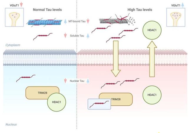 Fig. 1 Graphical model of Tau interplay with TRIM28/HDAC1 complex.1
Key structural properties of HDAC1:
- N-terminal nuclear localization signals are guided into the nucleus
- Highly conserved deacetylase domains
- A Zn²⁺-dependent catalytic center
Functions of HDAC1
The core function of HDAC1 is to regulate gene transcription. It achieves this by deacetylating histones and other non-histone substrates, thereby influencing the chromatin structure and cell fate.
| Function | Description |
| Transcription inhibition | Removes the acetyl groups of histone H3/H4, compresses chromatin, and silences downstream target genes. |
| Cell cycle regulation | Interacts with Rb protein and transcription factor E2F, inhibits the expression of S-phase genes, and blocks the G1 phase progression. |
| DNA damage repair | Recruits to the damage site, assists in the recruitment of 53BP1, and promotes homologous recombination repair. |
| Stem cell maintenance | In embryonic stem cells, cooperates with factors such as Oct4 to maintain self-renewal and inhibit differentiation genes. |
| Tumor occurrence | Abnormally highly expressed in various cancer types, promotes proliferation and metastasis by silencing tumor suppressor genes. |
The enzymatic activity of HDAC1 depends on Zn²⁺rather than NAD⁺. Its substrate spectrum is far beyond histones and has been identified as dozens of non-histone substrates, including key signaling molecules such as p53 and STAT3.
Applications of HDAC1 and HDAC1 Antibody in Literature
1. Siano, Giacomo, et al. "Tau-dependent HDAC1 nuclear reduction is associated with altered VGluT1 expression." Frontiers in Cell and Developmental Biology 11 (2023): 1151223. https://doi.org/10.3389/fcell.2023.1151223
The article indicates that abnormal elevation of Tau protein leads to a decrease in the nuclear localization of HDAC1, and a reduction in its activity, which in turn regulates the expression of synaptic genes. This mechanism reveals a new pathway through which Tau pathology affects synaptic dysfunction by influencing epigenetic regulatory factors.
2. Pao, Ping-Chieh, et al. "HDAC1 modulates OGG1-initiated oxidative DNA damage repair in the aging brain and Alzheimer's disease." Nature communications 11.1 (2020): 2484. https://doi.org/10.1038/s41467-020-16361-y
The article indicates that HDAC1 can activate OGG1 to repair oxidative DNA damage (8-oxoG). The loss of this function will lead to inhibition of neural gene expression and cognitive impairment; while activating HDAC1 can alleviate the related pathological changes in aging and Alzheimer's disease models.
3. Nieto-Estevez, Vanesa, et al. "HDAC1 regulates neuronal differentiation." Frontiers in molecular neuroscience 14 (2022): 815808. https://doi.org/10.3389/fnmol.2021.815808
The research found that during the neurogenesis process in adult hippocampus, HDAC1 is widely expressed at all cell stages. In vitro experiments showed that knocking out HDAC1 would impair the differentiation ability of neural stem cells into neurons, suggesting that it plays an important role in neuronal regeneration.
4. Jiang, Min, et al. "MARCKSL1–2 reverses docetaxel-resistance of lung adenocarcinoma cells by recruiting SUZ12 to suppress HDAC1 and elevate miR-200b." Molecular cancer 21.1 (2022): 150. https://doi.org/10.1186/s12943-022-01605-w
The study found that LncRNA MARCKSL1-2 inhibits the expression of HDAC1 by recruiting SUZ12, thereby relieving the inhibition of HDAC1 on miR-200b, and thereby alleviating the resistance of lung adenocarcinoma to docetaxel. This provides a potential target for improving chemotherapy.
5. Bahl, Sonali, et al. "EGFR phosphorylates HDAC1 to regulate its expression and anti-apoptotic function." Cell death & disease 12.5 (2021): 469. https://doi.org/10.1038/s41419-021-03697-6
This study reveals that EGFR can maintain the protein stability of HDAC1 by promoting its tyrosine phosphorylation (especially at the Tyr72 site), thereby regulating cell apoptosis. This provides a new strategy for cancer treatment targeting HDAC1 overexpression and EGFR activity.
Creative Biolabs: HDAC1 Antibodies for Research
Creative Biolabs specializes in the production of high-quality HDAC1 antibodies for research and industrial applications. Our portfolio includes monoclonal and polyclonal antibodies tailored for ELISA, Flow Cytometry, Western blot, immunohistochemistry, and other diagnostic methodologies.
- Custom HDAC1 Antibody Development: Tailor-made solutions to meet specific research requirements.
- Bulk Production: Large-scale antibody manufacturing for industry partners.
- Technical Support: Expert consultation for protocol optimization and troubleshooting.
- Aliquoting Services: Conveniently sized aliquots for long-term storage and consistent experimental outcomes.
For more details on our HDAC1 antibodies, custom preparations, or technical support, contact us at email.
Reference
- Siano, Giacomo, et al. "Tau-dependent HDAC1 nuclear reduction is associated with altered VGluT1 expression." Frontiers in Cell and Developmental Biology 11 (2023): 1151223. Distributed under Open Access license CC BY 4.0, without modification. https://doi.org/10.3389/fcell.2023.1151223
Anti-HDAC1 antibodies
 Loading...
 Compare




Hot products 
-
Mouse Anti-BIRC7 Recombinant Antibody (88C570) (CBMAB-L0261-YJ)

-
Mouse Anti-ACKR3 Recombinant Antibody (V2-261265) (CBMAB-C1023-LY)

-
Mouse Anti-CCT6A/B Recombinant Antibody (CBXC-0168) (CBMAB-C5570-CQ)

-
Mouse Anti-ATP1B3 Recombinant Antibody (1E9) (CBMAB-A4021-YC)

-
Rabbit Anti-ENO2 Recombinant Antibody (BA0013) (CBMAB-0272CQ)

-
Mouse Anti-APP Recombinant Antibody (DE2B4) (CBMAB-1122-CN)

-
Mouse Anti-FOSB Recombinant Antibody (CBXF-3593) (CBMAB-F2522-CQ)

-
Mouse Anti-Acetyl SMC3 (K105/K106) Recombinant Antibody (V2-634053) (CBMAB-AP052LY)

-
Mouse Anti-CDK7 Recombinant Antibody (CBYY-C1783) (CBMAB-C3221-YY)

-
Mouse Anti-dsDNA Recombinant Antibody (22) (CBMAB-AP1954LY)

-
Mouse Anti-ASTN1 Recombinant Antibody (H-9) (CBMAB-1154-CN)

-
Mouse Anti-GGT1 Recombinant Antibody (1F9) (CBMAB-G3273-LY)

-
Mouse Anti-ADGRE2 Recombinant Antibody (V2-261270) (CBMAB-C0813-LY)

-
Mouse Anti-GFAP Recombinant Antibody (24) (CBMAB-G2927-LY)

-
Mouse Anti-A2M Recombinant Antibody (V2-178822) (CBMAB-A0036-YC)

-
Mouse Anti-FPR2 Recombinant Antibody (1D6) (CBMAB-F2628-CQ)

-
Mouse Anti-AAV-5 Recombinant Antibody (V2-503416) (CBMAB-V208-1402-FY)

-
Mouse Anti-CD33 Recombinant Antibody (P67.6) (CBMAB-C10189-LY)

-
Mouse Anti-ARG1 Recombinant Antibody (CBYCL-103) (CBMAB-L0004-YC)

-
Mouse Anti-dsRNA Recombinant Antibody (2) (CBMAB-D1807-YC)

- AActivation
- AGAgonist
- APApoptosis
- BBlocking
- BABioassay
- BIBioimaging
- CImmunohistochemistry-Frozen Sections
- CIChromatin Immunoprecipitation
- CTCytotoxicity
- CSCostimulation
- DDepletion
- DBDot Blot
- EELISA
- ECELISA(Cap)
- EDELISA(Det)
- ESELISpot
- EMElectron Microscopy
- FFlow Cytometry
- FNFunction Assay
- GSGel Supershift
- IInhibition
- IAEnzyme Immunoassay
- ICImmunocytochemistry
- IDImmunodiffusion
- IEImmunoelectrophoresis
- IFImmunofluorescence
- IGImmunochromatography
- IHImmunohistochemistry
- IMImmunomicroscopy
- IOImmunoassay
- IPImmunoprecipitation
- ISIntracellular Staining for Flow Cytometry
- LALuminex Assay
- LFLateral Flow Immunoassay
- MMicroarray
- MCMass Cytometry/CyTOF
- MDMeDIP
- MSElectrophoretic Mobility Shift Assay
- NNeutralization
- PImmunohistologyp-Paraffin Sections
- PAPeptide Array
- PEPeptide ELISA
- PLProximity Ligation Assay
- RRadioimmunoassay
- SStimulation
- SESandwich ELISA
- SHIn situ hybridization
- TCTissue Culture
- WBWestern Blot
