IL7R Antibodies
Background
The IL7R gene encodes the alpha chain of the interleukin 7 receptor, and is mainly expressed on the cell membrane surface of lymphocytes. This receptor activates downstream signaling pathways by binding to interleukin 7, thereby regulating the development, proliferation, and survival of lymphocytes, and is crucial for the normal function of the adaptive immune system. This gene was cloned and identified in the early 1990s, and its function has deepened our understanding of immune cell differentiation and homeostasis mechanisms. Notably, specific variations in the IL7R gene have been proven to be significantly associated with the risk of autoimmune diseases such as multiple sclerosis, making it an important molecular target for research on immune-related diseases. Continuous research on this gene and its signaling network is constantly driving the development of basic immunological theories and clinical treatment strategies.
Structure of IL7R
The protein encoded by the IL7R gene is a typical type I transmembrane glycoprotein, with a molecular weight of approximately 40-45 kDa (in its mature form). Its molecular weight may vary slightly due to the degree of glycosylation modification and species differences.
| Species | Human | Mouse | Rat |
| Molecular Weight (kDa) | 44 | 42 | 43 |
| Primary Structural Differences | Two fibronectin type III domains in the extracellular region; WSXWS motif is conserved | Highly similar to humans and with interchangeable functions | Consistent sequence, signal transduction mechanism |
This protein is composed of 449 amino acids and its primary structure can be divided into the extracellular region, the transmembrane region and the intracellular region. The core function depends on the conserved WSXWS motif in the extracellular region, which is crucial for the correct folding of the receptor and signal transduction. The secondary structure is mainly composed of β-sheet, jointly forming the spatial conformation of the fibronectin type III domain. The transmembrane region consists of a single α-helix, while the intracellular region itself lacks kinase activity and requires non-covalent binding to the γc chain (common γ chain) to form a functional high-affinity receptor complex, thereby initiating the downstream JAK-STAT signaling pathway and regulating gene expression.
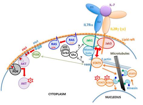 Fig. 1 Schematic of the IL7R structure and signaling mechanism.1
Key structural properties of IL7R:
- Type I transmembrane protein topology structure
- Extracellular region including conservative WSXWS motif and fibronectin III structure domain
- Dependent on forming heterodimers with the γc chain to construct a functional receptor
- Intracellular area through JAK kinase coupling league start STAT signal pathway
Functions of IL7R
The IL7R gene encodes the α chain of the interleukin 7 receptor. Its core function is to mediate the signal transduction of IL-7, thereby precisely regulating the development, homeostasis and survival of lymphocytes. However, this receptor is also involved in a variety of crucial immune pathological physiological processes.
| Function | Description |
| Lymphocyte Development | In the thymus, it transmits survival and proliferation signals, driving T-cell precursors to differentiate from the DN stage to the DP stage, which is essential for the differentiation of the T-cell lineage. |
| Peripheral T Cell Homeostasis | Maintaining the long-term survival and stability of mature and immature T cells in peripheral immune organs is achieved through continuous signaling that inhibits cell apoptosis. |
| Early Development of B Cells | In the bone marrow, other cytokines work together to support the proliferation and differentiation of B-lineage progenitor cells (pro-B, pre-B stages). |
| Autoimmune Regulation | The abnormality in the intensity and duration of its signals is closely related to the occurrence and development of autoimmune diseases such as multiple sclerosis and rheumatoid arthritis. |
| Immune memory formation | By promoting the survival of memory T cell precursors, it participates in the establishment and maintenance of adaptive immune memory. |
The activation of the IL-7R signaling pathway strictly depends on its formation of an heterodimer with the common γ chain (γc). This structural characteristic endows its signal with high cell type specificity. Compared to many rapidly activated cytokine receptors, IL-7R mediates a persistent and stable signaling process, which determines its unique and irreplaceable role in maintaining the lifelong functional capacity of the immune system. Dysregulation of its signal can directly lead to severe immunodeficiency or autoimmune pathological conditions.
Applications of IL7R and IL7R Antibody in Literature
1. Campos, Lívia Weijenborg, Leonardo Granato Pissinato, and José Andrés Yunes. "Deleterious and Oncogenic Mutations in the IL7RA." Cancers 11.12 (2019): 1952. https://doi.org/10.3390/cancers11121952
The article indicates that mutations or variations in the interleukin 7 receptor alpha (IL7Rα) are associated with immunodeficiency, inflammation, and leukemia. Inactivating mutations are mostly located in the extracellular region, while oncogenic mutations are concentrated in the extracellular near-membrane or transmembrane region, which can cause abnormal dimerization or changes in affinity, leading to continuous activation of the signaling pathway. This article conducts a classified review based on its structural influence and biological activity.
2. Li, Jin, et al. "IL7R, GZMA and CD8A serve as potential molecular biomarkers for sepsis based on bioinformatics analysis." Frontiers in Immunology 15 (2024): 1445858. https://doi.org/10.3389/fimmu.2024.1445858
The article indicates that by integrating the database with sequencing analysis, ten key genes related to sepsis were identified through the research. Among them, IL7R, GZMA and CD8A showed significant differences in expression and were interrelated, suggesting their potential value as molecular markers.
3. Xu, Nan, et al. "Low IL7R Expression at Diagnosis Predicted Relapse in Adult Acute Myeloid Leukemia Patients With t (8; 21)." Frontiers in Immunology 13 (2022): 909104. https://doi.org/10.3389/fimmu.2022.909104
The article indicates that in t(8;21) acute myeloid leukemia, low expression of IL7R in the bone marrow at the time of diagnosis predicts a higher risk of recurrence and is associated with KIT mutations. The mechanism may be related to the impairment of T cell quantity and function, suggesting that IL7R can serve as a prognostic marker.
4. Russell, Jamie, et al. "Lrp10 suppresses IL7R limiting CD8 T cell homeostatic expansion and anti-tumor immunity." EMBO reports 25.8 (2024): 3601-3626. https://doi.org/10.1038/s44319-024-00191-w
The research has found that the Lrp10 protein negatively regulates the homeostasis and memory formation of CD8+ T cells after T cell activation by inhibiting the expression of IL7R. The absence of Lrp10 can enhance the IL7R signal, promote the proliferation of T cell homeostasis, improve anti-tumor ability and enhance the immune treatment response.
5. Oliveira, Mariana L., et al. "Mutant IL7R collaborates with MYC to induce T-cell acute lymphoblastic leukemia." Leukemia 36.6 (2022): 1533-1540. https://doi.org/10.1038/s41375-022-01590-5
The research has found that the mutation of IL7R itself can induce T-cell acute lymphoblastic leukemia, and it works synergistically with MYC to significantly accelerate the onset of the disease. The mechanism involves the activation of the STAT5/AKT pathway, inhibition of cell apoptosis, and the ability to enrich leukemia proliferating cells and enhance the potential for transplantation and recurrence.
Creative Biolabs: IL7R Antibodies for Research
Creative Biolabs specializes in the production of high-quality IL7R antibodies for research and industrial applications. Our portfolio includes monoclonal antibodies tailored for ELISA, Flow Cytometry, Western blot, immunohistochemistry, and other diagnostic methodologies.
- Custom IL7R Antibody Development: Tailor-made solutions to meet specific research requirements.
- Bulk Production: Large-scale antibody manufacturing for industry partners.
- Technical Support: Expert consultation for protocol optimization and troubleshooting.
- Aliquoting Services: Conveniently sized aliquots for long-term storage and consistent experimental outcomes.
For more details on our IL7R antibodies, custom preparations, or technical support, contact us at email.
Reference
- Campos, Lívia Weijenborg, Leonardo Granato Pissinato, and José Andrés Yunes. "Deleterious and Oncogenic Mutations in the IL7RA." Cancers 11.12 (2019): 1952. Distributed under Open Access license CC BY 4.0, without modification. https://doi.org/10.3390/cancers11121952
Anti-IL7R antibodies
 Loading...


Hot products 
-
Mouse Anti-ACO2 Recombinant Antibody (V2-179329) (CBMAB-A0627-YC)

-
Mouse Anti-DMD Recombinant Antibody (D1190) (CBMAB-D1190-YC)

-
Mouse Anti-EPO Recombinant Antibody (CBFYR0196) (CBMAB-R0196-FY)

-
Rat Anti-ABCC11 Recombinant Antibody (V2-179001) (CBMAB-A0236-YC)

-
Mouse Anti-ALX1 Recombinant Antibody (96k) (CBMAB-C0616-FY)

-
Mouse Anti-C5b-9 Recombinant Antibody (aE11) (CBMAB-AO138LY)

-
Mouse Anti-GGT1 Recombinant Antibody (1F9) (CBMAB-G3273-LY)

-
Mouse Anti-ESR1 Recombinant Antibody (Y31) (CBMAB-1208-YC)

-
Mouse Anti-BRCA2 Recombinant Antibody (CBYY-1728) (CBMAB-2077-YY)

-
Mouse Anti-AGK Recombinant Antibody (V2-258056) (CBMAB-M0989-FY)

-
Mouse Anti-ATG5 Recombinant Antibody (9H197) (CBMAB-A3945-YC)

-
Mouse Anti-ADGRE2 Recombinant Antibody (V2-261270) (CBMAB-C0813-LY)

-
Mouse Anti-BZLF1 Recombinant Antibody (BZ.1) (CBMAB-AP705LY)

-
Mouse Anti-FLT1 Recombinant Antibody (11) (CBMAB-V0154-LY)

-
Mouse Anti-ARG1 Recombinant Antibody (CBYCL-103) (CBMAB-L0004-YC)

-
Rat Anti-EPO Recombinant Antibody (16) (CBMAB-E1578-FY)

-
Mouse Anti-FPR2 Recombinant Antibody (1D6) (CBMAB-F2628-CQ)

-
Mouse Anti-ARIH1 Recombinant Antibody (C-7) (CBMAB-A3563-YC)

-
Mouse Anti-AMACR Recombinant Antibody (CB34A) (CBMAB-CA034LY)

-
Mouse Anti-DHFR Recombinant Antibody (D0821) (CBMAB-D0821-YC)

- AActivation
- AGAgonist
- APApoptosis
- BBlocking
- BABioassay
- BIBioimaging
- CImmunohistochemistry-Frozen Sections
- CIChromatin Immunoprecipitation
- CTCytotoxicity
- CSCostimulation
- DDepletion
- DBDot Blot
- EELISA
- ECELISA(Cap)
- EDELISA(Det)
- ESELISpot
- EMElectron Microscopy
- FFlow Cytometry
- FNFunction Assay
- GSGel Supershift
- IInhibition
- IAEnzyme Immunoassay
- ICImmunocytochemistry
- IDImmunodiffusion
- IEImmunoelectrophoresis
- IFImmunofluorescence
- IGImmunochromatography
- IHImmunohistochemistry
- IMImmunomicroscopy
- IOImmunoassay
- IPImmunoprecipitation
- ISIntracellular Staining for Flow Cytometry
- LALuminex Assay
- LFLateral Flow Immunoassay
- MMicroarray
- MCMass Cytometry/CyTOF
- MDMeDIP
- MSElectrophoretic Mobility Shift Assay
- NNeutralization
- PImmunohistologyp-Paraffin Sections
- PAPeptide Array
- PEPeptide ELISA
- PLProximity Ligation Assay
- RRadioimmunoassay
- SStimulation
- SESandwich ELISA
- SHIn situ hybridization
- TCTissue Culture
- WBWestern Blot
