
Lambda Light Chain Antibodies
Background
Lambda light chain is a polypeptide subunit in immunoglobulin molecules, mainly existing in B lymphocytes and plasma cells of vertebrates. It combines with other immunoglobulin components to form functional antibodies, participating in antigen recognition and immune response processes, thereby ensuring the adaptive immune function of the body. This gene undergoes a V(D)J rearrangement mechanism during B cell development, and this specific gene recombination ensures the generation of antibody diversity. In 1956, the λ light chain gene was first discovered in the myeloma protein by scientists, laying an important foundation for the study of immunoglobulin genetics. Its ingenious rearrangement mechanism and expression regulation have become a classic model in immunogenetics research, greatly promoting our understanding of antibody diversity production, B-cell differentiation and related disease mechanisms.
Structure of Lambda Light Chain
Lambda light chain is a protein with a molecular weight of approximately 23 kDa. Its precise molecular weight varies slightly among different species, mainly due to changes in the amino acid sequences of the constant and variable regions. The following is a comparison of the molecular weight and structural characteristics of Lambda light chains in common species:
| Species | Human | Mouse | Rabbit | Bovine |
| Molecular Weight (kDa) | 23.0 | 22.8 | 23.2 | 22.9 |
| Primary Structural Differences | With VJ - C gene rearrangement structure | The variable region sequence is relatively short | The hinge area has a special structure | The constant zone is highly conservative |
The Lambda light chain is composed of approximately 210 amino acids, and its three-dimensional structure contains two immunoglobulin domains: the variable domain (VL) at the N-terminal and the constant domain (CL) at the C-terminal. Each domain adopts a typical immunoglobulin folding pattern, composed of two reverse-parallel β sheets stably linked by disulfide bonds. The complementary determinant (CDR) in the variable domain is responsible for antigen-specific binding, and its conformational diversity is the structural basis for the functional diversity of antibodies. Light chains and heavy chains form complete antibody molecules through disulfide bonds and hydrophobic interactions between the CL and CH1 domains.
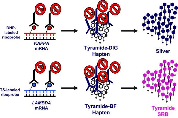 Fig. 1 Lambda Light Chain-Restricted Hematogones: A Diagnostic Pitfall in Bone Marrow Analysis.1
Key structural properties of Lambda Light Chain:
- Typical immunoglobulin folding structure (β -sandwich configuration)
- The variable region (VL) and the constant region (CL) are stably connected by disulfide bonds
- Complementary determinant regions (CDRS) mediate antigen-specific recognition and binding
Functions of Lambda Light Chain
The main function of Lambda light chain is to serve as an important component of antibodies, participating in immune recognition and antigen binding. In addition, it also involves the development of B cells, signal transduction and the regulation of various pathological processes.
| Function | Description |
| Antibody assembly | Pairing with the heavy chain forms a functional antibody molecule that forms part of the antigen binding site. |
| Antigen recognition and binding | Antigenic epitopes are specifically recognized and bound to the complementary determinant (CDR) in the variable region (VL). |
| B-cell receptor signal transduction | As a component of B-cell receptors (BCRS), it participates in cell activation and the initiation of immune responses. |
| Immune regulation | Affect antibody type conversion and affinity mature, adjust the accuracy and diversity of humoral immunity. |
| Disease markers | Abnormal levels of free light chains are closely related to diseases such as multiple myeloma and light chain amyloidosis. |
The ratio of Lambda light chain to κ light chain in the human body is approximately 1:2. This relatively constant ratio (light chain ratio) is often used in clinical diagnosis and immunohistochemical analysis to detect B-cell clonal proliferation and related diseases.
Applications of Lambda Light Chain and Lambda Light Chain Antibody in Literature
1. Lan, Mingfu, et al. "Lambda light chain-restricted non-crystalline proximal tubulopathy with cast nephropathy in multiple myeloma: a case report and literature review." BMC nephrology 25.1 (2024): 324. https://doi.org/10.1186/s12882-024-03721-9
This article reports a rare case of IgD-λ multiple myeloma complicated with amorphous λ light chain proximal renal tubular lesions and light chain tubular nephropathy. Through clinicopathological analysis, its characteristics are explored to enhance the understanding of the diagnosis of this disease.
2. Rimsza, Lisa M., et al. "Kappa and lambda light chain mRNA in situ hybridization compared to flow cytometry and immunohistochemistry in B cell lymphomas." Diagnostic pathology 9.1 (2014): 144. https://doi.org/10.1186/1746-1596-9-144
This study evaluated the novel dual-color CISH technology, which can detect κ/LAMBDA light chain mRNA with high sensitivity. In the clonal assessment of B-cell lymphoma, it was highly consistent with the results of flow cytometry /IHC (with a coincidence rate of 98.6%), significantly enhancing the detection ability of light chains in tissue sections.
3. Guillory, Tesha, et al. "Hematogones With Lambda Light Chain Restriction in a 4-Year-Old Boy With Burkitt Lymphoma: A Potential Diagnostic Pitfall." Laboratory Medicine 47.2 (2016): 163-170. https://doi.org/10.1093/labmed/lmw009
This article reports a rare case: abnormal hematopoietic precursor cells (hematogones) appeared in the bone marrow of a 4-year-old child during treatment. Although they had a typical phenotype, they abnormally expressed λ light chain restriction, and molecular testing showed no clonicity, suggesting the need to be vigilant about potential misunderstandings in the assessment of B-cell lymphoma.
4. Raybould, Matthew IJ, et al. "Contextualising the developability risk of antibodies with lambda light chains using enhanced therapeutic antibody profiling." Communications Biology 7.1 (2024): 62. https://doi.org/10.1038/s42003-023-05744-8
This study, through the analysis of the machine learning tool TAP, found that although the overall developability risk of λ light chain antibodies is higher than that of κ antibodies, a considerable number of λ antibodies still have a relatively low risk and can be used as high-quality candidate drugs. It is called for a rational increase in the diversity of λ antibodies in immunotherapy.
5. Berghaus, Natalie, et al. "Analysis of the complete lambda light chain germline usage in patients with AL amyloidosis and dominant heart or kidney involvement." PLoS One 17.2 (2022): e0264407. https://doi.org/10.1371/journal.pone.0264407
This study analyzed 85 patients with λ light amyloidosis and found that the type of organ involved was significantly associated with specific IGLV gene subtypes: the cardiodominant type was mostly associated with IGLV3-21, the kidney-dominant type was mostly associated with IGLV1-44, and the cardiorenal co-affected type was mainly associated with IGLV6-57.
Creative Biolabs: Lambda Light Chain Antibodies for Research
Creative Biolabs specializes in the production of high-quality Lambda Light Chain antibodies for research and industrial applications. Our portfolio includes monoclonal antibodies tailored for ELISA, Flow Cytometry, Western blot, immunohistochemistry, and other diagnostic methodologies.
- Custom Lambda Light Chain Antibody Development: Tailor-made solutions to meet specific research requirements.
- Bulk Production: Large-scale antibody manufacturing for industry partners.
- Technical Support: Expert consultation for protocol optimization and troubleshooting.
- Aliquoting Services: Conveniently sized aliquots for long-term storage and consistent experimental outcomes.
For more details on our Lambda Light Chain antibodies, custom preparations, or technical support, contact us at email.
Reference
- Rimsza, Lisa M., et al. "Kappa and lambda light chain mRNA in situ hybridization compared to flow cytometry and immunohistochemistry in B cell lymphomas." Diagnostic pathology 9.1 (2014): 144. https://doi.org/10.1186/1746-1596-9-144
Anti-Lambda Light Chain antibodies
 Loading...
 Compare




Hot products 
-
Rat Anti-ADGRE4 Recombinant Antibody (V2-160163) (CBMAB-F0011-CQ)

-
Mouse Anti-CRTAM Recombinant Antibody (CBFYC-2235) (CBMAB-C2305-FY)

-
Mouse Anti-ADV Recombinant Antibody (V2-503423) (CBMAB-V208-1364-FY)

-
Mouse Anti-ARHGDIA Recombinant Antibody (CBCNA-009) (CBMAB-R0415-CN)

-
Mouse Anti-ESR1 Recombinant Antibody (Y31) (CBMAB-1208-YC)

-
Mouse Anti-AAV8 Recombinant Antibody (V2-634028) (CBMAB-AP022LY)

-
Mouse Anti-GFAP Recombinant Antibody (20) (CBMAB-G2914-LY)

-
Rabbit Anti-ABL1 (Phosphorylated Y245) Recombinant Antibody (V2-505716) (PTM-CBMAB-0465LY)

-
Mouse Anti-CARTPT Recombinant Antibody (113612) (CBMAB-C2450-LY)

-
Mouse Anti-C5b-9 Recombinant Antibody (aE11) (CBMAB-AO138LY)

-
Rat Anti-ABCC11 Recombinant Antibody (V2-179001) (CBMAB-A0236-YC)

-
Mouse Anti-CCND2 Recombinant Antibody (DCS-3) (CBMAB-G1318-LY)

-
Mouse Anti-AK4 Recombinant Antibody (V2-180419) (CBMAB-A1891-YC)

-
Rat Anti-CD34 Recombinant Antibody (MEC 14.7) (CBMAB-C10196-LY)

-
Mouse Anti-ARSA Recombinant Antibody (CBYC-A799) (CBMAB-A3679-YC)

-
Mouse Anti-ABIN2 Recombinant Antibody (V2-179106) (CBMAB-A0349-YC)

-
Mouse Anti-CD1C Recombinant Antibody (L161) (CBMAB-C2173-CQ)

-
Mouse Anti-CGAS Recombinant Antibody (CBFYM-0995) (CBMAB-M1146-FY)

-
Mouse Anti-AMACR Recombinant Antibody (CB34A) (CBMAB-CA034LY)

-
Mouse Anti-GST Recombinant Antibody (CBLG1-1938) (CBMAB-G5371-LY)

- AActivation
- AGAgonist
- APApoptosis
- BBlocking
- BABioassay
- BIBioimaging
- CImmunohistochemistry-Frozen Sections
- CIChromatin Immunoprecipitation
- CTCytotoxicity
- CSCostimulation
- DDepletion
- DBDot Blot
- EELISA
- ECELISA(Cap)
- EDELISA(Det)
- ESELISpot
- EMElectron Microscopy
- FFlow Cytometry
- FNFunction Assay
- GSGel Supershift
- IInhibition
- IAEnzyme Immunoassay
- ICImmunocytochemistry
- IDImmunodiffusion
- IEImmunoelectrophoresis
- IFImmunofluorescence
- IGImmunochromatography
- IHImmunohistochemistry
- IMImmunomicroscopy
- IOImmunoassay
- IPImmunoprecipitation
- ISIntracellular Staining for Flow Cytometry
- LALuminex Assay
- LFLateral Flow Immunoassay
- MMicroarray
- MCMass Cytometry/CyTOF
- MDMeDIP
- MSElectrophoretic Mobility Shift Assay
- NNeutralization
- PImmunohistologyp-Paraffin Sections
- PAPeptide Array
- PEPeptide ELISA
- PLProximity Ligation Assay
- RRadioimmunoassay
- SStimulation
- SESandwich ELISA
- SHIn situ hybridization
- TCTissue Culture
- WBWestern Blot
