LGALS1 Antibodies
Background
The LGALS1 gene encodes galectin-1, a β-galactoside-binding protein that mainly exists in a homodimeric form. It is widely expressed in various tissues and cell types. This protein participates in regulating various physiological processes such as cell adhesion, proliferation, apoptosis, and immune responses by recognizing sugar conjugates on the cell surface and in the extracellular matrix. In the tumor microenvironment, LGALS1 can promote tumor immune escape by regulating the function of immune cells, and changes in its expression level are also closely related to inflammatory responses and tissue fibrosis. Since its first isolation and identification from the electric eel tissue in 1975, scientists have gradually clarified its multiple functions in maintaining cellular homeostasis. The crystal structure of this protein reveals its unique β-triangular folding pattern, providing an important model for understanding the molecular mechanism of the lectin family's recognition of sugar ligands.
Structure of LGALS1
The LGALS1 gene encodes a galectin-1 protein with a molecular weight of approximately 14.5 kDa, which is a homodimeric protein. The monomer of this protein consists of 135 amino acids. This protein is highly conserved in evolution, and the amino acid sequence similarity among different species can reach over 80%.
| Species | Human | Mouse | Rat | Pig | Bovine |
| Molecular Weight (kDa) | 14.5 | 14.7 | 14.6 | 14.5 | 14.8 |
| Primary Structural Differences | Contains 6 cysteines, forming 3 disulfide bonds | There are two amino acid substitutions in the C-terminal region | It has a 97% homology with mice | The sugar-binding domain is completely conserved | There is an amino acid insertion at the N-terminus |
The three-dimensional structure of this protein exhibits a typical β-triangular folding pattern, consisting of two barrel-shaped domains formed by six antiparallel β-strands. Each monomer contains a conserved sugar recognition domain (CRD), which is composed of β-sheet and loop regions and forms a hydrophobic core. The highly conserved amino acid residues in the CRD (such as His44, Asn46, Arg48, Val59, and Asn61) are mediated by a hydrogen bond network for specific binding to β-galactosides. The two monomers form a functional dimer with two sugar binding sites through hydrophobic interactions, and this dimerization state is crucial for its cross-linking of sugar ligands and induction of cell signal transduction.
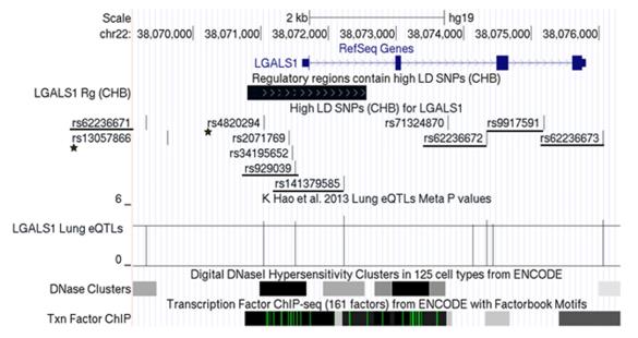 Fig. 1 The genetic architecture of LGALS1 gene.1
Key structural properties of LGALS1:
- Typical β-triangular sandwich folding structure
- Hydrophobic cores formed by two antiparallel β-folds
- Each subunit contains a conserved sugar recognition domain (CRD)
- Highly conserved dimer interfaces mediate homologous dimerization
Functions of LGALS1
The LGALS1 gene encodes the galectin-1 protein, which primarily functions in recognizing β-galactosides and mediating interactions between cells and between cells and the extracellular matrix. This protein is involved in regulating cell adhesion, proliferation, and apoptosis, and also plays a role in maintaining immune homeostasis.
| Function | Description |
| Cell Adhesion Regulation | Regulates cell-matrix and cell-cell adhesion by forming cross-links with cell surface glycoproteins. |
| Apoptosis Regulation | Has a pro-apoptotic effect on activated T cells and certain tumor cell lines. |
| Immune Regulation | Negatively regulates T cell activation and participates in the establishment of an immune-privileged microenvironment. |
| Inflammation Regulation | Affects the adhesion and exudation of neutrophils and regulates the intensity of the inflammatory response. |
| Tumor-related Function | Promotes immune escape and angiogenesis in the tumor microenvironment. |
This protein regulates signal transduction in a dose-dependent manner by recognizing the polyglucosamine structure on the cell surface and in the extracellular matrix. Its biological effects are cell type-specific and concentration-dependent. At low concentrations, it promotes cell adhesion, while at high concentrations, it induces apoptosis.
Applications of LGALS1 and LGALS1 Antibody in Literature
1. Chen, Yu, et al. "Functional variants regulating LGALS1 (Galectin 1) expression affect human susceptibility to influenza A (H7N9)." Scientific reports 5.1 (2015): 8517. https://doi.org/10.1038/srep08517
This study, through genome-wide association analysis, discovered that functional variations in the LGALS1 gene affect its expression level, thereby leading to differences in individual susceptibility to the H7N9 avian influenza virus. Among them, the haplotype GG of rs4820294/rs2899292 was significantly associated with a lower infection risk, and was also correlated with increased mRNA and protein expression levels of this gene.
2. Qin, Huanhuan, et al. "A novel LGALS1-depended and immune-associated fatty acid metabolism risk model in acute myeloid leukemia stem cells." Cell Death & Disease 15.7 (2024): 482. https://doi.org/10.1038/s41419-024-06865-6
The study found that LGALS1 is highly expressed in leukemia stem cells and is associated with poor prognosis in patients with acute myeloid leukemia. Inhibiting LGALS1 can reduce lipid accumulation, inhibit cancer cell proliferation and promote apoptosis, providing a potential target for clinical prognosis assessment and individualized treatment.
3. Finotto, Lise, et al. "Single‐cell profiling and zebrafish avatars reveal LGALS1 as immunomodulating target in glioblastoma." EMBO molecular medicine 15.11 (2023): EMMM202318144. https://doi.org/10.15252/emmm.202318144
The study, through single-cell sequencing and zebrafish models, revealed that glioblastoma stem cells regulate tumor-associated macrophages via LGALS1, inducing an immunosuppressive microenvironment. LGALS1 is a crucial immune regulatory factor, and its expression level is associated with the patient's survival period.
4. Triguero-Martínez, Ana, et al. "Genetic LGALS1 variants are associated with heterogeneity in galectin-1 serum levels in patients with early arthritis." International Journal of Molecular Sciences 23.13 (2022): 7181. https://doi.org/10.3390/ijms23137181
Genotyping studies on patients with early arthritis revealed that specific genetic variations of LGALS1 (such as rs9622682) were associated with elevated levels of galectin-1 and decreased levels of IL-6. Patients carrying the specific genotype showed higher expression of Gal1 after treatment, suggesting that this gene regulates the inflammatory response.
5. Sun, Huanhuan, et al. "NCAPG promotes the oncogenesis and progression of non-small cell lung cancer cells through upregulating LGALS1 expression." Molecular cancer 21.1 (2022): 55. https://doi.org/10.1186/s12943-022-01533-9
The study found that NCAPG is a new oncogenic driver gene for non-small cell lung cancer, and its high expression is associated with poor prognosis of patients. Mechanistically, the function of NCAPG is closely related to LGALS1, which is also upregulated in lung cancer and may directly interact with it.
Creative Biolabs: LGALS1 Antibodies for Research
Creative Biolabs specializes in the production of high-quality LGALS1 antibodies for research and industrial applications. Our portfolio includes monoclonal and polyclonal antibodies tailored for ELISA, Flow Cytometry, Western blot, immunohistochemistry, and other diagnostic methodologies.
- Custom LGALS1 Antibody Development: Tailor-made solutions to meet specific research requirements.
- Bulk Production: Large-scale antibody manufacturing for industry partners.
- Technical Support: Expert consultation for protocol optimization and troubleshooting.
- Aliquoting Services: Conveniently sized aliquots for long-term storage and consistent experimental outcomes.
For more details on our LGALS1 antibodies, custom preparations, or technical support, contact us at email.
Reference
- Chen, Yu, et al. "Functional variants regulating LGALS1 (Galectin 1) expression affect human susceptibility to influenza A (H7N9)." Scientific reports 5.1 (2015): 8517. Distributed under Open Access license CC BY 4.0, without modification. https://doi.org/10.1038/srep08517
Anti-LGALS1 antibodies
 Loading...


Hot products 
-
Human Anti-SARS-CoV-2 Spike Recombinant Antibody (CR3022) (CBMAB-CR014LY)

-
Mouse Anti-ARG1 Recombinant Antibody (CBYCL-103) (CBMAB-L0004-YC)

-
Mouse Anti-C4B Recombinant Antibody (CBYY-C2996) (CBMAB-C4439-YY)

-
Rabbit Anti-Acetyl-Histone H3 (Lys36) Recombinant Antibody (V2-623395) (CBMAB-CP0994-LY)

-
Mouse Anti-ACO2 Recombinant Antibody (V2-179329) (CBMAB-A0627-YC)

-
Human Anti-SARS-CoV-2 Spike Recombinant Antibody (CBC05) (CBMAB-CR005LY)

-
Mouse Anti-Acetyl SMC3 (K105/K106) Recombinant Antibody (V2-634053) (CBMAB-AP052LY)

-
Mouse Anti-GIPC2 Recombinant Antibody (10) (CBMAB-G0476-LY)

-
Mouse Anti-AP4E1 Recombinant Antibody (32) (CBMAB-A2996-YC)

-
Mouse Anti-8-oxoguanine Recombinant Antibody (V2-7697) (CBMAB-1869CQ)

-
Mouse Anti-ELAVL4 Recombinant Antibody (6B9) (CBMAB-1132-YC)

-
Rat Anti-ABCC11 Recombinant Antibody (V2-179001) (CBMAB-A0236-YC)

-
Mouse Anti-ATM Recombinant Antibody (2C1) (CBMAB-A3970-YC)

-
Mouse Anti-AAV-5 Recombinant Antibody (V2-503416) (CBMAB-V208-1402-FY)

-
Mouse Anti-ARHGAP5 Recombinant Antibody (54/P190-B) (CBMAB-P0070-YC)

-
Armenian hamster Anti-CD40 Recombinant Antibody (HM40-3) (CBMAB-C10365-LY)

-
Mouse Anti-BCL2L1 Recombinant Antibody (H5) (CBMAB-1025CQ)

-
Mouse Anti-ARID3A Antibody (A4) (CBMAB-0128-YC)

-
Mouse Anti-CD24 Recombinant Antibody (2Q1282) (CBMAB-C1624-CN)

-
Mouse Anti-ABL2 Recombinant Antibody (V2-179121) (CBMAB-A0364-YC)

- AActivation
- AGAgonist
- APApoptosis
- BBlocking
- BABioassay
- BIBioimaging
- CImmunohistochemistry-Frozen Sections
- CIChromatin Immunoprecipitation
- CTCytotoxicity
- CSCostimulation
- DDepletion
- DBDot Blot
- EELISA
- ECELISA(Cap)
- EDELISA(Det)
- ESELISpot
- EMElectron Microscopy
- FFlow Cytometry
- FNFunction Assay
- GSGel Supershift
- IInhibition
- IAEnzyme Immunoassay
- ICImmunocytochemistry
- IDImmunodiffusion
- IEImmunoelectrophoresis
- IFImmunofluorescence
- IGImmunochromatography
- IHImmunohistochemistry
- IMImmunomicroscopy
- IOImmunoassay
- IPImmunoprecipitation
- ISIntracellular Staining for Flow Cytometry
- LALuminex Assay
- LFLateral Flow Immunoassay
- MMicroarray
- MCMass Cytometry/CyTOF
- MDMeDIP
- MSElectrophoretic Mobility Shift Assay
- NNeutralization
- PImmunohistologyp-Paraffin Sections
- PAPeptide Array
- PEPeptide ELISA
- PLProximity Ligation Assay
- RRadioimmunoassay
- SStimulation
- SESandwich ELISA
- SHIn situ hybridization
- TCTissue Culture
- WBWestern Blot
