MMP3 Antibodies
Background
MMP3 is a zinc-dependent protease mainly secreted by connective tissue cells and belongs to the matrix metalloproteinase family. This protein can degrade various components in the extracellular matrix, including collagen, proteoglycans, and laminin, thereby playing a crucial role in physiological processes such as tissue remodeling, wound repair, and inflammatory responses. Its activity is strictly regulated by specific inhibitors (such as TIMP) to prevent excessive degradation and prevent pathological damage. Studies have shown that abnormal expression of MMP3 is closely related to various diseases such as arthritis, cardiovascular diseases, and tumor metastasis, making it an important target in biomedical research. Through continuous exploration of the structure and function of MMP3, scientists have been able to deeply reveal the regulatory mechanism of the dynamic balance of the extracellular matrix and provide new directions for the diagnosis and treatment of related diseases.
Structure of MMP3
MMP3 (Matrix Metalloproteinase-3) is a zinc-dependent protease with a molecular weight of approximately 54 kDa. The precise molecular weight may vary slightly depending on different splicing variants or post-translational modifications.
| Species | Human | Mouse | Rat |
| Molecular Weight (kDa) | About 54 | About 53 | About 53.5 |
| Primary Structural Differences | Before the signal peptide, peptide, catalytic domain, hinge sample area and the heme binding protein domains | Catalytic domain highly conservative, species differences hinge region length and sequence | Similar to the previous two, the overall domain composition is conserved. |
This protein is composed of approximately 477 amino acids and its primary structure encodes a multi-domain protein. The core of its three-dimensional structure is a conserved catalytic domain, which contains a zinc ion binding motif (HEXXHXXGXXH) and a crucial active site glutamate, which is crucial for the hydrolytic activity. The catalytic domain is connected to the carboxyl-terminal heme-binding protein-like domain through a flexible hinge region, and this domain is involved in substrate recognition and binding. This structure enables MMP3 to specifically degrade various substrates in the extracellular matrix, such as collagen, proteoglycans, and laminin, playing a central role in tissue remodeling.
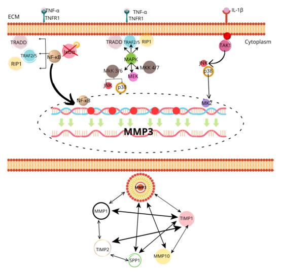 Fig. 1 Regulatory Pathways of MMP-3 Expression and Activation in ECM Degradation.1
Key structural properties of MMP3:
- Multi-domain organization
- Zinc ion-dependent catalytic center
- Substrate binding pocket and specificity
- Flexibility of the hinge area
Functions of MMP3
The main function of MMP3 (Matrix Metalloproteinase-3) is to degrade the extracellular matrix and participate in tissue remodeling. However, it is also involved in a variety of important physiological and pathological processes, including inflammation regulation and signal transduction.
| Function | Description |
| Matrix Degradation | It can specifically break down various components in the extracellular matrix, such as collagen (types III, IV, V), proteoglycans, laminin, and fibronectin, creating conditions for cell migration and proliferation. |
| Organ Reorganization | During physiological tissue repair (such as wound healing) and organ development (such as breast regression), controlled matrix degradation supports the reconstruction of the structure. |
| Inflammation Regulation | At the site of inflammation, activated stromal cells (such as fibroblasts) and immune cells secrete in large quantities, which, by disrupting the basement membrane and releasing cytokine precursors, amplify and regulate the inflammatory response. |
| Signal Activation | By cleaving and activating specific precursor molecules (such as pro-TNF-α) and other protease precursors (such as MMP-1, -7, -9), a crucial proteinase cascade amplification effect is achieved. |
| Disease Association | Its abnormal overexpression is closely related to the progression of various diseases, such as degrading joint cartilage in rheumatoid arthritis, destroying the fibrous cap in atherosclerosis, and promoting invasion and angiogenesis in tumor metastasis. |
Unlike other highly specialized members within the family, the substrate spectrum of MMP3 is relatively broad, making it a crucial "upstream regulator" that can initiate and coordinate multiple degradation and signaling events in the complex tissue microenvironment.
Applications of MMP3 and MMP3 Antibody in Literature
1. Roa-Bruzón, Iliannis Y., et al. "MMP3 as a Molecular Link: Unraveling the Connection Between Ankylosing Spondylitis and Acute Coronary Syndrome." Cells 14.8 (2025): 597. https://doi.org/10.3390/cells14080597
The research has found that the chronic inflammation in patients with ankylosing spondylitis is associated with an increased risk of cardiovascular diseases. Studies have shown that matrix metalloproteinase 3 (MMP-3) degrades the extracellular matrix, leading to the instability of atherosclerotic plaques, and interacts with inflammatory pathways such as TNF-α, potentially becoming a new therapeutic target for reducing the cardiovascular risk in such patients.
2. Seehawer, Marco, et al. "Loss of Kmt2c or Kmt2d drives brain metastasis via KDM6A-dependent upregulation of MMP3." Nature Cell Biology 26.7 (2024): 1165-1175. https://doi.org/10.1038/s41556-024-01446-3
The study found that in triple-negative breast cancer, the absence of KMT2C or KMT2D would upregulate the expression of matrix metalloproteinase 3 (MMP-3), thereby driving brain metastasis of the tumor. This process is mediated by the histone demethylase KDM6A, and inhibiting this pathway can reduce the risk of metastasis.
3. Rivera-Serrano, Mariela, et al. "Upregulation of MMP3 Promotes Cisplatin Resistance in Ovarian Cancer." International Journal of Molecular Sciences 26.9 (2025): 4012. https://doi.org/10.3390/ijms26094012
The study found that MMP3 is highly expressed in cisplatin-resistant ovarian cancer cells. Knockdown of MMP3 can inhibit the proliferation and invasion of the resistant cells, and synergistically inhibit tumor growth in vivo with cisplatin, suggesting that it participates in the resistance process through multiple pathways such as regulating the cell cycle, but the direct drug inhibitory effect is limited.
4. Kollar, Branislav, et al. "MMP3 is a non-invasive biomarker of rejection in skin-bearing vascularized composite allotransplantation: a multicenter validation study." Frontiers in immunology 10 (2019): 2771. https://doi.org/10.3389/fimmu.2019.02771
This study verified that serum MMP3 can serve as a non-invasive monitoring marker for acute rejection reactions in vascularized composite tissue transplantation. The results showed that serum MMP3 significantly increased after the operation, and further increased during severe rejection. A serum MMP3 level ≥ 5 times the baseline can effectively distinguish severe rejection from non-severe rejection, with a sensitivity of 76% and a specificity of 81%.
5. Yin, Yanling, et al. "Association of MMP3, MMP14, and MMP25 gene polymorphisms with cerebral stroke risk: a case-control study." BMC Medical Genomics 16.1 (2023): 297. https://doi.org/10.1186/s12920-023-01734-1
This study found in the Han Chinese population that the polymorphisms of MMP3 gene at rs520540 and rs679620 were significantly associated with the risk of stroke. The rs520540-A allele and rs679620-T allele could reduce the risk of ischemic stroke, but increase the risk of hemorrhagic stroke, and were related to indicators such as uric acid.
Creative Biolabs: MMP3 Antibodies for Research
Creative Biolabs specializes in the production of high-quality MMP3 antibodies for research and industrial applications. Our portfolio includes monoclonal antibodies tailored for ELISA, Flow Cytometry, Western blot, immunohistochemistry, and other diagnostic methodologies.
- Custom MMP3 Antibody Development: Tailor-made solutions to meet specific research requirements.
- Bulk Production: Large-scale antibody manufacturing for industry partners.
- Technical Support: Expert consultation for protocol optimization and troubleshooting.
- Aliquoting Services: Conveniently sized aliquots for long-term storage and consistent experimental outcomes.
For more details on our MMP3 antibodies, custom preparations, or technical support, contact us at email.
Reference
- Roa-Bruzón, Iliannis Y., et al. "MMP3 as a Molecular Link: Unraveling the Connection Between Ankylosing Spondylitis and Acute Coronary Syndrome." Cells 14.8 (2025): 597. https://doi.org/10.3390/cells14080597
Anti-MMP3 antibodies
 Loading...


Hot products 
-
Mouse Anti-ATG5 Recombinant Antibody (9H197) (CBMAB-A3945-YC)

-
Rabbit Anti-AKT3 Recombinant Antibody (V2-12567) (CBMAB-1057-CN)

-
Mouse Anti-ALB Recombinant Antibody (V2-180650) (CBMAB-A2186-YC)

-
Mouse Anti-APOA1 Monoclonal Antibody (CBFYR0637) (CBMAB-R0637-FY)

-
Mouse Anti-CTNND1 Recombinant Antibody (CBFYC-2414) (CBMAB-C2487-FY)

-
Mouse Anti-ALX1 Recombinant Antibody (96k) (CBMAB-C0616-FY)

-
Mouse Anti-AQP2 Recombinant Antibody (E-2) (CBMAB-A3358-YC)

-
Mouse Anti-ARID3A Antibody (A4) (CBMAB-0128-YC)

-
Mouse Anti-BACE1 Recombinant Antibody (61-3E7) (CBMAB-1183-CN)

-
Mouse Anti-B2M Recombinant Antibody (CBYY-0050) (CBMAB-0050-YY)

-
Mouse Anti-CGAS Recombinant Antibody (CBFYM-0995) (CBMAB-M1146-FY)

-
Mouse Anti-CD24 Recombinant Antibody (HIS50) (CBMAB-C10123-LY)

-
Mouse Anti-ALOX5 Recombinant Antibody (33) (CBMAB-1890CQ)

-
Mouse Anti-dsDNA Recombinant Antibody (22) (CBMAB-AP1954LY)

-
Rabbit Anti-ALK (Phosphorylated Y1278) Recombinant Antibody (D59G10) (PTM-CBMAB-0035YC)

-
Mouse Anti-CAT Recombinant Antibody (724810) (CBMAB-C8431-LY)

-
Mouse Anti-ALDOA Recombinant Antibody (A2) (CBMAB-A2316-YC)

-
Mouse Anti-ALB Recombinant Antibody (V2-363290) (CBMAB-S0173-CQ)

-
Rat Anti-CD63 Recombinant Antibody (7G4.2E8) (CBMAB-C8725-LY)

-
Mouse Anti-AQP2 Recombinant Antibody (G-3) (CBMAB-A3359-YC)

- AActivation
- AGAgonist
- APApoptosis
- BBlocking
- BABioassay
- BIBioimaging
- CImmunohistochemistry-Frozen Sections
- CIChromatin Immunoprecipitation
- CTCytotoxicity
- CSCostimulation
- DDepletion
- DBDot Blot
- EELISA
- ECELISA(Cap)
- EDELISA(Det)
- ESELISpot
- EMElectron Microscopy
- FFlow Cytometry
- FNFunction Assay
- GSGel Supershift
- IInhibition
- IAEnzyme Immunoassay
- ICImmunocytochemistry
- IDImmunodiffusion
- IEImmunoelectrophoresis
- IFImmunofluorescence
- IGImmunochromatography
- IHImmunohistochemistry
- IMImmunomicroscopy
- IOImmunoassay
- IPImmunoprecipitation
- ISIntracellular Staining for Flow Cytometry
- LALuminex Assay
- LFLateral Flow Immunoassay
- MMicroarray
- MCMass Cytometry/CyTOF
- MDMeDIP
- MSElectrophoretic Mobility Shift Assay
- NNeutralization
- PImmunohistologyp-Paraffin Sections
- PAPeptide Array
- PEPeptide ELISA
- PLProximity Ligation Assay
- RRadioimmunoassay
- SStimulation
- SESandwich ELISA
- SHIn situ hybridization
- TCTissue Culture
- WBWestern Blot
