
PDGFRB Antibodies
Background
The PDGFRB gene encodes platelet-derived growth factor receptor β protein, which is a tyrosine kinase receptor located on the cell membrane and mainly distributed in vascular smooth muscle cells, pericytes and some mesenchymal cells. This receptor is activated by specifically binding to ligands such as PDGF-BB, thereby regulating cell proliferation, migration and survival, and playing a core role in angiogenesis, embryonic development and tissue repair. This gene was first identified in the 1980s. Its abnormal activation is closely related to a variety of diseases, especially having a clear association with adolescent myelomonocytic leukemia caused by somatic mutations and primary familial cerebral calcification related to germline mutations. Due to its crucial role in cellular signal transduction and pathological mechanisms, PDGFRB has become an important molecular target for tumor-targeted therapy and research on rare genetic diseases, deepening people's understanding of the function of receptor tyrosine kinases and the pathogenic mechanisms of abnormal signaling pathways.
Structure of PDGFRB
The protein encoded by the PDGFRB gene is a transmembrane tyrosine kinase receptor with a molecular weight of approximately 123 kDa. This value is highly conserved among different species because its protein structure and function are of vital importance.
| Species | Human | Mouse | Rat |
| Molecular Weight (kDa) | 123 | 122 | 123 |
| Primary Structural Differences | Regulate vascular development and cell proliferation | Highly homologous to humans, it is used for model research | Functions and signaling pathways are highly conserved |
This protein is composed of 1106 amino acids, and its primary structure folds to form characteristic extracellular ligand-binding domains (five immunoglobulin-like domains), transmembrane regions, and intracellular tyrosine kinase domains. In its three-dimensional structure, the kinase domain presents a self-inhibitory conformation in the non-activated state, and specific amino acids in the near-membrane region (such as human Asn666) are crucial for maintaining this conformation. Ligand binding triggers receptor dimerization, leading to auto-phosphorylation of tyrosine residues (such as Tyr857 and Tyr1021), thereby opening the kinase active center and recruiting downstream signaling molecules to drive cellular responses.
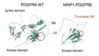 Fig. 1 Structures of WT PDGFRB and NRIP1::PDGFRB predicted with AlphaFold2.1
Key structural properties of PDGFRB:
- Extracellular immunoglobulin-like domains
- Proximal membrane autoinhibitory domain
- Split tyrosine kinase domain
Functions of PDGFRB
The receptor protein encoded by the PDGFRB gene plays a core role in cell signal transduction, with its main function being to regulate cell proliferation, survival and migration. However, it has also been found to be involved in a variety of physiological and pathological processes, including angiogenesis, tissue repair and tumorigenesis.
| Function | Description |
| Regulation of cell proliferation | After activation, it initiates downstream signaling pathways (such as RAS/MAPK), driving cell cycle progression and division. |
| Cell migration and chemotaxis | By reorganizing the cytoskeleton, it guides the directional migration of vascular smooth muscle cells and pericytes to participate in angiogenesis. |
| Cell survival inhibits apoptosis | Activate pro-survival pathways such as PI3K/AKT to maintain the vitality of cells under stressful conditions. |
| Vascular development and stability | In the embryonic and adult angiogenesis is critical, especially in weeks cell raise to stabilize the new blood vessels. |
| Tissue damage repair | In the process of wound healing and so on, mediated between the fibroblasts and mesenchymal cells repair responses. |
Like most single-pass transmembrane receptors, the activation of PDGFRB strictly depends on ligand-induced dimerization, and its signal transduction is typically spatiotemporal specific. Its function-acquired mutations can disrupt this regulation, leading to the continuous activation of receptors, and are key drivers of various myeloproliferative neoplasms and stromal tumors.
Applications of PDGFRB and PDGFRB Antibody in Literature
- Hao, Li, et al. "Somatic PDGFRB activating variants promote smooth muscle cell phenotype modulation in intracranial fusiform aneurysm." Journal of Biomedical Science 31.1 (2024): 51. https://doi.org/10.1186/s12929-024-01040-7
Research reveals that somatic mutations in PDGFRB activate the JAK-STAT pathway, leading to inflammatory phenotypic transformation in vascular smooth muscle cells and thereby driving the formation of fusiform aneurysms. Experiments have confirmed that the JAK inhibitor ruxolitinib can reverse this process, providing a new direction for treatment.
- Mathorne, Stine Westergaard, et al. "A novel PDGFRB sequence variant in a family with a mild form of primary familial brain calcification: a case report and a review of the literature." BMC neurology 19.1 (2019): 60. https://doi.org/10.1186/s12883-019-1292-8
A novel PDGFRB mutation (p.Gly612Arg) associated with cerebral calcification has been identified through research. This mutation is located in the tyrosine kinase domain. The symptoms and calcification degree of patients are usually mild. CT scans can detect asymptomatic mutation carriers.
- Li, Zhaoming, et al. "Recurrent PDGFRB mutations in unicentric Castleman disease." Leukemia 33.4 (2019): 1035-1038. https://doi.org/10.1038/s41375-018-0323-6
Whole exome sequencing studies have found that 17% of patients with single-center Castleman disease (UCD) have the PDGFRB p.asen666SER mutation. This mutation has the highest incidence in UCD among related cancers and is disease-specific, providing new clues for its pathogenesis.
- Sadras, Teresa, et al. "Unusual PDGFRB fusion reveals novel mechanism of kinase activation in Ph-like B-ALL." Leukemia 37.4 (2023): 905-909. https://doi.org/10.1038/s41375-023-01843-x
The study found that a novel CD74::PDGFRB fusion variant was discovered in children with Ph-like B-ALL. Although this variation does not form chimeric proteins, it can activate PDGFRB tyrosine kinases, drive leukemia occurrence, and provide a basis for targeted therapy.
- Nédélec, Audrey, et al. "Penttinen syndrome‐associated PDGFRB Val665Ala variant causes aberrant constitutive STAT1 signaling." Journal of cellular and molecular medicine 26.14 (2022): 3902-3912. https://doi.org/10.1111/jcmm.17427
Research has found that the PDGFRB p.Val665Ala mutation that causes Penttinen progeria continuously activates STAT1, triggering a unique interferon-like response that is not inhibited by ruxolitinib. This characteristic differs from other carcinogenic mutations and may explain its unique symptoms.
Creative Biolabs: PDGFRB Antibodies for Research
Creative Biolabs specializes in the production of high-quality PDGFRB antibodies for research and industrial applications. Our portfolio includes monoclonal antibodies tailored for ELISA, Flow Cytometry, Western blot, immunohistochemistry, and other diagnostic methodologies.
- Custom PDGFRB Antibody Development: Tailor-made solutions to meet specific research requirements.
- Bulk Production: Large-scale antibody manufacturing for industry partners.
- Technical Support: Expert consultation for protocol optimization and troubleshooting.
- Aliquoting Services: Conveniently sized aliquots for long-term storage and consistent experimental outcomes.
For more details on our PDGFRB antibodies, custom preparations, or technical support, contact us at email.
Reference
- Miyazaki, B., Ueno, T., Sugiyama. "Promoter swapping of truncated PDGFRB drives Ph-like acute lymphoblastic leukemia." npj Precision Oncology 7 (2023). https://doi.org/10.1038/s41698-023-00485-7
Anti-PDGFRB antibodies
 Loading...
 Compare




Hot products 
-
Mouse Anti-GGT1 Recombinant Antibody (1F9) (CBMAB-G3273-LY)

-
Rat Anti-CCR2 Recombinant Antibody (475301) (CBMAB-C1338-LY)

-
Mouse Anti-ADIPOR2 Recombinant Antibody (V2-179983) (CBMAB-A1369-YC)

-
Mouse Anti-AFDN Recombinant Antibody (V2-58751) (CBMAB-L0408-YJ)

-
Mouse Anti-FOXL1 Recombinant Antibody (CBXF-0845) (CBMAB-F0462-CQ)

-
Mouse Anti-CFL1 (Phospho-Ser3) Recombinant Antibody (CBFYC-1770) (CBMAB-C1832-FY)

-
Mouse Anti-14-3-3 Pan Recombinant Antibody (V2-9272) (CBMAB-1181-LY)

-
Mouse Anti-AP4E1 Recombinant Antibody (32) (CBMAB-A2996-YC)

-
Mouse Anti-CD24 Recombinant Antibody (HIS50) (CBMAB-C10123-LY)

-
Rabbit Anti-CCN1 Recombinant Antibody (CBWJC-3580) (CBMAB-C4816WJ)

-
Mouse Anti-APP Recombinant Antibody (DE2B4) (CBMAB-1122-CN)

-
Mouse Anti-ACTG1 Recombinant Antibody (V2-179597) (CBMAB-A0916-YC)

-
Mouse Anti-BrdU Recombinant Antibody (IIB5) (CBMAB-1038CQ)

-
Mouse Anti-AAV9 Recombinant Antibody (V2-634029) (CBMAB-AP023LY)

-
Mouse Anti-BCL2L1 Recombinant Antibody (H5) (CBMAB-1025CQ)

-
Mouse Anti-ARHGAP5 Recombinant Antibody (54/P190-B) (CBMAB-P0070-YC)

-
Mouse Anti-BIRC3 Recombinant Antibody (315304) (CBMAB-1214-CN)

-
Mouse Anti-CD59 Recombinant Antibody (CBXC-2097) (CBMAB-C4421-CQ)

-
Mouse Anti-CGAS Recombinant Antibody (CBFYM-0995) (CBMAB-M1146-FY)

-
Mouse Anti-CD83 Recombinant Antibody (HB15) (CBMAB-C1765-CQ)

- AActivation
- AGAgonist
- APApoptosis
- BBlocking
- BABioassay
- BIBioimaging
- CImmunohistochemistry-Frozen Sections
- CIChromatin Immunoprecipitation
- CTCytotoxicity
- CSCostimulation
- DDepletion
- DBDot Blot
- EELISA
- ECELISA(Cap)
- EDELISA(Det)
- ESELISpot
- EMElectron Microscopy
- FFlow Cytometry
- FNFunction Assay
- GSGel Supershift
- IInhibition
- IAEnzyme Immunoassay
- ICImmunocytochemistry
- IDImmunodiffusion
- IEImmunoelectrophoresis
- IFImmunofluorescence
- IGImmunochromatography
- IHImmunohistochemistry
- IMImmunomicroscopy
- IOImmunoassay
- IPImmunoprecipitation
- ISIntracellular Staining for Flow Cytometry
- LALuminex Assay
- LFLateral Flow Immunoassay
- MMicroarray
- MCMass Cytometry/CyTOF
- MDMeDIP
- MSElectrophoretic Mobility Shift Assay
- NNeutralization
- PImmunohistologyp-Paraffin Sections
- PAPeptide Array
- PEPeptide ELISA
- PLProximity Ligation Assay
- RRadioimmunoassay
- SStimulation
- SESandwich ELISA
- SHIn situ hybridization
- TCTissue Culture
- WBWestern Blot
