PFKFB3 Antibodies
Background
PFKFB3 is a crucial bifunctional enzyme protein that is mainly present in various mammalian cells. This enzyme regulates the synthesis and hydrolysis of fructose-2,6-diphosphate, thereby controlling the flux of glycolysis and influencing the energy metabolism and proliferation process of cells. Tumor cells often rely on the high expression of PFKFB3 to maintain their Warburg effect, which means that they still prefer glycolysis under aerobic conditions to support rapid growth. This gene was first identified in the 1990s, and its unique bifunctional domain and allosteric regulation mechanism have become an important model in metabolic research. In recent years, the development of inhibitors targeting PFKFB3 has provided a new direction for tumor metabolic therapy, and the study of its molecular mechanism continues to promote a deeper understanding of cellular metabolic plasticity and disease associations.
Structure of PFKFB3
The molecular weight of the PFKFB3 protein varies among different subtypes. The main isozyme, PFKFB3 protein, has a molecular weight of approximately 55 kDa. This difference mainly results from the selective splicing of its gene, generating different variants.
| Species | PFKFB3 (General Type) | PFKFB3 (splicing variant) |
| Molecular Weight (kDa) | ~55 | Slightly floating, ~53-57 |
| Main structural differences | Contains complete kinase and phosphatase domains | Specific exon deletions or insertions may occur, affecting the local functional regions. |
| Functional tendency | Typical dual-function enzyme activity, but kinase activity is significantly dominant | The activity ratio of specific variants or their subcellular localization may change. |
This protein is mainly composed of two key domains: the N-terminal kinase domain is responsible for synthesizing fructose-2,6-diphosphate, while the C-terminal phosphatase domain is responsible for its hydrolysis. Its tertiary structure forms a compact bifunctional unit, where the active site of the kinase domain contains a crucial ATP-binding pocket, and the phosphatase domain relies on a conserved histidine residue for catalysis. The two domains are connected by a flexible linker region, and this conformation allows its activity to be finely regulated by allosteric modulation and post-translational modifications (such as phosphorylation), thereby playing a central role in cellular metabolism and signal transduction.
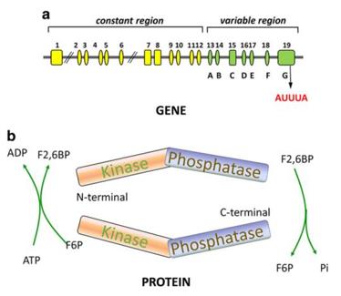 Fig. 1 General structure of the PFKFB3 gene and protein.1
Key structural properties of PFKFB3:
- Bifunctional domain configuration
- Conformational regulatory pocket
- Conservative catalytic site
- Translation modification site
Functions of PFKFB3
The core function of the PFKFB3 protein is to regulate the flux of glycolysis, but it is also involved in various pathological and physiological processes such as cell cycle, angiogenesis, and oxidative stress response.
| Function | Description |
| Glucose Oxidation Regulation | By generating fructose-2,6-bisphosphate (F2,6BP) as a co-activator, it activates phosphofructokinase-1 (PFK-1), thereby driving the glycolysis process and providing rapid energy supply to the cell. |
| Cell Cycle Promotion | It is expressed at an elevated level during the G1/S transition. Its metabolites provide precursors for nucleic acid and lipid synthesis, supporting cell proliferation. |
| Angiogenesis Support | Induced by HIF-1α under hypoxic conditions, it enhances the glycolysis of endothelial cells, providing energy and biosynthetic materials for angiogenesis. |
| Oxidative Stress Balance | The increased glycolytic flux mediated by it helps generate NADPH, which supports the antioxidant system (such as the glutathione cycle) to mitigate oxidative damage. |
| Survival Signal Integration | As the convergence point of cellular metabolism and growth factor signals (such as the PI3K/Akt pathway), it coordinates energy metabolism with cell growth and survival. |
The enzymatic activity of PFKFB3 is dominated by its kinase function (its kinase activity is approximately 700 times that of the phosphatase activity), which determines that its main role is to continuously promote the net synthesis of F2,6BP, thereby setting the cellular metabolism to an "oxidative glycolysis" state dominated by glycolysis, which is particularly crucial in tumor cells (Warburg effect) and activated immune cells.
Applications of PFKFB3 and PFKFB3 Antibody in Literature
1. Shi, Linlin, et al. "Roles of PFKFB3 in cancer." Signal transduction and targeted therapy 2.1 (2017): 1-10. https://doi.org/10.1038/sigtrans.2017.44
The article indicates that PFKFB3 is a key regulatory enzyme in glycolysis. The research has found that it plays a significant role in various processes such as cancer occurrence, proliferation, vascular invasion, drug resistance, and tumor microenvironment, and has become a potential anti-tumor treatment target.
2. Zhou, Zi-Yi, et al. "PFKFB3: a potential key to ocular angiogenesis." Frontiers in Cell and Developmental Biology 9 (2021): 628317. https://doi.org/10.3389/fcell.2021.628317
The article indicates that current anti-angiogenic treatments in ophthalmology mainly involve anti-VEGF therapy, but it has side effects and efficiency limitations. The regulation of glycolytic metabolism in endothelial cells has been found to affect angiogenesis, and the key enzyme PFKFB3 is expected to become a new target for treating pathological angiogenesis in eye diseases.
3. Liu, Peiyu, et al. "PFKFB3 in neovascular eye disease: unraveling mechanisms and exploring therapeutic strategies." Cell & Bioscience 14.1 (2024): 21. https://doi.org/10.1186/s13578-024-01205-9
The article indicates that current anti-VEGF therapy is the mainstream treatment for ocular neovascular diseases, but it has limitations. The key enzyme in glycolysis, PFKFB3, exhibits anti-angiogenic and multiple protective effects, and its targeted drugs provide a new potential direction for the treatment of this disease.
4. Zeng, Hao, et al. "Suppression of PFKFB3-driven glycolysis restrains endothelial-to-mesenchymal transition and fibrotic response." Signal Transduction and Targeted Therapy 7.1 (2022): 303. https://doi.org/10.1038/s41392-022-01097-6
The research has found that the abnormal glycolysis driven by PFKFB3 is a key mechanism for inducing endothelial-mesenchymal transition and can damage mitochondrial function. The inhibitor Danshen Polypeptide Acid C can counteract this process and cardiac fibrosis by disrupting the stability of PFKFB3, suggesting that metabolic intervention targeting PFKFB3 has therapeutic potential.
5. Xiao, Min, et al. "Role of PFKFB3-driven glycolysis in sepsis." Annals of medicine 55.1 (2023): 1278-1289. https://doi.org/10.1080/07853890.2023.2191217
The research has found that in sepsis, the key enzyme PFKFB3 of glycolysis is overly activated in both immune and non-immune cells, driving inflammatory responses and cell damage, resulting in a high mortality rate. Inhibiting PFKFB3 shows therapeutic potential, and the development of targeted drugs for this enzyme opens up new combined treatment strategies for sepsis.
Creative Biolabs: PFKFB3 Antibodies for Research
Creative Biolabs specializes in the production of high-quality PFKFB3 antibodies for research and industrial applications. Our portfolio includes monoclonal and polyclonal antibodies tailored for ELISA, Flow Cytometry, Western blot, immunohistochemistry, and other diagnostic methodologies.
- Custom PFKFB3 Antibody Development: Tailor-made solutions to meet specific research requirements.
- Bulk Production: Large-scale antibody manufacturing for industry partners.
- Technical Support: Expert consultation for protocol optimization and troubleshooting.
- Aliquoting Services: Conveniently sized aliquots for long-term storage and consistent experimental outcomes.
For more details on our PFKFB3 antibodies, custom preparations, or technical support, contact us at email.
Reference
- Shi, Linlin, et al. "Roles of PFKFB3 in cancer." Signal transduction and targeted therapy 2.1 (2017): 1-10. Distributed under Open Access license CC BY 4.0, without modification. https://doi.org/10.1038/sigtrans.2017.44
Anti-PFKFB3 antibodies
 Loading...


Hot products 
-
Mouse Anti-G6PD Recombinant Antibody (13B331) (CBMAB-G1553-LY)

-
Mouse Anti-ADIPOR1 Recombinant Antibody (V2-179982) (CBMAB-A1368-YC)

-
Mouse Anti-ENO1 Recombinant Antibody (8G8) (CBMAB-E1329-FY)

-
Mouse Anti-HTLV-1 gp46 Recombinant Antibody (CBMW-H1006) (CBMAB-V208-1154-FY)

-
Mouse Anti-BPGM Recombinant Antibody (CBYY-1806) (CBMAB-2155-YY)

-
Mouse Anti-ALB Recombinant Antibody (V2-55272) (CBMAB-H0819-FY)

-
Mouse Anti-CD59 Recombinant Antibody (CBXC-2097) (CBMAB-C4421-CQ)

-
Mouse Anti-EGR1 Recombinant Antibody (CBWJZ-100) (CBMAB-Z0289-WJ)

-
Mouse Anti-AFDN Recombinant Antibody (V2-58751) (CBMAB-L0408-YJ)

-
Mouse Anti-GLP1R Recombinant Antibody (4F3) (CBMAB-G0521-LY)

-
Human Anti-SARS-CoV-2 S1 Monoclonal Antibody (CBFYR-0120) (CBMAB-R0120-FY)

-
Rabbit Anti-ALOX5AP Recombinant Antibody (CBXF-1219) (CBMAB-F0750-CQ)

-
Mouse Anti-ALDOA Recombinant Antibody (A2) (CBMAB-A2316-YC)

-
Mouse Anti-COL1A2 Recombinant Antibody (CF108) (V2LY-1206-LY626)

-
Rat Anti-AChR Recombinant Antibody (V2-12500) (CBMAB-0990-CN)

-
Mouse Anti-BAX Recombinant Antibody (CBYY-0216) (CBMAB-0217-YY)

-
Mouse Anti-CFL1 Recombinant Antibody (CBFYC-1771) (CBMAB-C1833-FY)

-
Mouse Anti-ALB Recombinant Antibody (V2-363290) (CBMAB-S0173-CQ)

-
Mouse Anti-14-3-3 Pan Recombinant Antibody (V2-9272) (CBMAB-1181-LY)

-
Rabbit Anti-AKT3 Recombinant Antibody (V2-12567) (CBMAB-1057-CN)

- AActivation
- AGAgonist
- APApoptosis
- BBlocking
- BABioassay
- BIBioimaging
- CImmunohistochemistry-Frozen Sections
- CIChromatin Immunoprecipitation
- CTCytotoxicity
- CSCostimulation
- DDepletion
- DBDot Blot
- EELISA
- ECELISA(Cap)
- EDELISA(Det)
- ESELISpot
- EMElectron Microscopy
- FFlow Cytometry
- FNFunction Assay
- GSGel Supershift
- IInhibition
- IAEnzyme Immunoassay
- ICImmunocytochemistry
- IDImmunodiffusion
- IEImmunoelectrophoresis
- IFImmunofluorescence
- IGImmunochromatography
- IHImmunohistochemistry
- IMImmunomicroscopy
- IOImmunoassay
- IPImmunoprecipitation
- ISIntracellular Staining for Flow Cytometry
- LALuminex Assay
- LFLateral Flow Immunoassay
- MMicroarray
- MCMass Cytometry/CyTOF
- MDMeDIP
- MSElectrophoretic Mobility Shift Assay
- NNeutralization
- PImmunohistologyp-Paraffin Sections
- PAPeptide Array
- PEPeptide ELISA
- PLProximity Ligation Assay
- RRadioimmunoassay
- SStimulation
- SESandwich ELISA
- SHIn situ hybridization
- TCTissue Culture
- WBWestern Blot
