SARS2 Antibodies
Background
The SARS2 gene serves as the complete genetic material encoding the novel coronavirus pathogen and mainly exists within the nucleocapsid of the virus particle. This gene replicates through RNA polymerase and guides the synthesis of viral structural and non-structural proteins, thereby mediating key biological processes such as viral invasion of host cells and immune escape. Global scientific research teams rely on the sequencing and analysis of the SARS2 gene to track the virus's mutation trajectory, as its sequence characteristics continuously evolve with transmission. The genome of this virus was sequenced and made public by Chinese scientists in January 2020. Subsequently, the three-dimensional structure of its spike protein was rapidly resolved through cryo-electron microscopy technology, laying a decisive foundation for global vaccine research and development. Its highly modular gene organization and rapid evolution mechanism have become the core research objects in the fields of molecular epidemiology, RNA virology and new vaccine design, greatly promoting the systematic understanding of the evolution laws and prevention and control strategies of emerging zoonotic viruses by humans.
Structure of SARS2
The SARS2 (Severe Acute Respiratory Syndrome Coronavirus 2) gene is a single-stranded justice RNA genome of approximately 29.9 kb. There are slight differences in this length among different variants, mainly due to mutations and insertions and deletions in regions such as the spike protein.
| Species | Original plant | Delta variant | Omicron BA.1 | Omicron BA.2 |
| Molecular Weight (kDa) | 29,903 | About 29,892 | About 29,866 | About 29,872 |
| Primary Structural Differences | Original reference sequence | Mutations such as L452R and P681R | Numerous spike protein mutations (e.g., G339D, S371L) | Deletion of partial characteristic mutations in BA.1, such as deletion restoration of 69-70 |
The primary structure of the viral genome is composed of a series of open reading frames (ORFs), encoding structural proteins (such as spike protein S, nucleocapsid protein N) and non-structural proteins (such as RNA-dependent RNA polymerase RdRp). Its secondary structure forms various stem loops and pseudoknots through complex RNA folding, and these structures play a crucial role in viral genome replication, subgenomic mRNA synthesis, and evasion of host immune responses. For instance, conserved elements such as transcriptional regulatory sequences (TRS) and frame transition elements (FSE) regulate the expression of viral proteins through specific spatial conformations.
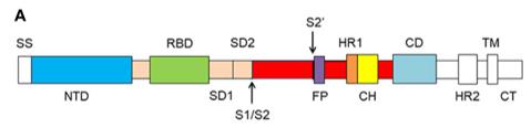 Fig. 1 Schematic of SARS-CoV-2 spike protein primary structure.1
Key structural properties of SARS2:
- Single-stranded justice RNA structure
- 5 'end cap structure and 3' end poly real (A) tail
- Contains multiple open reading frames (ORF1a/b, etc.)
Functions of SARS2
The main function of the SARS2 gene is to encode the structural and non-structural proteins of the novel coronavirus, guiding viral replication and infection. In addition, it also determines the virus's transmissibility, pathogenicity and immune escape ability.
| Function | Description |
| Virus replication | The genome serves as a template to replicate the progeny viral RNA through RNA-dependent RNA polymerase (RdRp) and transcribe it into subgenomic mRNA. |
| Protein coding | Encode structural proteins such as spike protein (S) and nucleocapsid protein (N), as well as non-structural proteins such as protease (PLpro) and helicase, and assemble them into complete viral particles. |
| Cell infection | The spike protein binds to the ACE2 receptor of the host cell, mediating the entry of the virus into the cell and initiating the infection cycle. |
| Immune regulation | Some proteins (such as ORF3a and ORF6) can inhibit the host interferon signaling pathway, helping viruses evade immune recognition. |
| Mutation and Evolution | The genome is prone to mutation and recombination during replication, generating different variants that affect the speed of transmission, the severity of the disease and the effectiveness of vaccines. |
The replication fidelity of the SARS2 gene is relatively low, and its RNA polymerase lacks proofreading function, resulting in a high mutation rate, which is similar to RNA viruses such as influenza viruses. However, SARS2 also has a replication correction mechanism, which to some extent limits the mutation rate, enabling it to adapt to the host during transmission while maintaining the stability of its basic functions.
Applications of SARS2 and SARS2 Antibody in Literature
- Wang, Mei-Yue, et al. "SARS-CoV-2: structure, biology, and structure-based therapeutics development." Frontiers in cellular and infection microbiology 10 (2020): 587269. https://doi.org/10.3389/fcimb.2020.587269
The article indicates that SARS-CoV-2 continues to threaten the world and there is still a lack of effective treatments and vaccines at present. This article reviews the epidemic characteristics, evolutionary origin, receptor recognition mechanism and protein structure of the virus, and discusses the research and development progress of antibodies, drugs and vaccines based on structure, with the aim of providing references for prevention and control practices.
- Ren, Jiang, et al. "TRIM28-mediated nucleocapsid protein SUMOylation enhances SARS-CoV-2 virulence." Nature Communications 15.1 (2024): 244. https://doi.org/10.1038/s41467-023-44502-6
This study found that the nucleocapsid protein of SARS-CoV-2 (SARS2-NP) underwent SUMOylation modification at lysine at position 65, and this modification enhanced its oligomerization, phase separation and immunosuppressive abilities. The SUMO ligase TRIM28 catalyzes this process, and its inhibitors can effectively suppress viral replication and restore the host's immune response.
- Wu, Zhengliang L., and James M. Ertelt. "Fluorescent glycan fingerprinting of SARS2 spike proteins." Scientific Reports 11.1 (2021): 20428. https://doi.org/10.1038/s41598-021-98919-4
This study established a new method for glucose fingerprinting based on enzymatic fluorescence labeling and gel electrophoresis, which can rapidly analyze the N-glycosylation and O-glycosylation patterns of the SARS-CoV-2 spike protein, providing a convenient technique for the global evaluation of glycoproteins.
- Wang, Youchun. "SARS-CoV-2 Neutralizing Antibodies 2.0." Viruses 16.11 (2024): 1791. https://doi.org/10.3390/v16111791
The article indicates that in the face of the continuously mutating SARS-CoV-2, this special issue brings together research covering broad-spectrum neutralizing antibodies, optimized therapeutic antibodies, vaccine booster shots, and new detection technologies, providing a variety of effective prevention and control strategies and tools for dealing with the current prevalent variants.
- Ariumi, Yasuo. "Host cellular RNA helicases regulate SARS-CoV-2 infection." Journal of Virology 96.6 (2022): e00002-22. https://doi.org/10.1128/jvi.00002-22
This study reveals the complex regulatory role of host cell RNA helicases in SARS-CoV-2 infection: DDX1/5/6 promotes viral replication, while DDX21 and MOV10 have inhibitory functions. Viral nucleocapsid proteins hijack the host mechanism by binding and interfering with the formation of P-bodies.
Creative Biolabs: SARS2 Antibodies for Research
Creative Biolabs specializes in the production of high-quality SARS2 antibodies for research and industrial applications. Our portfolio includes monoclonal antibodies tailored for ELISA, Flow Cytometry, Western blot, immunohistochemistry, and other diagnostic methodologies.
- Custom SARS2 Antibody Development: Tailor-made solutions to meet specific research requirements.
- Bulk Production: Large-scale antibody manufacturing for industry partners.
- Technical Support: Expert consultation for protocol optimization and troubleshooting.
- Aliquoting Services: Conveniently sized aliquots for long-term storage and consistent experimental outcomes.
For more details on our SARS2 antibodies, custom preparations, or technical support, contact us at email.
Reference
- Wang, Mei-Yue, et al. "SARS-CoV-2: structure, biology, and structure-based therapeutics development." Frontiers in cellular and infection microbiology 10 (2020): 587269. https://doi.org/10.3389/fcimb.2020.587269
Anti-SARS2 antibodies
 Loading...


Hot products 
-
Rat Anti-FABP3 Recombinant Antibody (CBXF-2299) (CBMAB-F1612-CQ)

-
Mouse Anti-C5AR1 Recombinant Antibody (R63) (CBMAB-C9553-LY)

-
Mouse Anti-CFL1 (Phospho-Ser3) Recombinant Antibody (CBFYC-1770) (CBMAB-C1832-FY)

-
Mouse Anti-CASQ1 Recombinant Antibody (CBFYC-0863) (CBMAB-C0918-FY)

-
Mouse Anti-ARIH1 Recombinant Antibody (C-7) (CBMAB-A3563-YC)

-
Mouse Anti-NSUN6 Recombinant Antibody (D-5) (CBMAB-N3674-WJ)

-
Mouse Anti-AHCYL1 Recombinant Antibody (V2-180270) (CBMAB-A1703-YC)

-
Mouse Anti-DLG1 Monolconal Antibody (4F3) (CBMAB-0225-CN)

-
Mouse Anti-G6PD Recombinant Antibody (13B331) (CBMAB-G1553-LY)

-
Mouse Anti-CD33 Recombinant Antibody (6C5/2) (CBMAB-C8126-LY)

-
Mouse Anti-AMOT Recombinant Antibody (CBYC-A564) (CBMAB-A2552-YC)

-
Mouse Anti-CD8 Recombinant Antibody (C1083) (CBMAB-C1083-LY)

-
Mouse Anti-APP Recombinant Antibody (DE2B4) (CBMAB-1122-CN)

-
Mouse Anti-ASB9 Recombinant Antibody (1D8) (CBMAB-A0529-LY)

-
Mouse Anti-ADIPOR2 Recombinant Antibody (V2-179983) (CBMAB-A1369-YC)

-
Mouse Anti-BSN Recombinant Antibody (219E1) (CBMAB-1228-CN)

-
Mouse Anti-ATP5F1A Recombinant Antibody (51) (CBMAB-A4043-YC)

-
Mouse Anti-BCL6 Recombinant Antibody (CBYY-0435) (CBMAB-0437-YY)

-
Mouse Anti-EMP3 Recombinant Antibody (CBFYE-0100) (CBMAB-E0207-FY)

-
Mouse Anti-FYN Recombinant Antibody (10) (CBMAB-S6332-CQ)

- AActivation
- AGAgonist
- APApoptosis
- BBlocking
- BABioassay
- BIBioimaging
- CImmunohistochemistry-Frozen Sections
- CIChromatin Immunoprecipitation
- CTCytotoxicity
- CSCostimulation
- DDepletion
- DBDot Blot
- EELISA
- ECELISA(Cap)
- EDELISA(Det)
- ESELISpot
- EMElectron Microscopy
- FFlow Cytometry
- FNFunction Assay
- GSGel Supershift
- IInhibition
- IAEnzyme Immunoassay
- ICImmunocytochemistry
- IDImmunodiffusion
- IEImmunoelectrophoresis
- IFImmunofluorescence
- IGImmunochromatography
- IHImmunohistochemistry
- IMImmunomicroscopy
- IOImmunoassay
- IPImmunoprecipitation
- ISIntracellular Staining for Flow Cytometry
- LALuminex Assay
- LFLateral Flow Immunoassay
- MMicroarray
- MCMass Cytometry/CyTOF
- MDMeDIP
- MSElectrophoretic Mobility Shift Assay
- NNeutralization
- PImmunohistologyp-Paraffin Sections
- PAPeptide Array
- PEPeptide ELISA
- PLProximity Ligation Assay
- RRadioimmunoassay
- SStimulation
- SESandwich ELISA
- SHIn situ hybridization
- TCTissue Culture
- WBWestern Blot
