TIM4 Antibodies
Background
TIM4 is an immunoglobulin superfamily receptor protein located on the cell membrane, mainly expressed on the surface of immune cells such as macrophages and dendritic cells. The protein encoded by this gene mediates the physiological process of phagocytosis and clearance of apoptotic cells by specifically recognizing and binding to phosphatidylserine on the surface of apoptotic cells, thereby maintaining tissue homeostasis and immune tolerance. Its function was jointly clarified by multiple research teams in 2003. As a member of the T-cell immunoglobulin domain and mucin domain protein family, TIM4 plays a key role in immune regulation. The extracellular immunoglobulin variable domain of this protein has been resolved into a typical β -sandwich folding conformation. This structural feature provides an important model for studying immune receptor-ligand interactions and greatly advances the understanding of programmed cell clearance mechanisms and molecular pathways related to autoimmune diseases.
Structure of TIM4
TIM4 is a type I transmembrane protein with a molecular weight of approximately 54 kDa. This value may vary slightly due to the degree of glycosylation modification in different species.
| Species | Human | Mouse | Rat | Rhesus monkey |
| Molecular Weight (kDa) | 54 | 53.5 | 53.8 | 54.2 |
| Primary Structural Differences | With the typical structure of IgV domain and mucins sample area | The intracellular region is relatively short, and there are differences in signal pathways | High homology with mouse | Highly similar to the human protein structure |
The TIM4 protein is composed of 302 amino acids, and its extracellular region contains an immunoglobulin variable region (IgV) domain, which recognizes and binds to phosphatidylserine on the surface of apoptotic cells through a specific stereoconformation. The secondary structure of the protein is mainly composed of β -folds, which form two reverse-parallel β -sheets and jointly shape the characteristic "sandwich" three-dimensional form of the IgV domain. A key metal ion binding site (typically a calcium ion) is located at the top of the domain and stabilizes the ligand binding interface by coordinating coordination bonds, which is crucial for maintaining its physiological functions.
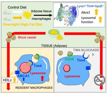 Fig. 1 High-Fat Diet Primes Tim4+ Adipose Macrophages via Abca1 and Lysosomes.1
Key structural properties of TIM4:
- Folding structure of extracellular immunoglobulin variable region (IgV)
- Apical metal ion binding ring stabilized ligand recognition interface
- Specific phosphatidylserine binding site
Functions of TIM4
The core function of the protein encoded by the TIM4 gene is to mediate the recognition and clearance of apoptotic cells. In addition, it is also involved in a variety of immune regulatory processes.
| Function | Description |
| Apoptosis cell clearance | Specifically recognizing and binding to phosphatidylserine on the surface of apoptotic cells, initiating phagocytosis and maintaining tissue homeostasis. |
| Regulation of immune tolerance | By eliminating its own apoptotic cells, it can prevent the exposure of internal antigens and avoid the occurrence of autoimmune reactions. |
| Regulation of inflammatory response | After the completion of cell burial, it can inhibit the release of pro-inflammatory factors and promote the resolution of inflammation. |
| Intercellular interactions | Cooperate with other receptors on the surface of immune cells (such as TIM1 and TIM3) to regulate cell-to-cell recognition and signal transmission. |
| Pathogen identification | In some cases, the phosphatidylserine mimic structure on the surface of the virus can be identified and participates in antiviral immunity. |
The affinity of TIM4 for phosphatidylserine is constant and single, which is different from some polyvalent receptors with synergistic effects. This is consistent with its physiological role as a rapid recognition and initial anchoring receptor.
Applications of TIM4 and TIM4 Antibody in Literature
1. Wang, Ziming, et al. "Tim4 deficiency reduces CD301b+ macrophage and aggravates periodontitis bone loss." International Journal of Oral Science 16.1 (2024): 20. https://doi.org/10.1038/s41368-023-00270-z
The article indicates that in periodontitis, Tim4 affects the phenotype of CD301b+ macrophages by regulating the p38 MAPK signaling pathway. Its absence reduces the number of these cells and exacerbates alveolar bone resorption, providing a new target for the immunotherapy of periodontitis.
2. Wang, Xinyue, et al. "Rescue RM/CS-AKI by blocking strategy with one-dose anti-myoglobin RabMAb." Nature Communications 16.1 (2025): 1044. https://doi.org/10.3389/frtra.2023.1176384
The article indicates that in liver transplantation ischemia-reperfusion injury (IRI), donor Tim4 deficiency can alleviate liver injury, while recipient Tim4 deficiency will exacerbate metabolic stress and apoptosis. Clinical data confirm that high expression of TIM4 is associated with aggravated liver injury and reduced graft survival rate. Tim4 plays a dual role in transplant immunity.
3. Caronni, Nicoletta, et al. "TIM4 expression by dendritic cells mediates uptake of tumor-associated antigens and anti-tumor responses." Nature communications 12.1 (2021): 2237. https://doi.org/10.1038/s41467-021-22535-z
The article indicates that in lung adenocarcinoma, TIM4 on the surface of dendritic cell cDC1 is the key to phagocytosis of tumor antigens and activation of CD8+ T cells. Tumor progression can inhibit the expression of TIM4, thereby weakening immune surveillance. Studies have confirmed that the level of TIM4 is an important indicator for evaluating prognosis and predicting the response to PD-1 treatment.
4. Wang, Liang, et al. "TIM4+ macrophages suppress the proinflammatory response to maintain the chronic alveolar echinococcosis infection." Frontiers in Cellular and Infection Microbiology 15 (2025): 1600686. https://doi.org/10.3389/fcimb.2025.1600686
The article indicates that in hepatic multilocular echinococcosis, Tim-4 is highly expressed in the lesion macrophages, maintains immune tolerance by inhibiting pro-inflammatory factors, promotes parasite immune escape and aggravates liver fibrosis. Inhibiting Tim-4 can restore the inflammatory response and reverse the fibrosis process.
5. Yang, Bo, et al. "Histone acetyltransferease p300 modulates TIM4 expression in dendritic cells." Scientific reports 6.1 (2016): 21336. https://doi.org/10.1038/srep21336
The article indicates that in the food allergy model, the expression of TIM4 in dendritic cells is regulated by p300 and STAT6. The microbial product CT increases p300 and then acts on STAT6, jointly inducing the expression of TIM4, thereby initiating the Th2 immune bias response.
Creative Biolabs: TIM4 Antibodies for Research
Creative Biolabs specializes in the production of high-quality TIM4 antibodies for research and industrial applications. Our portfolio includes monoclonal antibodies tailored for ELISA, Flow Cytometry, Western blot, immunohistochemistry, and other diagnostic methodologies.
- Custom TIM4 Antibody Development: Tailor-made solutions to meet specific research requirements.
- Bulk Production: Large-scale antibody manufacturing for industry partners.
- Technical Support: Expert consultation for protocol optimization and troubleshooting.
- Aliquoting Services: Conveniently sized aliquots for long-term storage and consistent experimental outcomes.
For more details on our TIM4 antibodies, custom preparations, or technical support, contact us at email.
Reference
- Magalhaes, Marlène Sophie, et al. "Role of Tim4 in the regulation of ABCA1+ adipose tissue macrophages and post-prandial cholesterol levels." Nature communications 12.1 (2021): 4434. https://doi.org/10.1038/s41467-021-24684-7
Anti-TIM4 antibodies
 Loading...


Hot products 
-
Mouse Anti-FOXL1 Recombinant Antibody (CBXF-0845) (CBMAB-F0462-CQ)

-
Rat Anti-ADGRE4 Recombinant Antibody (V2-160163) (CBMAB-F0011-CQ)

-
Mouse Anti-COL12A1 Recombinant Antibody (CBYY-C3117) (CBMAB-C4560-YY)

-
Mouse Anti-8-oxoguanine Recombinant Antibody (V2-7719) (CBMAB-1898CQ)

-
Mouse Anti-GDF5 Recombinant Antibody (1F4) (CBMAB-G2740-LY)

-
Mouse Anti-CAT Recombinant Antibody (724810) (CBMAB-C8431-LY)

-
Rat Anti-4-1BB Recombinant Antibody (V2-1558) (CBMAB-0953-LY)

-
Rabbit Anti-ABL1 (Phosphorylated Y245) Recombinant Antibody (V2-505716) (PTM-CBMAB-0465LY)

-
Rabbit Anti-AKT2 (Phosphorylated S474) Recombinant Antibody (V2-556130) (PTM-CBMAB-0605LY)

-
Mouse Anti-DLG1 Monolconal Antibody (4F3) (CBMAB-0225-CN)

-
Mouse Anti-C1QC Recombinant Antibody (CBFYC-0600) (CBMAB-C0654-FY)

-
Rat Anti-EPO Recombinant Antibody (16) (CBMAB-E1578-FY)

-
Mouse Anti-CAPZB Recombinant Antibody (CBYY-C0944) (CBMAB-C2381-YY)

-
Mouse Anti-ABCA3 Recombinant Antibody (V2-178911) (CBMAB-A0145-YC)

-
Mouse Anti-CARD11 Recombinant Antibody (CBFYC-0811) (CBMAB-C0866-FY)

-
Mouse Anti-ANXA7 Recombinant Antibody (A-1) (CBMAB-A2941-YC)

-
Mouse Anti-ADV Recombinant Antibody (V2-503423) (CBMAB-V208-1364-FY)

-
Mouse Anti-ENPP1 Recombinant Antibody (CBFYE-0159) (CBMAB-E0375-FY)

-
Rat Anti-CD63 Recombinant Antibody (7G4.2E8) (CBMAB-C8725-LY)

-
Mouse Anti-BrdU Recombinant Antibody (IIB5) (CBMAB-1038CQ)

- AActivation
- AGAgonist
- APApoptosis
- BBlocking
- BABioassay
- BIBioimaging
- CImmunohistochemistry-Frozen Sections
- CIChromatin Immunoprecipitation
- CTCytotoxicity
- CSCostimulation
- DDepletion
- DBDot Blot
- EELISA
- ECELISA(Cap)
- EDELISA(Det)
- ESELISpot
- EMElectron Microscopy
- FFlow Cytometry
- FNFunction Assay
- GSGel Supershift
- IInhibition
- IAEnzyme Immunoassay
- ICImmunocytochemistry
- IDImmunodiffusion
- IEImmunoelectrophoresis
- IFImmunofluorescence
- IGImmunochromatography
- IHImmunohistochemistry
- IMImmunomicroscopy
- IOImmunoassay
- IPImmunoprecipitation
- ISIntracellular Staining for Flow Cytometry
- LALuminex Assay
- LFLateral Flow Immunoassay
- MMicroarray
- MCMass Cytometry/CyTOF
- MDMeDIP
- MSElectrophoretic Mobility Shift Assay
- NNeutralization
- PImmunohistologyp-Paraffin Sections
- PAPeptide Array
- PEPeptide ELISA
- PLProximity Ligation Assay
- RRadioimmunoassay
- SStimulation
- SESandwich ELISA
- SHIn situ hybridization
- TCTissue Culture
- WBWestern Blot
