CD48 Antibodies
Background
CD48 is a glycosylphosphatidylinositol anchoring protein widely expressed on the surface of immune cells, mainly existing in hematopoietic cells such as lymphocytes, dendritic cells and macrophages. This protein, as an immunomodulatory molecule, participates in intercellular signal transduction through interactions with receptors such as CD244 and CD2, thereby regulating the activation and response of immune cells like natural killer cells and T cells. Since its identification in the 1980s, CD48 has received continuous attention due to its crucial role in immune synaptic formation, infection response and autoimmune diseases. The research on its structure and function has deepened people's understanding of the mechanisms of immune recognition, cell communication and inflammatory regulation, providing an important molecular basis for the development of related therapeutic strategies.
Structure of CD48
CD48 is a glycosylphosphatidylinositol anchored membrane protein with a molecular weight of approximately 40-45 kDa. Its exact molecular weight may fluctuate slightly among different species and tissues due to varying degrees of glycosylation modification. The following is a comparison of the typical characteristics of CD48 protein in some species:
| Species | Human | Mouse | Rat |
| Molecular Weight (kDa) | About 41 | About 43 | About 40 |
| Primary Structural Differences | With two immune globulin structure domain and C2 type (V), through the GPI anchor is due to the cell membrane | Structural homology with human height, glycosylation pattern | Conserved immunoglobulin-like folding, with similar extracellular domain structures |
This protein is composed of approximately 220 amino acid residues, and its extracellular region forms a typical folded structure of the immunoglobulin superfamily. CD48 does not directly participate in signal transduction, but as a ligand, it can play a key role in the regulation of adhesion and activation between immune cells through interactions with various receptors such as CD244 and CD2. Its structural stability mainly depends on conserved disulfide bonds and glycosylation modifications, which jointly maintain its functional conformation in the immune synapse.
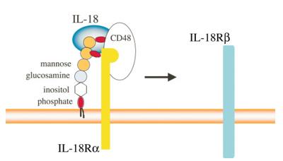 Fig. 1 Mechanism of IL-18 Binding Involving CD48.1
Key structural properties of CD48:
- Folding of the immunoglobulin superfamily (IgSF) domain
- Glycosylphosphatidylinositol (GPI) anchoring
- Highly glycosylated extracellular domains
- Conservative disulfide bond network
- Lack of transmembrane regions and intracellular domains
Functions of CD48
The main function of the CD48 protein is to act as a costimulatory molecule and adhesion molecule for immune regulation. However, it is also deeply involved in a variety of immunopathological processes, including infection defense, autoimmunity and tumor immune surveillance.
| Function | Description |
| Immune regulation and signal transduction | As a ligand, it binds to receptors such as CD244 and CD2, regulating the activation threshold and function of natural killer cells, T cells and B cells, and transmitting co-stimulatory or co-inhibitory signals. |
| Immune synaptic formation | Enriched at the contact interface between immune cells, it acts as a scaffold molecule to promote the stable formation of immune synapses and ensure the directional transmission of signal molecules. |
| Infection response | During bacterial or viral infections, its expression is upregulated and can act as a receptor for certain pathogens (such as the measles virus), while also helping to recruit and activate effector immune cells to clear the infection. |
| Autoimmune regulation | Abnormal signal transduction is associated with a variety of autoimmune diseases. For instance, in systemic lupus erythematosus and rheumatoid arthritis, its expression or dysfunction may lead to the disruption of immune tolerance. |
| Tumor immune surveillance | In the tumor microenvironment, the mediated interactions affect the ability of natural killer cells and cytotoxic T cells to recognize and clear tumor cells. |
The affinity of CD48 for interaction with receptors (such as CD244) is relatively low. This characteristic makes it more suitable for mediating transient and reversible intercellular contact, thereby precisely regulating the timing and intensity of immune activation. This forms a functional contrast with many high-affinity receptor-ligand systems that trigger continuous activation.
Applications of CD48 and CD48 Antibody in Literature
1. Liu, Jiye, et al. "Epigenetic regulation of CD38/CD48 by KDM6A mediates NK cell response in multiple myeloma." Nature Communications 15.1 (2024): 1367. https://doi.org/10.1038/s41467-024-45561-z
The article indicates that through CRISPR screening, it was found that the deletion of KDM6A up-regulates H3K27me3, inhibits the expression of CD38 and CD48, and leads to resistance to daratumumab. The use of EZH2 inhibitors can reverse this process, providing a new strategy for enhancing the therapeutic effect of multiple myeloma.
2. Martinez-Vicente, Pablo, et al. "Subversion of natural killer cell responses by a cytomegalovirus-encoded soluble CD48 decoy receptor." PLoS pathogens 15.4 (2019): e1007658. https://doi.org/10.1371/journal.ppat.1007658
Research has revealed that the owl monkey cytomegalovirus protein A43, as a soluble CD48 bait receptor, blocks the recognition and killing of NK cells by binding to host 2B4 with high affinity, revealing a new mechanism of viral immune escape.
3. Danquah, Bright D., et al. "Mass Spectrometric analysis of antibody—Epitope peptide complex dissociation: Theoretical concept and practical procedure of binding strength characterization." Molecules 25.20 (2020): 4776. https://doi.org/10.1155/2024/6908968
Research has revealed that through bioinformatics analysis, CCR7 and CD48 have been identified as key biomarkers of acute rejection after kidney transplantation, closely related to the immune response mediated by macrophages and NK cells, providing new targets for prediction and treatment.
4. Kotzur, Rebecca, et al. "Eradication of CD48-positive tumors by selectively enhanced YTS cells harnessing the lncRNA NeST." Iscience 26.8 (2023). https://doi.org/10.1016/j.isci.2023.107284
In this study, by enhancing the expression of lncRNA NeST, an NK-like cell line seYTS that secretes more IFNγ was constructed. This cell can still effectively kill tumors expressing CD48 after irradiation, providing a new strategy for universal tumor therapy targeting CD48.
5. Marchitto, Lorie, et al. "Impact of HIV-1 Vpu-mediated downregulation of CD48 on NK-cell-mediated antibody-dependent cellular cytotoxicity." Mbio 14.4 (2023): e00789-23. https://doi.org/10.1128/mbio.00789-23
The article indicates that HIV-1 down-regulates CD48 on the surface of infected cells through its Vpu protein to evade 2B4 receptor-mediated NK cell activation and antibody-dependent cytotoxicity. This mechanism, together with the down-regulation of NTB-A ligands, jointly weakens immune attack and is an important immune escape strategy of the virus.
Creative Biolabs: CD48 Antibodies for Research
Creative Biolabs specializes in the production of high-quality CD48 antibodies for research and industrial applications. Our portfolio includes monoclonal antibodies tailored for ELISA, Flow Cytometry, Western blot, immunohistochemistry, and other diagnostic methodologies.
- Custom CD48 Antibody Development: Tailor-made solutions to meet specific research requirements.
- Bulk Production: Large-scale antibody manufacturing for industry partners.
- Technical Support: Expert consultation for protocol optimization and troubleshooting.
- Aliquoting Services: Conveniently sized aliquots for long-term storage and consistent experimental outcomes.
For more details on our CD48 antibodies, custom preparations, or technical support, contact us at email.
Reference
- Fukushima, Keiko, Yukio Ikehara, and Katsuko Yamashita. "Functional role played by the glycosylphosphatidylinositol anchor glycan of CD48 in interleukin-18-induced interferon-γ production." Journal of Biological Chemistry 280.18 (2005): 18056-18062. https://doi.org/10.1074/jbc.M413297200
Anti-CD48 antibodies
 Loading...


Hot products 
-
Mouse Anti-ALPL Antibody (B4-78) (CBMAB-1009CQ)

-
Mouse Anti-FN1 Monoclonal Antibody (71) (CBMAB-1241CQ)

-
Rat Anti-ABCC11 Recombinant Antibody (V2-179001) (CBMAB-A0236-YC)

-
Mouse Anti-CASP8 Recombinant Antibody (CBYY-C0987) (CBMAB-C2424-YY)

-
Mouse Anti-FeLV g27 Recombinant Antibody (1) (CBMAB-V208-1714-FY)

-
Mouse Anti-CALR Recombinant Antibody (CBFYC-0763) (CBMAB-C0818-FY)

-
Mouse Anti-ATP5F1A Recombinant Antibody (51) (CBMAB-A4043-YC)

-
Human Anti-SARS-CoV-2 S1 Monoclonal Antibody (CBFYR-0120) (CBMAB-R0120-FY)

-
Mouse Anti-dsRNA Recombinant Antibody (2) (CBMAB-D1807-YC)

-
Mouse Anti-DHFR Recombinant Antibody (D0821) (CBMAB-D0821-YC)

-
Mouse Anti-ATM Recombinant Antibody (2C1) (CBMAB-A3970-YC)

-
Mouse Anti-ARSA Recombinant Antibody (CBYC-A799) (CBMAB-A3679-YC)

-
Mouse Anti-HTLV-1 gp46 Recombinant Antibody (CBMW-H1006) (CBMAB-V208-1154-FY)

-
Mouse Anti-ARG1 Recombinant Antibody (CBYCL-103) (CBMAB-L0004-YC)

-
Mouse Anti-CD2AP Recombinant Antibody (BR083) (CBMAB-BR083LY)

-
Mouse Anti-CD46 Recombinant Antibody (CBFYC-0076) (CBMAB-C0085-FY)

-
Rabbit Anti-CCN1 Recombinant Antibody (CBWJC-3580) (CBMAB-C4816WJ)

-
Mouse Anti-ALB Recombinant Antibody (V2-180650) (CBMAB-A2186-YC)

-
Mouse Anti-CDKL5 Recombinant Antibody (CBFYC-1629) (CBMAB-C1689-FY)

-
Mouse Anti-ASTN1 Recombinant Antibody (H-9) (CBMAB-1154-CN)

- AActivation
- AGAgonist
- APApoptosis
- BBlocking
- BABioassay
- BIBioimaging
- CImmunohistochemistry-Frozen Sections
- CIChromatin Immunoprecipitation
- CTCytotoxicity
- CSCostimulation
- DDepletion
- DBDot Blot
- EELISA
- ECELISA(Cap)
- EDELISA(Det)
- ESELISpot
- EMElectron Microscopy
- FFlow Cytometry
- FNFunction Assay
- GSGel Supershift
- IInhibition
- IAEnzyme Immunoassay
- ICImmunocytochemistry
- IDImmunodiffusion
- IEImmunoelectrophoresis
- IFImmunofluorescence
- IGImmunochromatography
- IHImmunohistochemistry
- IMImmunomicroscopy
- IOImmunoassay
- IPImmunoprecipitation
- ISIntracellular Staining for Flow Cytometry
- LALuminex Assay
- LFLateral Flow Immunoassay
- MMicroarray
- MCMass Cytometry/CyTOF
- MDMeDIP
- MSElectrophoretic Mobility Shift Assay
- NNeutralization
- PImmunohistologyp-Paraffin Sections
- PAPeptide Array
- PEPeptide ELISA
- PLProximity Ligation Assay
- RRadioimmunoassay
- SStimulation
- SESandwich ELISA
- SHIn situ hybridization
- TCTissue Culture
- WBWestern Blot
