CDH17 Antibodies
Background
CDH17 is a membrane glycoprotein mainly expressed in the epithelial cells of the gastrointestinal tract. It maintains the integrity of the intestinal barrier by mediating cell adhesion. This protein is abnormally highly expressed in various digestive tract tumors such as liver cancer, gastric cancer, and colorectal cancer, and has been confirmed to be a diagnostic marker. Studies have shown that CDH17 promotes tumor proliferation, metastasis, and chemotherapy resistance by activating the Wnt/β-catenin signaling pathway. Since it was first identified as a hepatocellular carcinoma-related antigen in 1998, therapeutic strategies targeting CDH17, such as CAR-NK cells and antibody-based immunotoxins, have demonstrated significant anti-tumor effects in preclinical models and have become important targets for precise treatment of digestive tract tumors.
Structure of CDH17
CDH17 is a membrane glycoprotein with a molecular weight of approximately 90-100 kDa. Its size varies slightly among different species due to glycosylation modifications.
| Species | Human | Mouse | Rat | Crab-eating Macaque |
| Molecular Weight (kDa) | 95 | 92 | 93 | 96 |
| Primary Structural Differences | Extracellular containing 7 cadherin domains | Homologous to humans, with conserved function | Highly similar calcium-binding sites | Highly homologous to humans |
This protein is composed of approximately 832 amino acids. Its structure includes a signal peptide, an extracellular region containing multiple cadherin repeat sequences (responsible for mediating calcium-dependent homotypic cell adhesion), a transmembrane region, and an intracellular tail. CDH17 forms dimers through its extracellular domain, and its intracellular segment does not bind to classical connexins but participates in cell signal transduction through unique interactions. This protein plays a crucial role in maintaining the integrity of the intestinal epithelial barrier, and its abnormal expression is closely related to the occurrence and development of various digestive tract tumors.
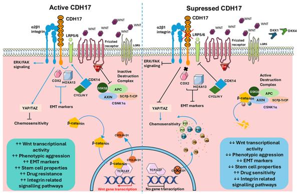 Fig. 1 Schematic diagramme of Wnt/β-catenin pathway with CDH17-mediated regulation in cancer biology.1
The core structural features of CDH17:
- Contains 7 extracellular cadherin repeat sequences, mediating calcium-dependent homophilic adhesion
- The transmembrane domain is anchored to the cell membrane, and the intracellular tail end independently conducts signals
- The extracellular domain forms parallel dimers, enhancing adhesion stability
- Unique catenin-independent signaling mechanism that regulates Wnt pathway activity
Functions of CDH17
The main function of CDH17 is to mediate calcium-dependent homotypic cell adhesion and maintain the integrity of intestinal epithelium. However, it is also involved in various pathological processes, including tumor occurrence and regulation of the Wnt signaling pathway.
| Function | Description |
| Cell adhesion | Mediated by extracellular cadherin repeat sequences, it enables homotypic cell adhesion and maintains the structure and polarity of the intestinal barrier. |
| Wnt signal activation | As an upstream regulatory factor, it activates the classical Wnt/β-catenin pathway and promotes the transcription of target genes. |
| Tumor proliferation promotion | Highly expressed in various digestive tract cancers, it drives the proliferation and survival of cancer cells through signal transduction. |
| Metastasis and invasion | Enhances the migration ability of tumor cells, and is closely related to lymph node metastasis and distant spread. |
| Stem cell characteristic maintenance | Participates in maintaining the stemness characteristics of tumor stem cells, and is associated with chemotherapy resistance and recurrence. |
Unlike other cadherins, the intracellular segment of CDH17 does not bind to the classical cadherins. Instead, it regulates downstream signals through a unique non-cadherin-dependent mechanism, playing a crucial role in tumor development.
Applications of CDH17 and CDH17 Antibody in Literature
1. Tha Shrestha, Bipusha, et al. "The role of cadherin 17 (CDH17) in cancer progression via Wnt/β-catenin signalling pathway: a systematic review and meta-analysis." International Journal of Molecular Sciences 26.20 (2025): 9838. https://doi.org/10.3390/ijms26209838
The review reveals that CDH17 drives the Wnt/β-catenin signaling pathway in multiple cancer types. Inhibiting CDH17 can reduce downstream activity and decrease tumor proliferation and metastasis. The inhibition rate of tumor growth in vivo reaches 80% - 95%, making it a potential broad-spectrum anti-cancer target.
2. Jacobsen, Frank, et al. "Cadherin-17 (CDH17) expression in human cancer: A tissue microarray study on 18,131 tumors." Pathology-Research and Practice 256 (2024): 155175. https://doi.org/10.1016/j.prp.2024.155175
Through the analysis of nearly 15,000 samples, the study confirmed that CDH17 is highly expressed in normal tissues such as intestinal epithelium and various digestive tract tumors. This finding is helpful in differentiating metastatic digestive tract tumors from primary lung adenocarcinoma and makes it a potential diagnostic marker.
3. Zheng, Liuhai, et al. "CDH17-targeting CAR-NK cells synergize with CD47 blockade for potent suppression of gastrointestinal cancers." Acta Pharmaceutica Sinica B 15.5 (2025): 2559-2574. https://doi.org/10.1016/j.apsb.2025.03.039
The research and development of CAR-NK cells targeting CDH17 can effectively eliminate digestive tract tumors. Combined with the CD47 inhibitor CV1, it can synergistically enhance the anti-tumor effect, providing a new strategy for treatment.
4. Ng, Lui, et al. "Tissue Cadherin 17 (CDH17): An Important Prognostic Determinant of Colorectal Cancer Using Digital Image Analysis." Cancer Reports 7.12 (2024): e70069. https://doi.org/10.1002/cnr2.70069
Studies have confirmed that CDH17 is highly expressed in colorectal cancer tissues and is significantly associated with tumor stage progression, metastasis, and poor prognosis. It is an independent prognostic marker for evaluating patient survival and recurrence.
5. Ma, Jingbo, et al. "CDH17 nanobodies facilitate rapid imaging of gastric cancer and efficient delivery of immunotoxin." Biomaterials research 26.1 (2022): 64. https://doi.org/10.1186/s40824-022-00312-3
The study has obtained highly specific nanobodies targeting CDH17, which can enable imaging of gastric cancer and toxin delivery. The immunotoxin significantly suppressed tumor growth and prolonged survival in animal models, demonstrating its potential for clinical application.
Creative Biolabs: CDH17 Antibodies for Research
Creative Biolabs specializes in the production of high-quality CDH17 antibodies for research and industrial applications. Our portfolio includes monoclonal and polyclonal antibodies tailored for ELISA, Flow Cytometry, Western blot, immunohistochemistry, and other diagnostic methodologies.
- Custom CDH17 Antibody Development: Tailor-made solutions to meet specific research requirements.
- Bulk Production: Large-scale antibody manufacturing for industry partners.
- Technical Support: Expert consultation for protocol optimization and troubleshooting.
- Aliquoting Services: Conveniently sized aliquots for long-term storage and consistent experimental outcomes.
For more details on our CDH17 antibodies, custom preparations, or technical support, contact us at email.
Reference
- Tha Shrestha, Bipusha, et al. "The role of cadherin 17 (CDH17) in cancer progression via Wnt/β-catenin signalling pathway: a systematic review and meta-analysis." International Journal of Molecular Sciences 26.20 (2025): 9838. Distributed under Open Access license CC BY 4.0, without modification.https://doi.org/10.3390/ijms26209838
Anti-CDH17 antibodies
 Loading...


Hot products 
-
Mouse Anti-BPGM Recombinant Antibody (CBYY-1806) (CBMAB-2155-YY)

-
Mouse Anti-AHCYL1 Recombinant Antibody (V2-180270) (CBMAB-A1703-YC)

-
Mouse Anti-BrdU Recombinant Antibody (IIB5) (CBMAB-1038CQ)

-
Mouse Anti-ADIPOR1 Recombinant Antibody (V2-179982) (CBMAB-A1368-YC)

-
Mouse Anti-FLT1 Recombinant Antibody (11) (CBMAB-V0154-LY)

-
Mouse Anti-ACTG1 Recombinant Antibody (V2-179597) (CBMAB-A0916-YC)

-
Mouse Anti-FYN Recombinant Antibody (10) (CBMAB-S6332-CQ)

-
Mouse Anti-CD164 Recombinant Antibody (CBFYC-0077) (CBMAB-C0086-FY)

-
Mouse Anti-ENO2 Recombinant Antibody (H14) (CBMAB-E1341-FY)

-
Rabbit Anti-Acetyl-Histone H4 (Lys16) Recombinant Antibody (V2-623415) (CBMAB-CP1021-LY)

-
Rabbit Anti-CBL Recombinant Antibody (D4E10) (CBMAB-CP0149-LY)

-
Mouse Anti-ADGRE5 Recombinant Antibody (V2-360335) (CBMAB-C2088-CQ)

-
Mouse Anti-ALB Recombinant Antibody (V2-363290) (CBMAB-S0173-CQ)

-
Mouse Anti-ACTN4 Recombinant Antibody (V2-6075) (CBMAB-0020CQ)

-
Mouse Anti-CD33 Recombinant Antibody (P67.6) (CBMAB-C10189-LY)

-
Mouse Anti-FOSB Recombinant Antibody (CBXF-3593) (CBMAB-F2522-CQ)

-
Rat Anti-CD34 Recombinant Antibody (MEC 14.7) (CBMAB-C10196-LY)

-
Mouse Anti-CCND2 Recombinant Antibody (DCS-3) (CBMAB-G1318-LY)

-
Mouse Anti-CTNND1 Recombinant Antibody (CBFYC-2414) (CBMAB-C2487-FY)

-
Rabbit Anti-AKT2 (Phosphorylated S474) Recombinant Antibody (V2-556130) (PTM-CBMAB-0605LY)

- AActivation
- AGAgonist
- APApoptosis
- BBlocking
- BABioassay
- BIBioimaging
- CImmunohistochemistry-Frozen Sections
- CIChromatin Immunoprecipitation
- CTCytotoxicity
- CSCostimulation
- DDepletion
- DBDot Blot
- EELISA
- ECELISA(Cap)
- EDELISA(Det)
- ESELISpot
- EMElectron Microscopy
- FFlow Cytometry
- FNFunction Assay
- GSGel Supershift
- IInhibition
- IAEnzyme Immunoassay
- ICImmunocytochemistry
- IDImmunodiffusion
- IEImmunoelectrophoresis
- IFImmunofluorescence
- IGImmunochromatography
- IHImmunohistochemistry
- IMImmunomicroscopy
- IOImmunoassay
- IPImmunoprecipitation
- ISIntracellular Staining for Flow Cytometry
- LALuminex Assay
- LFLateral Flow Immunoassay
- MMicroarray
- MCMass Cytometry/CyTOF
- MDMeDIP
- MSElectrophoretic Mobility Shift Assay
- NNeutralization
- PImmunohistologyp-Paraffin Sections
- PAPeptide Array
- PEPeptide ELISA
- PLProximity Ligation Assay
- RRadioimmunoassay
- SStimulation
- SESandwich ELISA
- SHIn situ hybridization
- TCTissue Culture
- WBWestern Blot
