
CNTF Antibodies
Background
Ciliary neurotrophic factor (CNTF) is a small molecule cytokine protein present in the neural tissues of vertebrates. It mainly activates downstream signaling pathways by binding to specific receptor complexes, thereby supporting neuronal survival, promoting axon regeneration and maintaining nerve cell function. Under conditions of neurological injury or lesion, CNTF can play an important protective and restorative role. This factor was first purified and identified in 1984, and its three-dimensional structure was subsequently resolved through techniques such as nuclear magnetic resonance, making it a key research object in the field of neurobiology. In-depth research on the structure and signaling mechanism of CNTF has greatly promoted scientific progress in the fields of neurotrophic factor theory, treatment strategies for neurodegenerative diseases, and cell signal transduction.
Structure of CNTF
Ciliary neurotrophic factor (CNTF) is a small cytokine protein with a molecular weight of approximately 22-24 kDa. Its exact molecular weight varies slightly among different species, mainly due to glycosylation modifications and subtle changes in amino acid sequences.
| Species | Human | Rat | Mouse |
| Molecular Weight (kDa) | About 22.8 | About 23.5 | About 23.0 |
| Primary Structural Differences | Composed of 200 amino acids, it is a no-signal peptide | Sequence is highly conserved, high homology with humans | Has a similar level 4 spiral beam structure |
This protein is composed of 200 amino acids, and its three-dimensional structure presents a typical "up-and-down" four-helix bundle folding pattern, which is a common feature of type I cytokines. This compact structure forms A hydrophobic core through four α -helices (A to D) and provides a key interface for binding to the ternary complex of its receptors (CNTFRα, gp130 and LIFRβ). Its functional activity is highly dependent on this unique spatial conformation to achieve signal transduction for the survival, differentiation and damage repair of neurons.
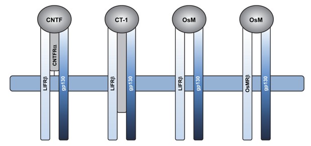 Fig. 1 Schematic diagram of CNTF, CT1 and OsM cytokine receptor complexes.1
Key structural properties of CNTF:
- Typical four-helix bundle folding structure (top down topology)
- Hydrophobic core to maintain the stability of spiral beam
- Secretory cytokines with no-signal peptide sequences
- Receptor binding sites are formed on specific helical surfaces
Functions of CNTF
The main function of ciliary neurotrophic factor (CNTF) is to promote the survival, differentiation and repair of neurons, and it also participates in regulating physiological processes such as energy metabolism and neuroinflammation.
| Function | Description |
| Neuron support | Support the survival of various central and peripheral neurons and prevent their degenerative death after development or injury. |
| Axon regeneration | After nerve injury, it induces the regeneration of damaged axons and promotes the recovery of nerve function. |
| Glial cell differentiation | Regulate the differentiation of oligodendrocyte precursor cells into mature oligodendrocytes and affect myelin formation. |
| Metabolic regulation | In the hypothalamus and other parts involved in body weight and the regulation of energy metabolism balance. |
| Regulation of neuroinflammation | Under conditions of nerve injury or disease, it regulates the activity of astrocytes and microglia and affects the inflammatory response. |
Unlike most classical secreted proteins, CNTF lacks a signal peptide sequence and is mainly released when cells are damaged. Its function is highly dependent on binding to the ternary receptor complex (CNTFRα-gp130-LIFRβ), thereby activating downstream signaling pathways such as JAK-STAT.
Applications of CNTF and CNTF Antibody in Literature
1. Yong, Jiawen, et al. "Ciliary neurotrophic factor (CNTF) and its receptors signal regulate cementoblasts apoptosis through a mechanism of ERK1/2 and caspases signaling." International Journal of Molecular Sciences 23.15 (2022): 8335. https://doi.org/10.3390/ijms23158335
The article indicates that CNTF exerts a bidirectional regulatory effect on the apoptosis of odontoblasts by up-regulating the receptor complex and activating the ERK1/2 signal, which is both early inhibitory and long-term promoting, and affects cell homeostasis.
2. Yong, Jiawen, et al. "Ciliary neurotrophic factor (CNTF) inhibits in vitro cementoblast mineralization and induces autophagy, in part by STAT3/ERK commitment." International Journal of Molecular Sciences 23.16 (2022): 9311. https://doi.org/10.3390/ijms23169311
The article indicates that exogenous CNTF inhibits the expression of mineralization and differentiation markers of odontoblasts and promotes autophagy by activating the ERK1/2 and STAT3 signaling pathways, suggesting that it may be involved in the inflammatory root absorption process during orthodontic tooth movement.
3. Li, Rong, et al. "CNTF mediates neurotrophic factor secretion and fluid absorption in human retinal pigment epithelium." PloS one 6.9 (2011): e23148. https://doi.org/10.1371/journal.pone.0023148
Studies have shown that CNTF significantly enhances the survival rate of RPE cells, alters the polarity secretion of cytokines, and promotes the regulation of chloride ion-dependent transepithelial homeostasis by activating the JAK/STAT3 pathway, which may have a protective effect on neurodegenerative diseases.
4. Jeong, Kyoung Hoon, et al. "Activation of CNTF/CNTFRα signaling pathway by hRheb (S16H) transduction of dopaminergic neurons in vivo." PLoS One 10.3 (2015): e0121803. https://doi.org/10.1371/journal.pone.0121803
Studies have confirmed that hRheb(S16H) gene therapy can upregulate the expression of CNTF and CNTFRα in dopaminergic neurons in the substantia nigra pars compact of Parkinson's disease, activate this signaling pathway to provide neuroprotective effects, and offer a new strategy for the treatment of PD.
5. Lin, Hsiao-Wen, et al. "Ciliary neurotrophic factor (CNTF) plus soluble CNTF receptor α increases cyclooxygenase-2 expression, PGE2release and interferon-γ-induced CD40 in murine microglia." Journal of Neuroinflammation 6.1 (2009): 7. https://doi.org/10.1186/1742-2094-6-7
Studies have shown that CNTF can activate microglia, but its mechanism of action is different from that of IL-6. It does not rely on the typical STAT-3/ERK pathway and gp130, and can synergically enhance CD40 expression and inflammatory response with exogenous sCNTFRα.
Creative Biolabs: CNTF Antibodies for Research
Creative Biolabs specializes in the production of high-quality CNTF antibodies for research and industrial applications. Our portfolio includes monoclonal antibodies tailored for ELISA, Flow Cytometry, Western blot, immunohistochemistry, and other diagnostic methodologies.
- Custom CNTF Antibody Development: Tailor-made solutions to meet specific research requirements.
- Bulk Production: Large-scale antibody manufacturing for industry partners.
- Technical Support: Expert consultation for protocol optimization and troubleshooting.
- Aliquoting Services: Conveniently sized aliquots for long-term storage and consistent experimental outcomes.
For more details on our CNTF antibodies, custom preparations, or technical support, contact us at email.
Reference
- Li, Rong, et al. "CNTF mediates neurotrophic factor secretion and fluid absorption in human retinal pigment epithelium." PloS one 6.9 (2011): e23148. https://doi.org/10.1371/journal.pone.0023148
Anti-CNTF antibodies
 Loading...
 Compare




Hot products 
-
Rabbit Anti-BRCA2 Recombinant Antibody (D9S6V) (CBMAB-CP0017-LY)

-
Mouse Anti-HTLV-1 gp46 Recombinant Antibody (CBMW-H1006) (CBMAB-V208-1154-FY)

-
Rat Anti-CCR2 Recombinant Antibody (475301) (CBMAB-C1338-LY)

-
Mouse Anti-FLT1 Recombinant Antibody (11) (CBMAB-V0154-LY)

-
Rat Anti-CD63 Recombinant Antibody (7G4.2E8) (CBMAB-C8725-LY)

-
Mouse Anti-ENO1 Recombinant Antibody (CBYC-A950) (CBMAB-A4388-YC)

-
Rabbit Anti-Acetyl-Histone H4 (Lys16) Recombinant Antibody (V2-623415) (CBMAB-CP1021-LY)

-
Mouse Anti-CD59 Recombinant Antibody (CBXC-2097) (CBMAB-C4421-CQ)

-
Mouse Anti-AAV9 Recombinant Antibody (V2-634029) (CBMAB-AP023LY)

-
Mouse Anti-CD33 Recombinant Antibody (P67.6) (CBMAB-C10189-LY)

-
Mouse Anti-FN1 Monoclonal Antibody (D6) (CBMAB-1240CQ)

-
Mouse Anti-EIF4G1 Recombinant Antibody (2A9) (CBMAB-A2544-LY)

-
Mouse Anti-Acetyl SMC3 (K105/K106) Recombinant Antibody (V2-634053) (CBMAB-AP052LY)

-
Mouse Anti-AMH Recombinant Antibody (5/6) (CBMAB-A2527-YC)

-
Mouse Anti-BCL6 Recombinant Antibody (CBYY-0435) (CBMAB-0437-YY)

-
Mouse Anti-ACLY Recombinant Antibody (V2-179314) (CBMAB-A0610-YC)

-
Mouse Anti-AAV-5 Recombinant Antibody (V2-503416) (CBMAB-V208-1402-FY)

-
Mouse Anti-ABCA3 Recombinant Antibody (V2-178911) (CBMAB-A0145-YC)

-
Rabbit Anti-ALOX5AP Recombinant Antibody (CBXF-1219) (CBMAB-F0750-CQ)

-
Human Anti-SARS-CoV-2 Spike Recombinant Antibody (CBC05) (CBMAB-CR005LY)

- AActivation
- AGAgonist
- APApoptosis
- BBlocking
- BABioassay
- BIBioimaging
- CImmunohistochemistry-Frozen Sections
- CIChromatin Immunoprecipitation
- CTCytotoxicity
- CSCostimulation
- DDepletion
- DBDot Blot
- EELISA
- ECELISA(Cap)
- EDELISA(Det)
- ESELISpot
- EMElectron Microscopy
- FFlow Cytometry
- FNFunction Assay
- GSGel Supershift
- IInhibition
- IAEnzyme Immunoassay
- ICImmunocytochemistry
- IDImmunodiffusion
- IEImmunoelectrophoresis
- IFImmunofluorescence
- IGImmunochromatography
- IHImmunohistochemistry
- IMImmunomicroscopy
- IOImmunoassay
- IPImmunoprecipitation
- ISIntracellular Staining for Flow Cytometry
- LALuminex Assay
- LFLateral Flow Immunoassay
- MMicroarray
- MCMass Cytometry/CyTOF
- MDMeDIP
- MSElectrophoretic Mobility Shift Assay
- NNeutralization
- PImmunohistologyp-Paraffin Sections
- PAPeptide Array
- PEPeptide ELISA
- PLProximity Ligation Assay
- RRadioimmunoassay
- SStimulation
- SESandwich ELISA
- SHIn situ hybridization
- TCTissue Culture
- WBWestern Blot
