
EIF4E Antibodies
Background
The EIF4E gene encodes eukaryotic translation initiation factor 4E, which serves as a core component of mRNA 5 'cap binding proteins and is mainly present in the cytoplasm. It regulates key physiological activities such as cell proliferation and differentiation by recognizing the 5 '-end methylated guanosine cap structure of mRNA, mediating the binding initiation translation process between ribosomes and mRNA. In cancer research, it has been found that the overexpression of EIF4E can activate the translation of various oncogenic proteins, leading to the occurrence and development of tumors. This gene was first identified in 1985, and its protein structure was analyzed by X-ray crystallography in 1997, revealing a unique cap-binding pocket conformation. Continuous research on EIF4E has deepened our understanding of gene expression regulation, cell signal transduction and tumorigenesis mechanisms, providing an important theoretical basis for targeted therapy.
Structure of EIF4E
EIF4E is an mRNA 5 'terminal cap-binding protein with a molecular weight of approximately 25 kDa. Its precise molecular weight varies slightly among different eukaryotes, mainly due to minor changes in the terminal of its amino acid sequence.
| Species | Human | Mouse | Fruit fly | Yeast |
| Molecular Weight (kDa) | 24.7 | 24.8 | 25.2 | 23.9 |
| Primary Structural Differences | Typical eukaryotic cap-binding protein | Highly homologous to humans | With functions of species specific domain | Simpler structure |
This protein is composed of 217 amino acid residues folded into a compact α/β domain. The core of its three-dimensional structure is a curved surface composed of 8 anti-parallel β -lamellar layers, surrounded by 3 α -helices on both sides, jointly forming a highly conserved, positively charged crack-like mRNA cap-binding pocket. At this functional center, tryptophan residues directly recognize and bind to the 7-methylguanosine cap structure at the 5' end of the mRNA through π-π stacking interactions, while the surrounding multiple positively charged residues are responsible for stabilizing the phosphate group. This precise molecular structure enables EIF4E to specifically recognize cap structures, thereby initiating the protein translation process.
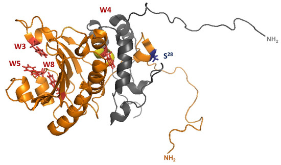 Fig. 1 3D structure (1RF8) of S. cerevisiae eIF4E (gold) in complex with eIF4G .1
Key structural properties of EIF4E:
- Conserved mRNA 5 'terminal cap binding pocket
- Composed of eight antiparallel beta folding bending lamella structure
- π-π stacking interaction is formed by tryptophan residues and 7-methylguanosine caps
Functions of EIF4E
The core function of the EIF4E gene is to initiate the biosynthesis of proteins. However, it is also deeply involved in a variety of key cellular physiological and pathological processes, including cell cycle regulation, tumorigenesis and virus-host interaction.
| Function | Description |
| Translation beginning | Specifically recognizing and binding to the 5' terminal cap structure of mRNA, mediating ribosome assembly, thereby initiating cap-dependent protein translation. |
| Growth regulation | Cell proliferation and growth are controlled by regulating the translation efficiency of specific growth-promoting mrnas (such as cyclins and transcription factors). |
| Tumorigenesis | Its overexpression selectively enhances the translation of various oncogenes, promotes the survival, invasion and angiogenesis of tumor cells, and is recognized as a proto-oncogene. |
| Virus utilization | Some viruses hijack the EIF4E of host cells through a "cap-snatching" mechanism to prioritize the translation of their own viral mRNA. |
| Synaptic plasticity | The local regulation of the synthesis of synaptic associated proteins in neurons is crucial for advanced neural functions such as learning and memory. |
Unlike the synergistic effect of hemoglobin, EIF4E functions in a monomer form. Its single high-affinity binding to the mRNA cap structure ensures precise and fundamental horizontal control of gene expression.
Applications of EIF4E and EIF4E Antibody in Literature
1. Ross-Kaschitza, Daniela, and Michael Altmann. "eIF4E and interactors from unicellular eukaryotes." International journal of molecular sciences 21.6 (2020): 2170. https://doi.org/10.3390/ijms21062170
This article mainly reviews the functions and diversity of eukaryotic initiation factor eIF4E and its interacting proteins in single-celled organisms such as yeast, trypanosoma and dinoflagellates. Research has revealed that different species evolve to form unique eIF4E variants and their interaction mechanisms to adapt to their specific translation initiation requirements.
2. Roiuk, Mykola, Marilena Neff, and Aurelio A. Teleman. "eIF4E-independent translation is largely eIF3d-dependent." Nature communications 15.1 (2024): 6692. https://doi.org/10.1038/s41467-024-51027-z
The article indicates that under stress conditions, after eIF4E1 is inactivated, cells recognize the mRNA cap structure by replacing eIF4E1 with eIF3d, thereby initiating the translation of specific mRNA. This study reveals a new translation mechanism that does not rely on eIF4E.
3. Montero, Hilda, Rebeca García-Román, and Silvia I. Mora. "eIF4E as a control target for viruses." Viruses 7.2 (2015): 739-750. https://doi.org/10.3390/v7020739
This article reviews how viruses manipulate the translation initiation factor eIF4E and its regulatory proteins in host cells to facilitate the translation of their own mRNA and viral replication, revealing the key strategies by which viruses hijack the host's translation mechanism.
4. Pugsley, Lauren, et al. "C8ORF88: a novel eIF4E-binding protein." Genes 14.11 (2023): 2076. https://doi.org/10.3390/genes14112076
This study identified a novel EIF4E-binding protein, C8ORF88, which is specifically expressed in early sperm cells and interacts with eIF4E through a classical binding motif. Its regulatory mechanism is different from the known 4E-BP1, expanding the understanding of the eIF4E regulatory network.
5. Chen X, Huang J, et al. "A noncanonical function of EIF4E limits ALDH1B1 activity and increases susceptibility to ferroptosis." Nature communications 13.1 (2022): 6318. https://doi.org/10.1038/s41467-022-34096-w
Research has found that eIF4E can regulate ferroptosis in a translation-independent manner. It promotes ferroptosis by inhibiting mitochondrial aldehyde dehydrogenase ALDH1B1, leading to the accumulation of toxic aldehydes and activating lipid peroxidation. This mechanism enhances the anti-cancer effect of ferroptosis inducers.
Creative Biolabs: EIF4E Antibodies for Research
Creative Biolabs specializes in the production of high-quality EIF4E antibodies for research and industrial applications. Our portfolio includes monoclonal antibodies tailored for ELISA, Flow Cytometry, Western blot, immunohistochemistry, and other diagnostic methodologies.
- Custom EIF4E Antibody Development: Tailor-made solutions to meet specific research requirements.
- Bulk Production: Large-scale antibody manufacturing for industry partners.
- Technical Support: Expert consultation for protocol optimization and troubleshooting.
- Aliquoting Services: Conveniently sized aliquots for long-term storage and consistent experimental outcomes.
For more details on our EIF4E antibodies, custom preparations, or technical support, contact us at email.
Reference
- Ross-Kaschitza, Daniela, and Michael Altmann. "eIF4E and interactors from unicellular eukaryotes." International journal of molecular sciences 21.6 (2020): 2170. https://doi.org/10.3390/ijms21062170
Anti-EIF4E antibodies
 Loading...
 Compare




Hot products 
- AActivation
- AGAgonist
- APApoptosis
- BBlocking
- BABioassay
- BIBioimaging
- CImmunohistochemistry-Frozen Sections
- CIChromatin Immunoprecipitation
- CTCytotoxicity
- CSCostimulation
- DDepletion
- DBDot Blot
- EELISA
- ECELISA(Cap)
- EDELISA(Det)
- ESELISpot
- EMElectron Microscopy
- FFlow Cytometry
- FNFunction Assay
- GSGel Supershift
- IInhibition
- IAEnzyme Immunoassay
- ICImmunocytochemistry
- IDImmunodiffusion
- IEImmunoelectrophoresis
- IFImmunofluorescence
- IGImmunochromatography
- IHImmunohistochemistry
- IMImmunomicroscopy
- IOImmunoassay
- IPImmunoprecipitation
- ISIntracellular Staining for Flow Cytometry
- LALuminex Assay
- LFLateral Flow Immunoassay
- MMicroarray
- MCMass Cytometry/CyTOF
- MDMeDIP
- MSElectrophoretic Mobility Shift Assay
- NNeutralization
- PImmunohistologyp-Paraffin Sections
- PAPeptide Array
- PEPeptide ELISA
- PLProximity Ligation Assay
- RRadioimmunoassay
- SStimulation
- SESandwich ELISA
- SHIn situ hybridization
- TCTissue Culture
- WBWestern Blot
