LAMA3 Antibodies
Background
The LAMA3 gene encodes laminin α3 chain, which is an important component of laminin -332 in the extracellular matrix. This protein maintains the stability and integrity of the epithelial tissue basement membrane by forming transmembrane junction complexes. Its main functions are reflected in promoting the adhesion and directional migration of keratinocytes and regulating the signal transduction between the epidermis and dermis, thereby supporting the barrier function and regenerative repair of the skin, mucous membranes and other tissues. Mutations in this gene can damage the basement membrane structure, leading to hereditary vesicular diseases such as junctional epidermolysis bullosa. The clinical manifestations are the appearance of blisters and erosion on the skin and mucous membranes with even slight friction. Since its discovery in the 1990s, research on the function of the LAMA3 gene has not only revealed the core mechanism of basement membrane assembly, but also provided a key molecular basis for the genetic diagnosis and targeted therapy of hereditary skin diseases.
Structure of LAMA3
The LAMA3 gene encodes the laminin α3 chain, a large glycoprotein with a molecular weight of approximately 200 kDa. The actual molecular weight of this protein varies depending on different splicing variants.
| Species | Human | Mouse | Rat |
| Molecular Weight (kDa) | 200 (Non-restoration) | 190 | 195 |
| Primary Structural Differences | Contains multiple EGF-like repeats and coiled-coil regions | High homology with human and similar domain composition | Arranged in accordance with basic human structure domain |
This protein is composed of multiple domains and presents as an extended fibrous molecule. Its core functional domains include the spherical domains at the N-terminal (LN, LEa) and the G-domain at the C-terminal, which mediate specific binding to cell surface receptors such as integrins. The α3 chain of laminin covalently binds to the β3 and γ2 chains through its coiled helical region, jointly assembling into the biologically active laminin -332 trimer. This unique structure enables it to act as a bridge, bidirectionally connecting epithelial cells with other stromal components in the basement membrane, thereby playing a crucial role in anchoring and signal transduction.
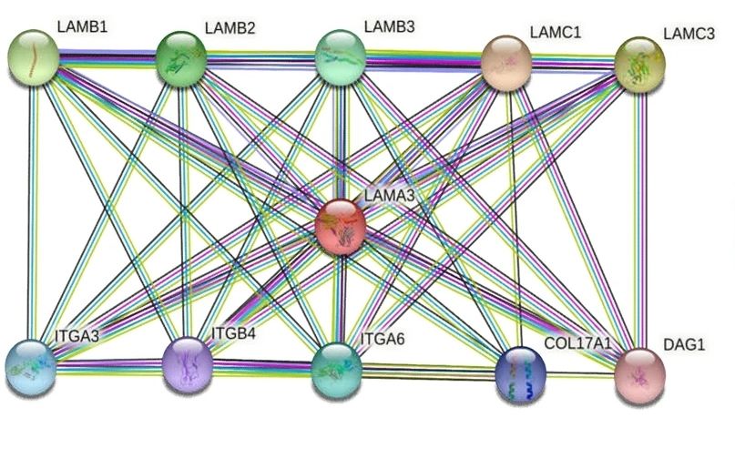 Fig. 1 Lama3 interaction genes (PPI).1
Key structural properties of LAMA3:
- Domains rich in epidermal growth factor-like (EGF-like) repeat sequences
- Form the C-terminal coiled coil region of the triple helix coil
- C-terminal laminin G-like domains that mediate cell adhesion
Functions of LAMA3
The LAMA3 gene encodes the laminin α3 chain, whose main function is to form and stabilize the basement membrane of epithelial tissues. However, it is also involved in a variety of complex physiological and pathological processes, including wound healing, cell signal transduction and tumor invasion.
| Function | Description |
| Substrate film assembly | As a core component of laminin -332, it connects with other basement membrane components (such as type IV collagen) to form a stable physical barrier. |
| Cell adhesion and migration | By binding to integrins on the cell surface (such as α6β4) through its G domain, it mediates the anchoring of epithelial cells and guides the directional migration of cells during development and repair. |
| Maintenance of organizational barriers | Maintain the structural integrity of the skin and mucosa epithelial tissue, its function as a direct result of border type epidermolysis bullosa sex disease. |
| Signal transduction regulation | Through integrin, two-way signals affect cell proliferation, differentiation and apoptosis, coordinate the communication between epithelium and stroma. |
| Promotion of wound healing | After skin damage, it accelerates the process of epithelial regeneration and wound closure by promoting the adhesion and migration of keratinocytes. |
Laminin -332 is assembled from three chains, α3, β3, and γ2, through covalent cross-linking in the coiled helical region. This unique heterotrimer structure determines its powerful network formation ability and biological function, which contrasts sharply with the oxygen storage function of myoglobin as an independent monomer.
Applications of LAMA3 and LAMA3 Antibody in Literature
1. Feng, Li-yuan, et al. "LAMA3 DNA methylation and transcriptome changes associated with chemotherapy resistance in ovarian cancer." Journal of ovarian research 14.1 (2021): 67. https://doi.org/10.1186/s13048-021-00807-y
This study confirmed through bioinformatics and experiments that hypermethylation of the LAMA3 gene leads to a decrease in its expression, which is associated with chemotherapy resistance and poor prognosis of ovarian cancer. LAMA3 can inhibit the proliferation of drug-resistant cells and promote their apoptosis, invasion and migration.
2. Islam, Kittiya, et al. "Upregulated LAMA3 modulates proliferation, adhesion, migration and epithelial‑to‑mesenchymal transition of cholangiocarcinoma cells." Scientific Reports 13.1 (2023): 22598. https://doi.org/10.1038/s41598-023-48798-8
Research has found that LAMA3 is the most significantly upregulated core laminin gene in cholangiocarcinoma, driving tumor proliferation, migration and invasion. LAMA3 has been revealed for the first time as a potential diagnostic marker and new therapeutic target for cholangiocarcinoma.
3. Ji, Zhong-Hao, et al. "A missense mutation in Lama3 causes androgen alopecia." Scientific Reports 13.1 (2023): 20818. https://doi.org/10.1038/s41598-023-48337-5
This study, through gene sequencing and CRISPR/Cas9 technology, confirmed that point mutations in the Lama3 gene (R217C) would cause mice to exhibit phenotypes similar to androgenetic alopecia, including hair loss and abnormal hair follicle structure. This mutant mouse model provides a new tool for the study of hair loss diseases.
4. Ullah, Asmat, Fibhaa Syed, and Shazia Khan. "c. 151dup variant in LAMA3 in Pakistani patients affected with Shabbir Syndrome but showing mild symptoms." Pakistan Journal of Medical Sciences 39.4 (2023): 1124. https://doi.org/10.12669/pjms.39.4.6926
This study found that a homozygous repeat mutation (c.151dup) of the LAMA3 gene in a Pakistani family is the cause of Shabbir syndrome. This mutation is relatively common among the Punjabi population in Pakistan. It is recommended to develop targeted detection technologies for this mutation to facilitate carrier screening and prenatal diagnosis.
5. Tian, Chengming, Xiyao Li, and Chunlin Ge. "High expression of LAMA3/AC245041. 2 gene pair associated with KRAS mutation and poor survival in pancreatic adenocarcinoma: a comprehensive TCGA analysis." Molecular Medicine 27.1 (2021): 62. https://doi.org/10.1186/s10020-021-00322-2
In this study, a prognostic model for KRAS-mutated pancreatic cancer was constructed. It was found that LAMA3 was highly correlated with the expression of lncRNA AC245041.2 and jointly participated in the regulatory network of transcription factors, providing new clues for exploring its molecular mechanism.
Creative Biolabs: LAMA3 Antibodies for Research
Creative Biolabs specializes in the production of high-quality LAMA3 antibodies for research and industrial applications. Our portfolio includes monoclonal antibodies tailored for ELISA, Flow Cytometry, Western blot, immunohistochemistry, and other diagnostic methodologies.
- Custom LAMA3 Antibody Development: Tailor-made solutions to meet specific research requirements.
- Bulk Production: Large-scale antibody manufacturing for industry partners.
- Technical Support: Expert consultation for protocol optimization and troubleshooting.
- Aliquoting Services: Conveniently sized aliquots for long-term storage and consistent experimental outcomes.
For more details on our LAMA3 antibodies, custom preparations, or technical support, contact us at email.
Reference
- Feng, Li-yuan, et al. "LAMA3 DNA methylation and transcriptome changes associated with chemotherapy resistance in ovarian cancer." Journal of ovarian research 14.1 (2021): 67. https://doi.org/10.1186/s13048-021-00807-y
Anti-LAMA3 antibodies
 Loading...


Hot products 
-
Mouse Anti-2C TCR Recombinant Antibody (V2-1556) (CBMAB-0951-LY)

-
Mouse Anti-CAPZB Recombinant Antibody (CBYY-C0944) (CBMAB-C2381-YY)

-
Mouse Anti-BIRC5 Recombinant Antibody (6E4) (CBMAB-CP2646-LY)

-
Rabbit Anti-ALOX5AP Recombinant Antibody (CBXF-1219) (CBMAB-F0750-CQ)

-
Mouse Anti-FOXA3 Recombinant Antibody (2A9) (CBMAB-0377-YC)

-
Rabbit Anti-AKT3 Recombinant Antibody (V2-12567) (CBMAB-1057-CN)

-
Rabbit Anti-CCL5 Recombinant Antibody (R0437) (CBMAB-R0437-CN)

-
Mouse Anti-BMI1 Recombinant Antibody (CBYC-P026) (CBMAB-P0108-YC)

-
Mouse Anti-CRTAM Recombinant Antibody (CBFYC-2235) (CBMAB-C2305-FY)

-
Mouse Anti-ACLY Recombinant Antibody (V2-179314) (CBMAB-A0610-YC)

-
Mouse Anti-8-oxoguanine Recombinant Antibody (V2-7697) (CBMAB-1869CQ)

-
Mouse Anti-DDC Recombinant Antibody (8E8) (CBMAB-0992-YC)

-
Rabbit Anti-Acetyl-Histone H4 (Lys16) Recombinant Antibody (V2-623415) (CBMAB-CP1021-LY)

-
Mouse Anti-CD24 Recombinant Antibody (ALB9) (CBMAB-0176CQ)

-
Mouse Anti-ABCA3 Recombinant Antibody (V2-178911) (CBMAB-A0145-YC)

-
Mouse Anti-DLG1 Monolconal Antibody (4F3) (CBMAB-0225-CN)

-
Mouse Anti-CARTPT Recombinant Antibody (113612) (CBMAB-C2450-LY)

-
Mouse Anti-CCNH Recombinant Antibody (CBFYC-1054) (CBMAB-C1111-FY)

-
Mouse Anti-CASP8 Recombinant Antibody (CBYY-C0987) (CBMAB-C2424-YY)

-
Mouse Anti-CALR Recombinant Antibody (CBFYC-0763) (CBMAB-C0818-FY)

- AActivation
- AGAgonist
- APApoptosis
- BBlocking
- BABioassay
- BIBioimaging
- CImmunohistochemistry-Frozen Sections
- CIChromatin Immunoprecipitation
- CTCytotoxicity
- CSCostimulation
- DDepletion
- DBDot Blot
- EELISA
- ECELISA(Cap)
- EDELISA(Det)
- ESELISpot
- EMElectron Microscopy
- FFlow Cytometry
- FNFunction Assay
- GSGel Supershift
- IInhibition
- IAEnzyme Immunoassay
- ICImmunocytochemistry
- IDImmunodiffusion
- IEImmunoelectrophoresis
- IFImmunofluorescence
- IGImmunochromatography
- IHImmunohistochemistry
- IMImmunomicroscopy
- IOImmunoassay
- IPImmunoprecipitation
- ISIntracellular Staining for Flow Cytometry
- LALuminex Assay
- LFLateral Flow Immunoassay
- MMicroarray
- MCMass Cytometry/CyTOF
- MDMeDIP
- MSElectrophoretic Mobility Shift Assay
- NNeutralization
- PImmunohistologyp-Paraffin Sections
- PAPeptide Array
- PEPeptide ELISA
- PLProximity Ligation Assay
- RRadioimmunoassay
- SStimulation
- SESandwich ELISA
- SHIn situ hybridization
- TCTissue Culture
- WBWestern Blot
