PDK1 Antibodies
Background
PDK1, as a serine/threonine protein kinase, is mainly distributed in various tissue cells of mammals. This gene regulates the glucose metabolism process by phosphorylating the pyruvate dehydrogenase complex, thereby controlling the conversion between energy supply and anabolism in cells. Tumor cells often utilize PDK1 to maintain the Warburg effect to adapt to hypoxic environments because it plays a core role in metabolic recombination. This gene was first identified by Sugden's team in 1998 while studying the insulin signaling pathway and was subsequently confirmed to be a key node in the PI3K/PKB signaling pathway that regulates cell growth. Its unique kinase domain and allosteric regulation mechanism have become important targets in the research of metabolic diseases and cancer treatment, providing a molecular basis for understanding cellular energy perception and metabolic adaptability.
Structure of PDK1
PDK1 (pyruvate dehydrogenase kinase 1) is a serine/threonine protein kinase with a molecular weight of approximately 49 kDa. Its molecular weight varies slightly among different mammals, mainly due to the differences in gene splicing variants and post-translational modifications.
| Species | Human | Mouse | Rat | Bovine |
| Molecular Weight (kDa) | 48.8 | 49.2 | 49.1 | 48.9 |
This protein is composed of 556 amino acids, forming a typical kinase folding structure: the N-terminal lobe is mainly responsible for ATP binding, while the C-terminal lobe undertakes substrate recognition function. A key structural feature in its three-dimensional structure is the PHI-Pocket binding domain located at the C-terminal of the kinase domain. This allosteric regulatory pocket regulates kinase activity by binding to a specific hydrophobic motif. The phosphorylation site Ser241 on the activation ring is crucial for catalytic activity, while the PH domain at the N-terminal is involved in protein-protein interactions and cellular localization, jointly maintaining its precise perception and regulation of the cellular metabolic state.
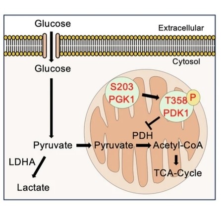 Fig. 1 Schematic showing glycolysis regulation by PGK1 and PDK1.1
Key structural properties of PDK1:
- Typical duplex kinase folding configuration
- Allosteric regulatory pockets located deep in catalytic fissure
- Phosphorylation site Ser241 on the activation ring maintains catalytic activity
- PH domains mediate protein interactions and subcellular localization
Functions of PDK1
The core function of the PDK1 gene is to serve as a key regulatory factor for cellular metabolism. It regulates the metabolic balance between glucose oxidation and glycolysis in cells by phosphorylating the E1α subunit of pyruvate dehydrogenase (PDH) and inhibiting the conversion of mitochondrial pyruvate to acetyl-CoA.
| Function | Description |
| Regulation of metabolic switches | Phosphorylating the E1α subunit of PDH inactivates it, blocking the aerobic oxidation pathway of glucose. |
| The Warburg effect promotes | Enhance the glycolytic activity of tumor cells and support the generation of biosynthetic precursors. |
| Adaptation to hypoxia | Maintain cellular redox balance and energy homeostasis in a hypoxic environment. |
| Insulin signal integration | Through the Akt/PKB pathways, growth factor reception coordination metabolism and proliferation. |
| Regulation of cell survival | Inhibit the excessive production of reactive oxygen species in mitochondria and support the survival of cells under metabolic stress. |
The inhibitory effect of PDK1 on PDH shows typical substrate specificity. Its kinase activity is regulated at multiple levels, including the ATP/ADP ratio, NADH/NAD+ ratio, and pyruvate concentration. This complex regulatory network makes PDK1 the core hub of cellular metabolic plasticity.
Applications of PDK1 and PDK1 Antibody in Literature
1. Zhang, Lei, et al. "PDK1 promotes epithelial ovarian cancer progression by upregulating BGN: PDK1/BGN axis facilitates EOC progression." Acta Biochimica et Biophysica Sinica 57.5 (2024): 712. https://doi.org/10.3724/abbs.2024186
This study reveals that PDK1 is highly expressed in ovarian cancer. It upregulates BGN by enhancing the stability of BGN mRNA, thereby activating the NF-κB pathway and promoting tumor growth and metastasis. The PDK1/BGN axis can serve as a potential therapeutic target.
2. Li, Yuebo, et al. "PIWIL2/PDK1 Axis Promotes the Progression of Cervical Epithelial Lesions via Metabolic Reprogramming to Maintain Tumor‐Initiating Cell Stemness." Advanced Science 11.48 (2024): 2410756. https://doi.org/10.1002/advs.202410756
This study reveals that in precancerous lesions of cervical cancer, PIWIL2 upregulates the expression of PDK1 through the LIN28/let-7 axis. PDK1 maintains the stemness and tumorigenicity of tumor-initiating cells by activating the PI3K/AKT/mTOR pathway and glycolysis, thereby driving disease progression.
3. Tokumura, Kazuya, et al. "PDK1-dependent metabolic reprogramming regulates stemness and tumorigenicity of osteosarcoma stem cells through ATF3." Cell Death & Disease 16.1 (2025): 574. https://doi.org/10.1038/s41419-025-07903-7
This study reveals that in osteosarcoma, PDK1 maintains the stemness and tumorigenicity of tumor stem cells by regulating the metabolic balance between glycolysis and OXPHOS, upregulating ATF3 and activating the TGF-β/Smad pathway, suggesting that it can serve as a potential therapeutic target.
4. Kim, Hyemin, Jinyoung Lee, and Yongcheol Cho. "PDK1 is a negative regulator of axon regeneration." Molecular brain 14.1 (2021): 31. https://doi.org/10.1186/s13041-021-00748-z
Research has found that PDK1 is a key negative regulatory factor for axon regeneration. Its expression is upregulated after injury of adult neurons and inhibits regeneration. Gene knockdown or chemical inhibition of PDK1 can effectively promote the regenerative capacity of axons both in vitro and in vivo, indicating that it can serve as a potential target for nerve repair therapy.
5. Wei, Yu, et al. "PDK1 promotes breast cancer progression by enhancing the stability and transcriptional activity of HIF-1α." Genes & Diseases 11.4 (2024): 101041. https://doi.org/10.1016/j.gendis.2023.06.013
This study reveals that PDK1 enhances the protein stability and transcriptional activity of HIF-1α in breast cancer by phosphorylating the Ser451 site, forming a positive feedback loop and jointly promoting tumor progression. PDK1 is a potential therapeutic target for breast cancer.
Creative Biolabs: PDK1 Antibodies for Research
Creative Biolabs specializes in the production of high-quality PDK1 antibodies for research and industrial applications. Our portfolio includes monoclonal antibodies tailored for ELISA, Flow Cytometry, Western blot, immunohistochemistry, and other diagnostic methodologies.
- Custom PDK1 Antibody Development: Tailor-made solutions to meet specific research requirements.
- Bulk Production: Large-scale antibody manufacturing for industry partners.
- Technical Support: Expert consultation for protocol optimization and troubleshooting.
- Aliquoting Services: Conveniently sized aliquots for long-term storage and consistent experimental outcomes.
For more details on our PDK1 antibodies, custom preparations, or technical support, contact us at email.
Reference
- Tokumura, Kazuya, et al. "PDK1-dependent metabolic reprogramming regulates stemness and tumorigenicity of osteosarcoma stem cells through ATF3." Cell Death & Disease 16.1 (2025): 574. https://doi.org/10.1038/s41419-025-07903-7
Anti-PDK1 antibodies
 Loading...


Hot products 
-
Rabbit Anti-ALOX5AP Recombinant Antibody (CBXF-1219) (CBMAB-F0750-CQ)

-
Mouse Anti-BAX Recombinant Antibody (CBYY-0216) (CBMAB-0217-YY)

-
Mouse Anti-AAV8 Recombinant Antibody (V2-634028) (CBMAB-AP022LY)

-
Mouse Anti-C5b-9 Recombinant Antibody (aE11) (CBMAB-AO138LY)

-
Mouse Anti-DMD Recombinant Antibody (D1190) (CBMAB-D1190-YC)

-
Mouse Anti-CCDC25 Recombinant Antibody (CBLC132-LY) (CBMAB-C9786-LY)

-
Mouse Anti-CCNH Recombinant Antibody (CBFYC-1054) (CBMAB-C1111-FY)

-
Mouse Anti-ASH1L Monoclonal Antibody (ASH5H03) (CBMAB-1372-YC)

-
Mouse Anti-ATP1B1 Recombinant Antibody (E4) (CBMAB-0463-LY)

-
Rabbit Anti-AP2M1 (Phosphorylated T156) Recombinant Antibody (D4F3) (PTM-CBMAB-0610LY)

-
Mouse Anti-ATP1B3 Recombinant Antibody (1E9) (CBMAB-A4021-YC)

-
Mouse Anti-CCS Recombinant Antibody (CBFYC-1093) (CBMAB-C1150-FY)

-
Rabbit Anti-ABL1 (Phosphorylated Y245) Recombinant Antibody (V2-505716) (PTM-CBMAB-0465LY)

-
Mouse Anti-DHFR Recombinant Antibody (D0821) (CBMAB-D0821-YC)

-
Mouse Anti-FOXL1 Recombinant Antibody (CBXF-0845) (CBMAB-F0462-CQ)

-
Mouse Anti-AQP2 Recombinant Antibody (E-2) (CBMAB-A3358-YC)

-
Mouse Anti-CAPZB Recombinant Antibody (CBYY-C0944) (CBMAB-C2381-YY)

-
Mouse Anti-AQP2 Recombinant Antibody (G-3) (CBMAB-A3359-YC)

-
Mouse Anti-ENO2 Recombinant Antibody (H14) (CBMAB-E1341-FY)

-
Mouse Anti-GGT1 Recombinant Antibody (1F9) (CBMAB-G3273-LY)

- AActivation
- AGAgonist
- APApoptosis
- BBlocking
- BABioassay
- BIBioimaging
- CImmunohistochemistry-Frozen Sections
- CIChromatin Immunoprecipitation
- CTCytotoxicity
- CSCostimulation
- DDepletion
- DBDot Blot
- EELISA
- ECELISA(Cap)
- EDELISA(Det)
- ESELISpot
- EMElectron Microscopy
- FFlow Cytometry
- FNFunction Assay
- GSGel Supershift
- IInhibition
- IAEnzyme Immunoassay
- ICImmunocytochemistry
- IDImmunodiffusion
- IEImmunoelectrophoresis
- IFImmunofluorescence
- IGImmunochromatography
- IHImmunohistochemistry
- IMImmunomicroscopy
- IOImmunoassay
- IPImmunoprecipitation
- ISIntracellular Staining for Flow Cytometry
- LALuminex Assay
- LFLateral Flow Immunoassay
- MMicroarray
- MCMass Cytometry/CyTOF
- MDMeDIP
- MSElectrophoretic Mobility Shift Assay
- NNeutralization
- PImmunohistologyp-Paraffin Sections
- PAPeptide Array
- PEPeptide ELISA
- PLProximity Ligation Assay
- RRadioimmunoassay
- SStimulation
- SESandwich ELISA
- SHIn situ hybridization
- TCTissue Culture
- WBWestern Blot
