
PHGDH Antibodies
Background
The PHGDH gene encodes a metabolic enzyme that catalyzes the first reaction in the glycolysis pathway. It is expressed in various tissues and cells of animals. This enzyme converts 3-phosphoglyceric acid into 3-phosphohydroxypropionic acid, providing a carbon skeleton for serine biosynthesis and thereby supporting nucleotide synthesis and cell proliferation. Rapidly dividing cells, especially certain malignant tumor cells, often rely on upregulating PHGDH activity to maintain redox balance and biosynthetic requirements. The dysfunction of this gene was first discovered in congenital serine deficiency, and subsequent studies have confirmed its amplification or high expression in breast cancer, melanoma, and other tumors. Therefore, the function of this gene has become an important research direction for targeting tumor metabolism. The development of its structure and inhibitors has greatly enriched our understanding of the relationship between metabolic reprogramming and diseases.
Structure of PHGDH
The protein encoded by PHGDH has a molecular weight of approximately 56.7 kDa and shows slight variations among different species.
| Species | Human | Mouse | Rat | Zebrafish | Fruit fly |
| Molecular Weight (kDa) | 56.7 | 56.5 | 56.6 | 56.3 | 57.0 |
| Primary Structural Differences | The total length is 533 amino acids | Homology 89% | Homology 88% | Domain conservation | The branch chain is relatively short |
PHGDH is composed of 533 amino acids and its active form is a dimer. Each subunit contains a NAD⁺binding domain, a substrate binding domain and an oligomerization domain.
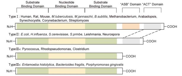 Fig. 1 Isoforms of PHGDH in various organisms.1
Key structural properties of PHGDH:
- NAD⁺binding domain, composed of Rossmann folding
- Substrate binding domain, containing a conserved catalytic loop
- Oligomerization interface, forming homodimers
- Active center Glu¹⁹⁷ and Arg⁵⁴ recognize the substrate
- Binding of NAD⁺/NADH cofactors for hydrogen ion transfer
Functions of PHGDH
The core function of PHGDH is to catalyze the first step reaction of serine biosynthesis. Additionally, this enzyme is also involved in processes such as maintaining redox homeostasis and regenerating NADH.
| Function | Description |
| Serine Synthesis | Catalyzes the conversion of 3-phosphoglyceric acid to 3-phosphohydroxypropionic acid, providing a carbon source for serine. |
| Nucleotide Synthesis Support | Serine metabolism enters the one-carbon unit cycle, providing precursors for purine and pyrimidine synthesis. |
| Redox Regulation | The reaction is accompanied by the reduction of NAD⁺to NADH, participating in the maintenance of intracellular reducing power balance. |
| Hypoxia Adaptation | Upregulated expression under hypoxic conditions, supporting tumor cell proliferation in low-oxygen environments. |
| Metabolic Reprogramming Support | Highly proliferative cells upregulate PHGDH to divert glycolytic intermediates to the synthetic metabolic pathway. |
The substrate affinity of PHGDH is relatively high. The flux of the branch metabolic pathway is mainly regulated by the transcriptional level and the concentration of the cofactor NAD⁺, which is different from the rapid response regulation of the main pathway of glycolysis.
Applications of PHGDH and PHGDH Antibody in Literature
1. Lee, Chae Min, et al. "PHGDH: a novel therapeutic target in cancer." Experimental & Molecular Medicine 56.7 (2024): 1513-1522. https://doi.org/10.1038/s12276-024-01268-1
The article indicates that serine is crucial for the proliferation of cancer cells. PHGDH, as the key enzyme for its synthesis, is activated and upregulated in various cancers, promoting tumor growth, metastasis and drug resistance. This paper, by analyzing its structure and metabolic function, proposes that PHGDH inhibitors may become a new type of anti-cancer strategy, capable of overcoming the drug resistance mechanism.
2. Shen, Liliang, et al. "PHGDH inhibits ferroptosis and promotes malignant progression by upregulating SLC7A11 in bladder cancer." International journal of biological sciences 18.14 (2022): 5459. https://doi.org/10.7150/ijbs.74546
The article indicates that PHGDH is highly expressed in bladder cancer. It stabilizes SLC7A11 mRNA by binding to PCBP2, inhibits ferroptosis and promotes tumor progression. The PHGDH inhibitor NCT-502 can promote ferroptosis and inhibit tumors, and its combined score with SLC7A11 can predict the prognosis of patients.
3. Lee, Chae Min, et al. "PHGDH: a novel therapeutic target in cancer." Experimental & Molecular Medicine 56.7 (2024): 1513-1522. https://doi.org/10.1038/s12276-024-01268-1
The article indicates that the rate-limiting enzyme for serine synthesis, PHGDH, is abnormally upregulated in various cancers, driving tumor growth, metastasis and drug resistance. This review analyzes its structure and metabolic function, and proposes that targeting PHGDH can become a new type of anti-cancer strategy to overcome drug resistance.
4. Cai, Zhengnan, et al. "Targeting PHGDH reverses the immunosuppressive phenotype of tumor-associated macrophages through α-ketoglutarate and mTORC1 signaling." Cellular & molecular immunology 21.5 (2024): 448-465. https://doi.org/10.1038/s41423-024-01134-0
The article indicates that in the tumor microenvironment, factors such as interleukin 4 can upregulate the expression of PHGDH in tumor-associated macrophages. This enzyme activates the mTORC1 signaling through serine metabolism, maintaining the M2 immunosuppressive phenotype of macrophages and thereby promoting tumor growth. Targeting PHGDH can reshape the immune microenvironment and inhibit tumor progression.
5. Wang, Kui, et al. "PHGDH arginine methylation by PRMT1 promotes serine synthesis and represents a therapeutic vulnerability in hepatocellular carcinoma." Nature communications 14.1 (2023): 1011. https://doi.org/10.1038/s41467-023-36708-5
The article indicates that in liver cancer, the arginine methylation at position 236 of PHGDH mediated by PRMT1 can enhance its catalytic activity, promote serine synthesis and alleviate oxidative stress, thereby driving tumor growth. This modification is associated with poor prognosis in patients, and targeting this methylation site can effectively inhibit tumors.
Creative Biolabs: PHGDH Antibodies for Research
Creative Biolabs specializes in the production of high-quality PHGDH antibodies for research and industrial applications. Our portfolio includes monoclonal and polyclonal antibodies tailored for ELISA, Flow Cytometry, Western blot, immunohistochemistry, and other diagnostic methodologies.
- Custom PHGDH Antibody Development: Tailor-made solutions to meet specific research requirements.
- Bulk Production: Large-scale antibody manufacturing for industry partners.
- Technical Support: Expert consultation for protocol optimization and troubleshooting.
- Aliquoting Services: Conveniently sized aliquots for long-term storage and consistent experimental outcomes.
For more details on our PHGDH antibodies, custom preparations, or technical support, contact us at email.
Reference
- Lee, Chae Min, et al. "PHGDH: a novel therapeutic target in cancer." Experimental & Molecular Medicine 56.7 (2024): 1513-1522. Distributed under Open Access license CC BY 4.0, without modification. https://doi.org/10.1038/s12276-024-01268-1
Anti-PHGDH antibodies
 Loading...
 Compare




Hot products 
-
Mouse Anti-CD24 Recombinant Antibody (SN3) (CBMAB-C1037-CQ)

-
Armenian hamster Anti-CD40 Recombinant Antibody (HM40-3) (CBMAB-C10365-LY)

-
Mouse Anti-AZGP1 Recombinant Antibody (CBWJZ-007) (CBMAB-Z0012-WJ)

-
Mouse Anti-GIPC2 Recombinant Antibody (10) (CBMAB-G0476-LY)

-
Mouse Anti-HTLV-1 gp46 Recombinant Antibody (CBMW-H1006) (CBMAB-V208-1154-FY)

-
Mouse Anti-AKT1 (Phosphorylated S473) Recombinant Antibody (V2-505430) (PTM-CBMAB-0067LY)

-
Mouse Anti-AKR1C3 Recombinant Antibody (V2-12560) (CBMAB-1050-CN)

-
Mouse Anti-CIITA Recombinant Antibody (CBLC160-LY) (CBMAB-C10987-LY)

-
Mouse Anti-ARID3A Antibody (A4) (CBMAB-0128-YC)

-
Rabbit Anti-ENO2 Recombinant Antibody (BA0013) (CBMAB-0272CQ)

-
Mouse Anti-BAX Recombinant Antibody (CBYY-0216) (CBMAB-0217-YY)

-
Mouse Anti-CASP7 Recombinant Antibody (10-01-62) (CBMAB-C2005-LY)

-
Mouse Anti-G6PD Recombinant Antibody (13B331) (CBMAB-G1553-LY)

-
Mouse Anti-ADRB2 Recombinant Antibody (V2-180026) (CBMAB-A1420-YC)

-
Mouse Anti-CCDC25 Recombinant Antibody (CBLC132-LY) (CBMAB-C9786-LY)

-
Mouse Anti-CRTAM Recombinant Antibody (CBFYC-2235) (CBMAB-C2305-FY)

-
Mouse Anti-GP1BA Recombinant Antibody (6D1) (CBMAB-0271-CN)

-
Mouse Anti-CA9 Recombinant Antibody (CBXC-2079) (CBMAB-C0131-CQ)

-
Mouse Anti-C5AR1 Recombinant Antibody (R63) (CBMAB-C9553-LY)

-
Rat Anti-CD34 Recombinant Antibody (MEC 14.7) (CBMAB-C10196-LY)

- AActivation
- AGAgonist
- APApoptosis
- BBlocking
- BABioassay
- BIBioimaging
- CImmunohistochemistry-Frozen Sections
- CIChromatin Immunoprecipitation
- CTCytotoxicity
- CSCostimulation
- DDepletion
- DBDot Blot
- EELISA
- ECELISA(Cap)
- EDELISA(Det)
- ESELISpot
- EMElectron Microscopy
- FFlow Cytometry
- FNFunction Assay
- GSGel Supershift
- IInhibition
- IAEnzyme Immunoassay
- ICImmunocytochemistry
- IDImmunodiffusion
- IEImmunoelectrophoresis
- IFImmunofluorescence
- IGImmunochromatography
- IHImmunohistochemistry
- IMImmunomicroscopy
- IOImmunoassay
- IPImmunoprecipitation
- ISIntracellular Staining for Flow Cytometry
- LALuminex Assay
- LFLateral Flow Immunoassay
- MMicroarray
- MCMass Cytometry/CyTOF
- MDMeDIP
- MSElectrophoretic Mobility Shift Assay
- NNeutralization
- PImmunohistologyp-Paraffin Sections
- PAPeptide Array
- PEPeptide ELISA
- PLProximity Ligation Assay
- RRadioimmunoassay
- SStimulation
- SESandwich ELISA
- SHIn situ hybridization
- TCTissue Culture
- WBWestern Blot
