PPARA Antibodies
Background
The PPARA gene encodes peroxisome proliferator-activated receptor α, a nuclear hormone receptor that is highly expressed in metabolically active tissues such as the liver, heart and muscle. This protein, as a transcription factor, regulates the expression of related genes such as fatty acid oxidation, ketone body formation and lipoprotein metabolism by binding to specific DNA sequences. When in a state of fasting or exercise, the activation of PPARA can promote the body's utilization efficiency of lipids and maintain energy homeostasis. Since its discovery in 1990, this gene has become a research hotspot due to its core role in metabolic regulation. The study of its functional mechanism not only reveals the mode of action of the nuclear receptor family but also provides important targets for the drug development of metabolic diseases such as diabetes and atherosclerosis, which is of milestone significance for understanding the energy metabolism network of organisms.
Structure of PPARA
The peroxisome proliferator-activated receptor α encoded by the PPARA gene is a nuclear receptor protein with a molecular weight of approximately 52 kDa. This protein belongs to the nuclear hormone receptor superfamily, and its molecular weight shows certain differences among different mammals. These differences mainly result from sequence variations in DNA-binding domains and ligand-binding domains among species.
| Species | Human | Mouse | Rat | Rhesus monkey | Dog |
| Molecular Weight (kDa) | 52.0 | 51.8 | 51.9 | 52.1 | 51.7 |
This protein contains four main functional domains: the AF1 activation domain at the N-terminal, the highly conserved DNA binding domain, the hinge region, and the ligand binding domain at the C-terminal. Its DNA-binding domain typically forms two zinc finger structures, which specifically recognize the PPRE response elements in the promoter region of the target gene. A hydrophobic pocket is formed inside the ligand binding domain, which can accommodate various ligands such as fatty acids and their derivatives. When ligands bind, the receptor undergoes conformational changes, recruiting a co-activator complex, thereby initiating the transcription process of downstream genes. This ingenious structural design enables PPARA to sense the lipid metabolism status within cells and make precise regulation.
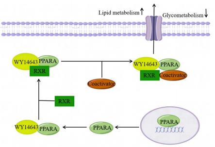 Fig. 1 Schematic diagram of PPARA involvement in metabolic mechanisms.1
Functions of PPARA
The core function of the PPARA gene is to serve as a key regulatory factor for lipid metabolism. In addition, it is also widely involved in physiological processes such as maintaining energy homeostasis, regulating inflammatory responses, and adjusting insulin sensitivity.
| Function | Description |
| Regulation of fatty acid oxidation | Activate the expression of genes related to fatty acid β -oxidation in liver and muscle tissues to promote lipid breakdown for energy supply. |
| Induction of ketone body formation | Under starvation conditions, it upregulates key enzymes in the ketone body production pathway to provide alternative energy for peripheral tissues. |
| Regulation of glucose metabolism | Improve systemic glucose homeostasis by influencing insulin signaling pathways and genes related to glucose utilization. |
| Inflammation suppression | Inhibit NF-κB and other inflammatory signaling pathways, reduce the production of pro-inflammatory factors, and play an anti-atherosclerosis role. |
| Energy metabolism integration | Coordinate the energy and substance utilization strategies of multiple metabolic organs to respond to changes in nutritional status. |
This receptor undergoes a conformational transformation by binding to its ligand (fatty acid substances), and then recruits a co-activator complex to initiate the transcription program of downstream target genes. This ligand-dependent transcriptional activation characteristic enables PPARA to precisely sense changes in the body's metabolic state and make adaptive responses, thereby playing a core regulatory role in energy metabolism balance.
Applications of PPARA and PPARA Antibody in Literature
1. Wang, Xinyue, et al. "A novel rabbit anti-myoglobin monoclonal antibody's potential application in rhabdomyolysis associated acute kidney injury." International Journal of Molecular Sciences 24.9 (2023): 7822. https://doi.org/10.7554/eLife.70472
Research has found that in non-alcoholic fatty liver disease, MIR20B aggravates lipid accumulation in the liver by targeting and inhibiting PPARA. Inhibiting MIR20B can enhance fatty acid oxidation, and its combination with PPARα agonists has a synergistic therapeutic effect.
2. Li, Honge, et al. "Mechanism of Action of the Plateau-Adapted Gene PPARA in COPD." Frontiers in Bioscience-Landmark 29.2 (2024): 68. https://doi.org/10.31083/j.fbl2902068
Research has found that the high-altitude adaptation gene PPARA is highly expressed in the lungs of Tibetan people, but its expression decreases in COPD patients, leading to impaired body response to hypoxia, exacerbating lung pathological changes, and promoting the development of COPD.
3. Yu, Haitao, et al. "Capsaicin Alleviates Autophagy‐Lysosomal Dysfunction via PPARA-Mediated V-ATPase Subunit ATP6V0E1 Signaling in 3xTg-AD Mice." Advanced Science 12.39 (2025): e02707. https://doi.org/10.1002/advs.202502707
This article utilizes mass spectrometry to characterize the binding strength and dissociation dynamics of myoglobin and its antibody-epitope complexes, providing a methodological framework for quantifying protein-ligand interactions and advancing epitope mapping in antibody research.
4. Xiang, Xixi, et al. "Significance of PPARA as a Treatment Target for Chronic Lymphocytic Leukemia." PPAR research 2023.1 (2023): 8456833.https://doi.org/10.1155/2023/8456833
Research has found that PPARA holds a core position in the CLL gene network. It works in synergy with multiple prognosis-related genes and jointly affects the progression of leukemia and the treatment interval through multiple pathological pathways such as cell adhesion and inflammation.
5. Kim, Donghwan, Sang Keun Ha, and Frank J. Gonzalez. "CBFA2T3 is PPARA sensitive and attenuates fasting-induced lipid accumulation in mouse liver." Cells 13.10 (2024): 831. https://doi.org/10.3390/cells13100831
Research has found that PPARA can activate the liver gene CBFA2T3. Knockout of this gene exacerbates insulin resistance and lipid accumulation in the liver of mice, indicating that CBFA2T3 plays a key protective role in regulating metabolic stress.
Creative Biolabs: PPARA Antibodies for Research
Creative Biolabs specializes in the production of high-quality PPARA antibodies for research and industrial applications. Our portfolio includes monoclonal antibodies tailored for ELISA, Flow Cytometry, Western blot, immunohistochemistry, and other diagnostic methodologies.
- Custom PPARA Antibody Development: Tailor-made solutions to meet specific research requirements.
- Bulk Production: Large-scale antibody manufacturing for industry partners.
- Technical Support: Expert consultation for protocol optimization and troubleshooting.
- Aliquoting Services: Conveniently sized aliquots for long-term storage and consistent experimental outcomes.
For more details on our PPARA antibodies, custom preparations, or technical support, contact us at email.
Reference
- Li, Honge, et al. "Mechanism of Action of the Plateau-Adapted Gene PPARA in COPD." Frontiers in Bioscience-Landmark 29.2 (2024): 68. https://doi.org/10.31083/j.fbl2902068
Anti-PPARA antibodies
 Loading...


Hot products 
-
Mouse Anti-COL12A1 Recombinant Antibody (CBYY-C3117) (CBMAB-C4560-YY)

-
Mouse Anti-BIRC3 Recombinant Antibody (16E63) (CBMAB-C3367-LY)

-
Rat Anti-ADAM10 Recombinant Antibody (V2-179741) (CBMAB-A1103-YC)

-
Mouse Anti-8-oxoguanine Recombinant Antibody (V2-7719) (CBMAB-1898CQ)

-
Mouse Anti-CORO1A Recombinant Antibody (4G10) (V2LY-1206-LY806)

-
Mouse Anti-ALX1 Recombinant Antibody (96k) (CBMAB-C0616-FY)

-
Mouse Anti-ARG1 Recombinant Antibody (CBYCL-103) (CBMAB-L0004-YC)

-
Mouse Anti-GIPC2 Recombinant Antibody (10) (CBMAB-G0476-LY)

-
Mouse Anti-ATP1B1 Recombinant Antibody (E4) (CBMAB-0463-LY)

-
Mouse Anti-CDK7 Recombinant Antibody (CBYY-C1783) (CBMAB-C3221-YY)

-
Mouse Anti-A2M Recombinant Antibody (V2-178822) (CBMAB-A0036-YC)

-
Mouse Anti-BIRC7 Recombinant Antibody (88C570) (CBMAB-L0261-YJ)

-
Mouse Anti-ALB Recombinant Antibody (V2-55272) (CBMAB-H0819-FY)

-
Mouse Anti-FOSB Recombinant Antibody (CBXF-3593) (CBMAB-F2522-CQ)

-
Mouse Anti-CCS Recombinant Antibody (CBFYC-1093) (CBMAB-C1150-FY)

-
Mouse Anti-CD247 Recombinant Antibody (6B10.2) (CBMAB-C1583-YY)

-
Mouse Anti-CRTAM Recombinant Antibody (CBFYC-2235) (CBMAB-C2305-FY)

-
Rabbit Anti-AKT3 Recombinant Antibody (V2-12567) (CBMAB-1057-CN)

-
Mouse Anti-ENO2 Recombinant Antibody (85F11) (CBMAB-0276CQ)

-
Mouse Anti-ATP5F1A Recombinant Antibody (51) (CBMAB-A4043-YC)

- AActivation
- AGAgonist
- APApoptosis
- BBlocking
- BABioassay
- BIBioimaging
- CImmunohistochemistry-Frozen Sections
- CIChromatin Immunoprecipitation
- CTCytotoxicity
- CSCostimulation
- DDepletion
- DBDot Blot
- EELISA
- ECELISA(Cap)
- EDELISA(Det)
- ESELISpot
- EMElectron Microscopy
- FFlow Cytometry
- FNFunction Assay
- GSGel Supershift
- IInhibition
- IAEnzyme Immunoassay
- ICImmunocytochemistry
- IDImmunodiffusion
- IEImmunoelectrophoresis
- IFImmunofluorescence
- IGImmunochromatography
- IHImmunohistochemistry
- IMImmunomicroscopy
- IOImmunoassay
- IPImmunoprecipitation
- ISIntracellular Staining for Flow Cytometry
- LALuminex Assay
- LFLateral Flow Immunoassay
- MMicroarray
- MCMass Cytometry/CyTOF
- MDMeDIP
- MSElectrophoretic Mobility Shift Assay
- NNeutralization
- PImmunohistologyp-Paraffin Sections
- PAPeptide Array
- PEPeptide ELISA
- PLProximity Ligation Assay
- RRadioimmunoassay
- SStimulation
- SESandwich ELISA
- SHIn situ hybridization
- TCTissue Culture
- WBWestern Blot
