XRN1 Antibodies
Background
The XRN1 gene encodes a nucleic acid exonuclease that is widely present in eukaryotic cells and is mainly located in the cytoplasm. This enzyme degrades RNA molecules from the 5' end to the 3' end and plays a central role in RNA metabolism, gene expression regulation, and maintenance of cellular homeostasis. Mutations or abnormal expression of this gene are closely associated with various human diseases such as neurodevelopmental disorders and cancer. Initially identified as a homologous homologous gene in yeast, the XRN1 protein was later proven to be an evolutionarily conserved key factor for RNA degradation in eukaryotes. The study of its structure and function has revealed the fine regulatory mechanism of RNA metabolism. As an important component of the RNA surveillance system, XRN1 continuously removes abnormal RNA molecules within the cell, playing an irreplaceable role in maintaining the accuracy of the post-transcriptional regulatory network.
Structure of XRN1
XRN1 is a large molecular protein with a molecular weight of approximately 170 kDa. The molecular weight of this protein varies slightly among different species, mainly due to the conservative changes in the amino acid sequence.
| Species | Human | Mouse | Zebrafish | Yeast | Arabidopsis |
| Molecular Weight (kDa) | 170 | 170 | 175 | 175 | 110 |
| Primary Structural Differences | Highly conservative, containing an exonuclease domain | Highly similar to humans | Retain the key functional domains | Classic research model | Plant representatives of homologous proteins |
The XRN1 protein consists of approximately 1700 amino acids and its spatial structure adopts a stretched conformation. This protein contains a characteristic N-terminal catalytic domain, which is responsible for performing 5'-3' nucleic acid exonuclease activity, and a C-terminal domain involved in protein-protein interactions. The secondary structure of XRN1 contains multiple conserved α-helices and β-sheets, jointly forming an efficient RNA degradation machine. Its activity depends on the cooperative action of two magnesium ions, and it achieves precise cutting of RNA substrates through an acid-base catalytic mechanism.
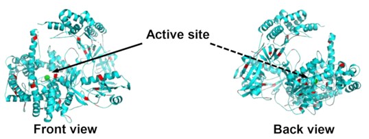 Fig. 1 Front and back view of XRN1 with lysine groups highlighted in red.1
Key structural properties of XRN1:
- Highly conserved N-terminal catalytic domain containing two magnesium ion binding sites
- The C-terminal domain mediates protein complex assembly and substrate recognition
- The characteristic helix-ring-helix conformation forms an RNA binding channel
- The conserved aromatic amino acid residues are involved in substrate capture and cleavage
Functions of XRN1
The main function of XRN1 is to participate in RNA metabolism and gene expression regulation. At the same time, it plays an important role in various cellular processes, including RNA surveillance and gene silencing, etc.
| Function | Description |
| RNA degradation | XRN1 degrades RNA molecules from 5' to 3', participating in the renewal and quality control of RNA within the cell. |
| Gene expression regulation | By regulating the half-life and abundance of mRNA, it affects the level of gene expression and protein synthesis. |
| RNA surveillance | XRN1 recognizes and degrades abnormal or defective RNA molecules, preventing the accumulation of toxic protein products. |
| Gene silencing participation | It plays a role in RNA interference and miRNA-mediated gene silencing pathways, assisting in the degradation of target RNA. |
| Maintenance of cellular homeostasis | By clearing specific RNA substrates, it participates in processes such as cell differentiation, development, and stress responses. |
The enzymatic activity of XRN1 depends on the cooperative action of two magnesium ions. Its catalytic efficiency is finely regulated by the structure of the substrate and binding proteins, ensuring the accuracy and plasticity of RNA metabolism.
Applications of XRN1 and XRN1 Antibody in Literature
1. Athapattu, Uditha S., et al. "Solid-phase XRN1 reactions for RNA cleavage: application in single-molecule sequencing." Nucleic acids research 49.7 (2021): e41-e41. https://doi.org/10.1093/nar/gkab001
In this study, XRN1 was covalently immobilized on a solid-phase carrier using EDC/NHS, and a novel RNA exonuclease sequencing reactor was constructed. The solid-phase enzyme cleavage efficiency reached 87.6%, which was higher than that of the solution phase (78.3%). Single-molecule fluorescence analysis showed that its cleavage rate was 26 nt/s, and its continuous synthesis capacity was >10.5 kb, providing a new tool for RNA modification detection.
2. Chen, Tianyong, et al. "Mn-XRN1 Has an Inhibitory Effect on Ovarian Reproduction in Macrobrachium nipponense." Genes 14.7 (2023): 1454. https://doi.org/10.3390/genes14071454
The article indicates that XRN1 is highly expressed in the ovaries of Japanese crayfish and negatively regulates the VASA gene to inhibit ovary maturation. After RNA interference targeting XRN1, VASA expression increases and ovary development accelerates significantly. The study shows that Mn-XRN1 is a key inhibitory factor for ovary maturation.
3. Sharma, Sunny, et al. "Xrn1 is a deNADding enzyme modulating mitochondrial NAD-capped RNA." Nature communications 13.1 (2022): 889. https://doi.org/10.1038/s41467-022-28555-7
The research has found that the yeast Xrn1 possesses both NAD cap removal and exonuclease functions. The mutant experiments have demonstrated that its cap removal activity affects the growth of non-fermentable sugars, regulates mitochondrial NAD-RNA levels, and plays a central role in the NAD metabolism of cells.
4. Piloto, Ana Margarida, et al. "Plastic antibodies tailored on quantum dots for an optical detection of myoglobin down to the femtomolar range." Scientific reports 8.1 (2018): 4944. https://doi.org/10.1038/s41467-019-09199-6
The research has found that Xrn1 promotes the translation of specific membrane protein mRNA in the endoplasmic reticulum, coordinating its transcription, translation and degradation. The study also discovered that this regulatory effect depends on the translation initiation complex and the secondary structure of the mRNA, revealing the new function of Xrn1 in multiple stages of gene expression.
5. Shanmugam, Renuka, et al. "Evidence that Xrn1 is in complex with Gcn1, and is required for full levels of eIF2α phosphorylation." Biochemical Journal 481.7 (2024): 481-498. https://doi.org/10.1042/BCJ20220531
The research has found that Xrn1 forms a complex with Gcn1 and Gcn2, and participates in the amino acid regulation pathway. The absence of Xrn1 will reduce the phosphorylation level of eIF2α and affect the growth of cells under starvation conditions. The study shows that Xrn1 is necessary for the efficient activation of Gcn2, connecting RNA metabolism with amino acid signal regulation.
Creative Biolabs: XRN1 Antibodies for Research
Creative Biolabs specializes in the production of high-quality XRN1 antibodies for research and industrial applications. Our portfolio includes monoclonal and polyclonal antibodies tailored for ELISA, Flow Cytometry, Western blot, immunohistochemistry, and other diagnostic methodologies.
- Custom XRN1 Antibody Development: Tailor-made solutions to meet specific research requirements.
- Bulk Production: Large-scale antibody manufacturing for industry partners.
- Technical Support: Expert consultation for protocol optimization and troubleshooting.
- Aliquoting Services: Conveniently sized aliquots for long-term storage and consistent experimental outcomes.
For more details on our XRN1 antibodies, custom preparations, or technical support, contact us at email.
Reference
- Athapattu, Uditha S., et al. "Solid-phase XRN1 reactions for RNA cleavage: application in single-molecule sequencing." Nucleic acids research 49.7 (2021): e41-e41. Distributed under Open Access license CC BY 4.0, without modification. https://doi.org/10.1093/nar/gkab001
Anti-XRN1 antibodies
 Loading...


Hot products 
-
Mouse Anti-8-oxoguanine Recombinant Antibody (V2-7697) (CBMAB-1869CQ)

-
Mouse Anti-DDC Recombinant Antibody (8E8) (CBMAB-0992-YC)

-
Rabbit Anti-ALK (Phosphorylated Y1278) Recombinant Antibody (D59G10) (PTM-CBMAB-0035YC)

-
Mouse Anti-AMH Recombinant Antibody (5/6) (CBMAB-A2527-YC)

-
Mouse Anti-ABCA3 Recombinant Antibody (V2-178911) (CBMAB-A0145-YC)

-
Rat Anti-C5AR1 Recombinant Antibody (8D6) (CBMAB-C9139-LY)

-
Rat Anti-4-1BB Recombinant Antibody (V2-1558) (CBMAB-0953-LY)

-
Mouse Anti-Acetyl-α-Tubulin (Lys40) Recombinant Antibody (V2-623485) (CBMAB-CP2897-LY)

-
Mouse Anti-AAV9 Recombinant Antibody (V2-634029) (CBMAB-AP023LY)

-
Mouse Anti-ACE2 Recombinant Antibody (V2-179293) (CBMAB-A0566-YC)

-
Mouse Anti-APP Recombinant Antibody (DE2B4) (CBMAB-1122-CN)

-
Mouse Anti-CTCF Recombinant Antibody (CBFYC-2371) (CBMAB-C2443-FY)

-
Mouse Anti-GGT1 Recombinant Antibody (1F9) (CBMAB-G3273-LY)

-
Mouse Anti-EMP3 Recombinant Antibody (CBFYE-0100) (CBMAB-E0207-FY)

-
Mouse Anti-AQP2 Recombinant Antibody (G-3) (CBMAB-A3359-YC)

-
Mouse Anti-CDKL5 Recombinant Antibody (CBFYC-1629) (CBMAB-C1689-FY)

-
Mouse Anti-APOE Recombinant Antibody (A1) (CBMAB-0078CQ)

-
Mouse Anti-ARHGAP5 Recombinant Antibody (54/P190-B) (CBMAB-P0070-YC)

-
Mouse Anti-ENO1 Recombinant Antibody (8G8) (CBMAB-E1329-FY)

-
Rabbit Anti-B2M Recombinant Antibody (CBYY-0059) (CBMAB-0059-YY)

- AActivation
- AGAgonist
- APApoptosis
- BBlocking
- BABioassay
- BIBioimaging
- CImmunohistochemistry-Frozen Sections
- CIChromatin Immunoprecipitation
- CTCytotoxicity
- CSCostimulation
- DDepletion
- DBDot Blot
- EELISA
- ECELISA(Cap)
- EDELISA(Det)
- ESELISpot
- EMElectron Microscopy
- FFlow Cytometry
- FNFunction Assay
- GSGel Supershift
- IInhibition
- IAEnzyme Immunoassay
- ICImmunocytochemistry
- IDImmunodiffusion
- IEImmunoelectrophoresis
- IFImmunofluorescence
- IGImmunochromatography
- IHImmunohistochemistry
- IMImmunomicroscopy
- IOImmunoassay
- IPImmunoprecipitation
- ISIntracellular Staining for Flow Cytometry
- LALuminex Assay
- LFLateral Flow Immunoassay
- MMicroarray
- MCMass Cytometry/CyTOF
- MDMeDIP
- MSElectrophoretic Mobility Shift Assay
- NNeutralization
- PImmunohistologyp-Paraffin Sections
- PAPeptide Array
- PEPeptide ELISA
- PLProximity Ligation Assay
- RRadioimmunoassay
- SStimulation
- SESandwich ELISA
- SHIn situ hybridization
- TCTissue Culture
- WBWestern Blot
