
B220 Antibodies
Background
B220 (also known as CD45) is a transmembrane glycoprotein widely expressed on the surface of hematopoietic cells in vertebrates, mainly functioning as a tyrosine phosphatase to regulate lymphocyte receptor signaling. This gene generates multiple isomers through selective splicing, which can activate or inhibit the signaling pathways of immune cells, thereby playing a core role in immune responses, cell differentiation and inflammatory reactions. B220 was first identified by the team of Roger Coffman and Irving Weissman in 1975 and is a key surface marker for the classification of lymphocyte subsets, especially commonly used in the recognition and research of B cells. Its complex regulatory mechanism and multi-functionality remain important models in immunology research to this day, providing a key theoretical basis for understanding the molecular mechanisms of autoimmune diseases, leukemia and immunodeficiency disorders.
Structure of B220
B220 (CD45) is a large type I transmembrane protein with a molecular weight of approximately 180-220 kDa. The specific molecular weight varies depending on the splicing isomer. This value is highly conserved in different species and is mainly contributed by the highly glycosylated domain of the extracellular segment.
| Species | Human | Mouse | Rat |
| Molecular Weight (kDa) | 180-220 | 180-220 | 180-220 |
| Primary Structural Differences | Contains multiple variable exons (such as RO exon) | Highly homologous to humans, the extracellular domain glycosylation pattern is slightly different | CD45 structure very similar to mice |
The B220 protein is composed of an extracellular region, a transmembrane region and an intracellular region. Its extracellular region contains multiple fibronectin type III domains and a large number of glycosylation modifications (the name "B220" is derived from the electrophoretic mobility caused by its glycosylation characteristics), which endows it with its unique antigenicity. The intracellular region has two tyrosine phosphatase domains (D1 and D2) with catalytic activity. Among them, the D1 domain is the functional core. By dephosphorylating the key regulatory sites of Src family kinases, it directly participates in the activation or inhibition of T cell and B cell receptor signals, thereby precisely regulating the immune response of lymphocytes.
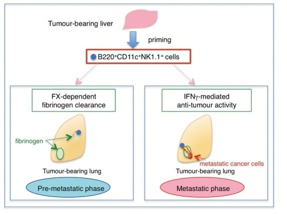 Fig. 1 Role of B220+CD11c+NK1.1+ HepELs in metastasis.1
Key structural properties of B220:
- Large type I transmembrane glycoproteins with multiple alternative splicing extracellular domains
- Extracellular region rich in cysteine and have a large number of glycosylation modification
- Intracellular area contains two series of tyrosine phosphatase domain structure (D1 and D2)
Functions of B220
The main function of B220 (CD45) is to act as a key regulatory molecule in lymphocyte signal transduction. However, it also extensively involves multiple aspects of the immune response, including cell activation, developmental differentiation, and immune tolerance.
| Function | Description |
| Immune signaling | As a tyrosine phosphatase, it positively regulates the T-cell and B-cell receptor signaling pathways by dephosphorylating the C-terminal inhibitory sites of Src family kinases (such as Lck and Fyn). |
| Lymphocyte activation and proliferation | Initiating antigen receptor-mediated activation signals is a core molecule essential for lymphocyte activation, clonal expansion, and effective immune response. |
| Lymphocyte development | In the thymus and bone marrow, regulating the positive and negative selection process of lymphocytes is crucial for the normal development and maturation of T cells and B cells. |
| Maintenance of immune tolerance | By precisely regulating signal intensity, it participates in the clearance of autoreactive lymphocytes and prevents the occurrence of autoimmune diseases. |
| Cytokine response regulation | Affect the JAK - STAT downstream signaling pathways, such as adjusting the lymphocyte response intensity and direction of cytokines. |
B220 has a high degree of specificity in signal regulation for different lymphocyte subsets, and its function depends on the cell type, developmental stage and the form of the isomer spliced. This complexity makes it a precise "regulatory hub" of the immune system.
Applications of B220 and B220 Antibody in Literature
1. Hiratsuka, Sachie, et al. "Hepato‐entrained B220+ CD 11c+ NK 1.1+ cells regulate pre‐metastatic niche formation in the lung." EMBO molecular medicine 10.7 (2018): e8643. https://doi.org/10.15252/emmm.201708643
The article indicates that the liver inhibits the formation of lung metastases by releasing B220+ CD11c+ NK1.1+ cells. This type of cells highly express coagulation factor X, which can clear fibrin deposits in lung tissue and exert anti-tumor effects by inducing IFNγ.
2. Vacani-Martins, Natalia, et al. "After Experimental Trypanosoma cruzi Infection, Dying Hepatic CD3+ TCRαβ+ B220+ T Lymphocytes Are Rescued from Death by Peripheral T Cells and Become Activated." Pathogens 9.9 (2020): 717. https://doi.org/10.3390/pathogens9090717
This study explored the role of liver CD3+B220+ T cells in Trypanosoma cruzii infection. It was found that this type of cells contained CD117− apoptotic subsets and CD117+ thymus-independent subsets, and the majority were 7AAD+ apoptotic cells. After infection, approximately 15% of the CD117+ population transformed into B220−7AAD−, expressing CD90.2 and activation markers, and was rescued and survived by peripheral activated T cells, indicating its potential for activation and immune regulation.
3. Benbijja, Mohcine, Amine Mellouk, and Pierre Bobé. "Sensitivity of leukemic T-cell lines to arsenic trioxide cytotoxicity is dependent on the induction of phosphatase B220/CD45R expression at the cell surface." Molecular cancer 13.1 (2014): 251. https://doi.org/10.1186/1476-4598-13-251
Research has found that the sensitivity of leukemia T cells to arsenic trioxide (As₂O₃) is closely related to the expression level of B220 (CD45 isomer). Treatment with As₂O₃ can dose-dependently induce membrane expression of B220 and inhibit nuclear translocation of NF-κB p50, thereby inducing apoptosis or mixed cell death. The expression level of B220 is strictly related to the degree and form of cell death, suggesting that it may serve as a key regulatory factor in the death pathway.
4. Wilson, Kyle A., et al. "Depletion of B220+ NK1. 1+ cells enhances the rejection of established melanoma by tumor-specific CD4+ T cells." Oncoimmunology 4.8 (2015): e1019196. https://doi.org/10.1080/2162402X.2015.1019196
This study found that in adoptive T-cell therapy (ACT) for melanoma, eliminating NK1.1+ cells can enhance the anti-tumor effect of CD4+ T cells. Among them, NK1.1+B220+ pre-mNK cells (i.e., IKDC) are the key population that inhibits the anti-tumor immune response. Targeting this group can increase the number of T cells, levels of inflammatory factors and survival rates, providing a new strategy for improving the efficacy of ACT.
5. Le Gall, Sylvain M., et al. "Loss of P2X7 receptor plasma membrane expression and function in pathogenic B220+ double-negative T lymphocytes of autoimmune MRL/lpr mice." PLoS One 7.12 (2012): e52161. https://doi.org/10.1371/journal.pone.0052161
In lupus model MRL/lpr mice, B220+ CD4-CD8-T cells accumulate abnormally. Research has found that although the B220+ T cell subset expresses the P2X7 receptor intracellularly, the P2X7R is absent on the membrane surface, resulting in no response to ATP/NAD stimulation and apoptotic signals. The surface expression of B220 is significantly correlated with the inhibition of the P2X7R pathway, suggesting its involvement in regulating abnormal T cell survival and autoimmunity.
Creative Biolabs: B220 Antibodies for Research
Creative Biolabs specializes in the production of high-quality B220 antibodies for research and industrial applications. Our portfolio includes monoclonal antibodies tailored for ELISA, Flow Cytometry, Western blot, immunohistochemistry, and other diagnostic methodologies.
- Custom B220 Antibody Development: Tailor-made solutions to meet specific research requirements.
- Bulk Production: Large-scale antibody manufacturing for industry partners.
- Technical Support: Expert consultation for protocol optimization and troubleshooting.
- Aliquoting Services: Conveniently sized aliquots for long-term storage and consistent experimental outcomes.
For more details on our B220 antibodies, custom preparations, or technical support, contact us at email.
Reference
- Hiratsuka, Sachie, et al. "Hepato‐entrained B220+ CD 11c+ NK 1.1+ cells regulate pre‐metastatic niche formation in the lung." EMBO molecular medicine 10.7 (2018): e8643. https://doi.org/10.15252/emmm.201708643
Anti-B220 antibodies
 Loading...
 Compare




Hot products 
-
Rabbit Anti-CBL Recombinant Antibody (D4E10) (CBMAB-CP0149-LY)

-
Rabbit Anti-BRCA2 Recombinant Antibody (D9S6V) (CBMAB-CP0017-LY)

-
Mouse Anti-ADGRL2 Recombinant Antibody (V2-58519) (CBMAB-L0166-YJ)

-
Mouse Anti-CFL1 Recombinant Antibody (CBFYC-1771) (CBMAB-C1833-FY)

-
Mouse Anti-AKT1/AKT2/AKT3 (Phosphorylated T308, T309, T305) Recombinant Antibody (V2-443454) (PTM-CBMAB-0030YC)

-
Mouse Anti-H1-0 Recombinant Antibody (CBFYH-0565) (CBMAB-H1451-FY)

-
Mouse Anti-ABCA3 Recombinant Antibody (V2-178911) (CBMAB-A0145-YC)

-
Mouse Anti-CCS Recombinant Antibody (CBFYC-1093) (CBMAB-C1150-FY)

-
Mouse Anti-CD8 Recombinant Antibody (C1083) (CBMAB-C1083-LY)

-
Mouse Anti-AQP2 Recombinant Antibody (G-3) (CBMAB-A3359-YC)

-
Mouse Anti-CD83 Recombinant Antibody (HB15) (CBMAB-C1765-CQ)

-
Mouse Anti-AMACR Recombinant Antibody (CB34A) (CBMAB-CA034LY)

-
Mouse Anti-CCDC25 Recombinant Antibody (CBLC132-LY) (CBMAB-C9786-LY)

-
Rat Anti-ADAM10 Recombinant Antibody (V2-179741) (CBMAB-A1103-YC)

-
Mouse Anti-GFAP Recombinant Antibody (24) (CBMAB-G2927-LY)

-
Rabbit Anti-Acetyl-Histone H3 (Lys36) Recombinant Antibody (V2-623395) (CBMAB-CP0994-LY)

-
Mouse Anti-AKT1 Recombinant Antibody (V2-180546) (CBMAB-A2070-YC)

-
Mouse Anti-BCL6 Recombinant Antibody (CBYY-0435) (CBMAB-0437-YY)

-
Mouse Anti-BMI1 Recombinant Antibody (CBYC-P026) (CBMAB-P0108-YC)

-
Mouse Anti-BACE1 Recombinant Antibody (61-3E7) (CBMAB-1183-CN)

- AActivation
- AGAgonist
- APApoptosis
- BBlocking
- BABioassay
- BIBioimaging
- CImmunohistochemistry-Frozen Sections
- CIChromatin Immunoprecipitation
- CTCytotoxicity
- CSCostimulation
- DDepletion
- DBDot Blot
- EELISA
- ECELISA(Cap)
- EDELISA(Det)
- ESELISpot
- EMElectron Microscopy
- FFlow Cytometry
- FNFunction Assay
- GSGel Supershift
- IInhibition
- IAEnzyme Immunoassay
- ICImmunocytochemistry
- IDImmunodiffusion
- IEImmunoelectrophoresis
- IFImmunofluorescence
- IGImmunochromatography
- IHImmunohistochemistry
- IMImmunomicroscopy
- IOImmunoassay
- IPImmunoprecipitation
- ISIntracellular Staining for Flow Cytometry
- LALuminex Assay
- LFLateral Flow Immunoassay
- MMicroarray
- MCMass Cytometry/CyTOF
- MDMeDIP
- MSElectrophoretic Mobility Shift Assay
- NNeutralization
- PImmunohistologyp-Paraffin Sections
- PAPeptide Array
- PEPeptide ELISA
- PLProximity Ligation Assay
- RRadioimmunoassay
- SStimulation
- SESandwich ELISA
- SHIn situ hybridization
- TCTissue Culture
- WBWestern Blot
