CD209 Antibodies
Background
CD209 is a transmembrane receptor protein mainly expressed on the surface of dendritic cells and macrophages, belonging to the C-type lectin family. This protein can specifically recognize and bind to the high-mannose structure on the surface of pathogens, thereby mediating the endocytosis and presentation of pathogens, and initiating the adaptive immune response of the body. During the infection of various pathogens such as HIV and Mycobacterium tuberculosis, CD209 can either function as a innate immune recognition receptor to exert defensive functions, or may be utilized by the pathogens to promote infection. It was first cloned and identified by Curtis et al. in 1992. Subsequent studies have revealed its multiple roles in immune cell migration, antigen presentation, and immune regulation. The structural and functional analysis of CD209 provides a key model for in-depth understanding of the interaction mechanism between pathogens and hosts, and lays an important foundation for the development of anti-infection vaccines and immunotherapy strategies.
Structure of CD209
The molecular weight of CD209 protein is approximately 45 kDa, and there are differences among different species. The extracellular segment of this protein contains a C-type lectin domain, which mediates pathogen recognition.
| Species | Human | Mouse | Rhesus monkey | Pig | Cow |
| Molecular Weight (kDa) | 45.1 | 44.8 | 45.3 | 44.5 | 44.9 |
| Primary Structural Differences | Cytoplasmic region contains endocytosis signal | Conserved in transmembrane region | Homology with humans reaches 93% | Different glycosylation sites | Variations in ligand binding domain |
The CD209 protein is composed of 404 amino acids and is a transmembrane receptor protein. Its structure consists of three main regions: the N-terminal cytoplasmic region, the transmembrane region, and the C-terminal extracellular region. The extracellular region contains a characteristic C-type lectin-like domain (CRD), which maintains its globular conformation through two disulfide bonds. The interior of this domain contains a conserved EPN motif (Glu-Pro-Asn), forming a ligand-binding site that specifically recognizes the mannose and fucose structures on the surface of pathogens. The CRD structure coordinates and binds two calcium ions, one to maintain structural stability and the other to directly participate in sugar recognition. Quaternization is the functional form of CD209 for recognizing multivalent ligands. Each subunit wraps around each other through the coiled-coil structure in the neck region to form a stalk-like structure, allowing the CRD to extend outward to capture the pathogen.
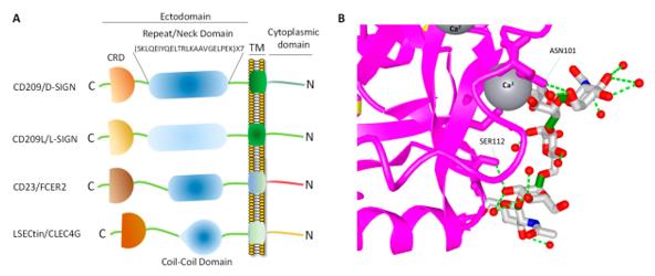 Fig. 1 CD209 family proteins.1
Key structural properties of CD209:
- Transmembrane receptor structure
- Extracellular region forms a tetramer
- Calcium ion-dependent recognition of pathogen surface carbohydrate structures
- Intracellular tail region contains an endocytosis motif, mediating antigen uptake and presentation
Functions of CD209
The main function of CD209 is to recognize the sugar structures on the surface of pathogens and trigger the immune response. It is also involved in various physiological processes such as immune regulation and cell migration.
| Function | Description |
| Pathogen Recognition | CD209 specifically binds to the high-mannose structures on the surfaces of viruses, bacteria, and fungi, mediating endocytosis of pathogens. |
| Antigen Presentation | The captured pathogens are internalized and then enter the MHC pathway, where they are processed and presented to T cells to initiate adaptive immunity . |
| HIV Infection Promotion | Utilized by the envelope protein gp120 of HIV-1, it mediates the entry of the virus into dendritic cells and its spread to T cells . |
| Immune Regulation | Binding to ICAM-3 mediates the initial contact between dendritic cells and T cells, regulating the intensity of the immune response. |
| Autoimmune Protection | Identifying self-glycosylated molecules, it participates in maintaining immune homeostasis and preventing excessive inflammatory responses. |
The ligand-binding characteristics of CD209 are calcium ion-dependent and exhibit a specific recognition pattern with the mannose-type structure, reflecting its crucial role in pathogen capture and its functional positioning for initiating rapid immune responses.
Applications of CD209 and CD209 Antibody in Literature
1. Rahimi, Nader. "C-type lectin CD209L/L-SIGN and CD209/DC-SIGN: cell adhesion molecules turned to pathogen recognition receptors." Biology 10.1 (2020): 1. https://doi.org/10.3390/biology10010001
During the COVID-19 pandemic, the CD209 family proteins have attracted much attention as viral receptors. They mediate invasion by recognizing the viral spike protein and are widely expressed in target organs with genetic polymorphisms. Studying these interactions is of great significance for understanding the pathogenic mechanism and developing vaccines.
2. Lin, Anqi, et al. "CD209 signaling pathway as a biomarker for cisplatin chemotherapy response in small cell lung cancer." Genes & Diseases 11.3 (2023): 101038. https://doi.org/10.1016/j.gendis.2023.06.011
The research has found that small cell lung cancer is prone to developing resistance to cisplatin. The high expression of CD209 in lung cancer indicates a better prognosis, and its signaling pathway may affect the chemotherapy outcome by regulating immunity. Studying the relationship between CD209 and the efficacy and prognosis of cisplatin treatment is of great significance for overcoming drug resistance.
3. Marzaioli, Viviana, et al. "CD209/CD14+ dendritic cells characterization in rheumatoid and psoriatic arthritis patients: activation, synovial infiltration, and therapeutic targeting." Frontiers in immunology 12 (2022): 722349. https://doi.org/10.3389/fimmu.2021.722349
The study found that there was an increase in CD209/CD14+ dendritic cells in the bodies of arthritis patients, which exhibited a unique inflammatory phenotype. Joint fluid can induce their formation, and the JAK/STAT pathway regulates their functions. Inhibiting this pathway may alleviate inflammation and provide a new strategy for treatment.
4. Chang, Kai, et al. "Association between CD209-336A/G and-871A/G polymorphisms and susceptibility of tuberculosis: a meta-analysis." PLoS One 7.7 (2012): e41519. https://doi.org/10.1371/journal.pone.0041519
The meta-analysis showed that the polymorphisms at positions -336 and -871 of the CD209 promoter were not associated with the overall risk of tuberculosis. However, the GG genotype at the -336 locus might increase the tuberculosis susceptibility in the Asian population, providing a basis for the genetic risk of specific populations.
5. Shadrina, Alexandra S., et al. "Mendelian randomization analysis of plasma levels of CD209 and MICB proteins and the risk of varicose veins of lower extremities." Plos one 17.5 (2022): e0268725. https://doi.org/10.1371/journal.pone.0268725
The Mendelian randomization study found that the elevated level of CD209 protein predicted by genes was significantly associated with an increased risk of lower extremity varicose veins (OR = 1.08). This result supports the possibility that CD209 may be involved in the pathogenesis of varicose veins, providing genetic evidence for subsequent mechanism studies.
Creative Biolabs: CD209 Antibodies for Research
Creative Biolabs specializes in the production of high-quality CD209 antibodies for research and industrial applications. Our portfolio includes monoclonal and polyclonal antibodies tailored for ELISA, Flow Cytometry, Western blot, immunohistochemistry, and other diagnostic methodologies.
- Custom CD209 Antibody Development: Tailor-made solutions to meet specific research requirements.
- Bulk Production: Large-scale antibody manufacturing for industry partners.
- Technical Support: Expert consultation for protocol optimization and troubleshooting.
- Aliquoting Services: Conveniently sized aliquots for long-term storage and consistent experimental outcomes.
For more details on our CD209 antibodies, custom preparations, or technical support, contact us at email.
Reference
- Rahimi, Nader. "C-type lectin CD209L/L-SIGN and CD209/DC-SIGN: cell adhesion molecules turned to pathogen recognition receptors." Biology 10.1 (2020): 1. Distributed under Open Access license CC BY 4.0, without modification. https://doi.org/10.3390/biology10010001
Anti-CD209 antibodies
 Loading...


Hot products 
-
Mouse Anti-COL1A2 Recombinant Antibody (CF108) (V2LY-1206-LY626)

-
Mouse Anti-FPR2 Recombinant Antibody (1D6) (CBMAB-F2628-CQ)

-
Mouse Anti-APP Recombinant Antibody (DE2B4) (CBMAB-1122-CN)

-
Mouse Anti-G6PD Recombinant Antibody (13B331) (CBMAB-G1553-LY)

-
Mouse Anti-FOXA3 Recombinant Antibody (2A9) (CBMAB-0377-YC)

-
Rat Anti-CD63 Recombinant Antibody (7G4.2E8) (CBMAB-C8725-LY)

-
Mouse Anti-2C TCR Recombinant Antibody (V2-1556) (CBMAB-0951-LY)

-
Rabbit Anti-AP2M1 (Phosphorylated T156) Recombinant Antibody (D4F3) (PTM-CBMAB-0610LY)

-
Mouse Anti-CGAS Recombinant Antibody (CBFYM-0995) (CBMAB-M1146-FY)

-
Mouse Anti-BRCA2 Recombinant Antibody (CBYY-1728) (CBMAB-2077-YY)

-
Mouse Anti-HTLV-1 gp46 Recombinant Antibody (CBMW-H1006) (CBMAB-V208-1154-FY)

-
Mouse Anti-ACE2 Recombinant Antibody (V2-179293) (CBMAB-A0566-YC)

-
Armenian hamster Anti-CD40 Recombinant Antibody (HM40-3) (CBMAB-C10365-LY)

-
Mouse Anti-COL12A1 Recombinant Antibody (CBYY-C3117) (CBMAB-C4560-YY)

-
Mouse Anti-AAV-5 Recombinant Antibody (V2-503417) (CBMAB-V208-1369-FY)

-
Mouse Anti-ATP1B3 Recombinant Antibody (1E9) (CBMAB-A4021-YC)

-
Mouse Anti-BRD3 Recombinant Antibody (CBYY-0801) (CBMAB-0804-YY)

-
Mouse Anti-BIRC3 Recombinant Antibody (315304) (CBMAB-1214-CN)

-
Mouse Anti-BCL2L1 Recombinant Antibody (H5) (CBMAB-1025CQ)

-
Mouse Anti-BCL6 Recombinant Antibody (CBYY-0435) (CBMAB-0437-YY)

- AActivation
- AGAgonist
- APApoptosis
- BBlocking
- BABioassay
- BIBioimaging
- CImmunohistochemistry-Frozen Sections
- CIChromatin Immunoprecipitation
- CTCytotoxicity
- CSCostimulation
- DDepletion
- DBDot Blot
- EELISA
- ECELISA(Cap)
- EDELISA(Det)
- ESELISpot
- EMElectron Microscopy
- FFlow Cytometry
- FNFunction Assay
- GSGel Supershift
- IInhibition
- IAEnzyme Immunoassay
- ICImmunocytochemistry
- IDImmunodiffusion
- IEImmunoelectrophoresis
- IFImmunofluorescence
- IGImmunochromatography
- IHImmunohistochemistry
- IMImmunomicroscopy
- IOImmunoassay
- IPImmunoprecipitation
- ISIntracellular Staining for Flow Cytometry
- LALuminex Assay
- LFLateral Flow Immunoassay
- MMicroarray
- MCMass Cytometry/CyTOF
- MDMeDIP
- MSElectrophoretic Mobility Shift Assay
- NNeutralization
- PImmunohistologyp-Paraffin Sections
- PAPeptide Array
- PEPeptide ELISA
- PLProximity Ligation Assay
- RRadioimmunoassay
- SStimulation
- SESandwich ELISA
- SHIn situ hybridization
- TCTissue Culture
- WBWestern Blot
