CHI3L1 Antibodies
Background
The CHI3L1 gene, also known as YKL-40, encodes a secretory glycoprotein belonging to the glycosylase family, but it does not have enzymatic activity itself. This protein is mainly secreted by activated macrophages, neutrophils, vascular smooth muscle cells and some tumor cells, and plays a role in processes such as cell proliferation, differentiation, inflammatory response and tissue remodeling. It is significantly elevated in the serum and lesion tissues of patients with various inflammatory diseases and tumors, and is regarded as an important marker of disease activity. By binding to its unknown receptors, CHI3L1 participates in regulating multiple signaling pathways including NF-κB and MAPK, thereby influencing cell survival and the production of inflammatory mediators. It is precisely because of its key role in the inflammatory and tumor microenvironment that CHI3L1 has become a research hotspot in disease diagnosis, prognosis assessment, and potential therapeutic targets. The in-depth analysis of its structure and function provides important clues for understanding the pathogenesis of chronic inflammatory diseases and cancer.
Structure of CHI3L1
The molecular weight of the CHI3L1 protein is approximately 40 kDa. Its size is relatively conserved among different species, but there are certain differences in its amino acid sequence, which are closely related to its biological functions.
| Species | Human | Mouse | Rat | Pig |
| Molecular Weight (kDa) | 42.6 | 44.3 | 43.8 | 42.0 |
| Primary Structural Differences | Composed of 383 amino acids, it contains heparin and chitin binding sites | About 72% homology with anthropogenic amino acid sequence | Structure is similar to that of mice | Sequence information is relatively scarce |
The molecular structure of CHI3L1 protein consists of 383 amino acids and presents a compact spherical conformation composed of multiple domains. The core of this protein is a characteristic TIM barrel-shaped domain, which is formed by 8 parallel α helices and 8 β sheets arranged alternately. This structure endows the protein with high stability. There is a conserved heparin-binding site distributed on its surface, which is the key region for its interaction with the extracellular matrix. Although CHI3L1 belongs to the glycosyl hydrolase family 18, a crucial mutation has occurred at the catalytic site, where glutamate has been replaced by leucine, resulting in the loss of chitinase activity. This inactivated deep fissure domain can still bind chitin oligosaccharides and other polysaccharides, thereby mediating cell signal transduction. Additionally, this protein contains a conserved α/β domain, which participates in protein-protein interactions and is crucial for its binding to membrane receptors and activation of downstream pathways.
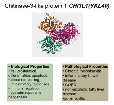 Fig. 1 CHI3L1’s (YKL40) biological and pathological characteristics.1
Key structural properties of CHI3L1:
- The characteristic TIM barrel-shaped domain
- The inactive chitinase catalytic site
- The heparin binding site, located on the protein surface
- The conserved cysteine residues
Functions of CHI3L1
The CHI3L1 protein has multiple biological functions, mainly involved in inflammatory responses, cell proliferation, and tissue remodeling processes.
| Function | Description |
| Inflammation Regulation | Secreted by activated macrophages and neutrophils, it promotes the chemotaxis of inflammatory cells and regulates the expression and release of inflammatory factors. |
| Cell Proliferation and Differentiation | Acting on various cell types such as fibroblasts and chondrocytes, it stimulates their proliferation and regulates the differentiation process. |
| Tissue Remodeling | Participating in the degradation and reconstruction of extracellular matrix, it plays a role in wound healing, fibrosis, and angiogenesis. |
| Anti-apoptotic Function | By activating signaling pathways such as PI3K/AKT, it inhibits cell apoptosis and promotes cell survival. |
| Tumor Progression Related | Highly expressed in various malignant tumors, it promotes tumor cell invasion, metastasis, and the formation of the tumor microenvironment. |
The expression of CHI3L1 is induced by various inflammatory factors such as IL-13 and TNF-α. Its signal transduction depends on the binding to transmembrane receptors such as TMEM219, which subsequently activates downstream pathways such as MAPK and NF-κB, thereby achieving transcriptional regulation of target genes.
Applications of CHI3L1 and CHI3L1 Antibody in Literature
1. Guo, Ling, et al. "Mechanistic studies on the role of CHI3L1 in eosinophilic inflammation in chronic sinusitis." Frontiers in Immunology 16 (2025): 1562546. https://doi.org/10.3389/fimmu.2025.1562546
The article indicates that over 10% of adults are affected by chronic rhinosinusitis (CRS), and the current treatments have a high recurrence rate. Chitinase 3-like protein 1 (CHI3L1) plays a crucial role in the activation and migration of eosinophils. This article reviews the biological characteristics and regulatory mechanisms of CHI3L1 in CRS, providing new ideas for precise targeted treatment of CRS.
2. Li, Tao, et al. "CHI3L1: a key driver in gastritis-to-cancer transformation." Journal of Translational Medicine 23.1 (2025): 349. https://doi.org/10.1186/s12967-025-06352-2
The research has found that CHI3L1 is a key driver gene for the transformation from gastritis to gastric cancer. It induces cellular malignant transformation by activating the CD44-β-catenin pathway. CHI3L1 is mainly secreted by fibroblasts and dendritic cells, and its high expression is associated with poor prognosis of gastric cancer, providing a new target for the early diagnosis and treatment of gastric cancer.
3. Jin, Guanghui, et al. "5-aminolevulinate and CHIL3/CHI3L1 treatment amid ischemia aids liver metabolism and reduces ischemia-reperfusion injury."Theranostics 13.14 (2023): 4802. https://doi.org/10.7150/thno.83163
The study found that supplementing with 5-ALA promotes M2 polarization of macrophages by down-regulating RelA and CX3CR1, and the CHIL3/CHI3L1 secreted by M2 improves the metabolism of liver cells. The combination of 5-ALA and CHIL3 can alleviate liver ischemia-reperfusion injury, providing a new strategy for clinical treatment.
4. Jatczak-Pawlik, Izabela, et al. "CHI3L1 in multiple sclerosis—From bench to clinic." Cells 13.24 (2024): 2086. https://doi.org/10.3390/cells13242086
The research has found that CHI3L1 is produced by central nervous system microglia and astrocytes, and is involved in the inflammatory and neurodegenerative processes of multiple sclerosis (MS). Its level can assist in the early diagnosis of MS, the assessment of disease progression, and the activation status of glial cells, providing a reference for optimizing treatment strategies.
5. Taifour, Tarek, et al. "The tumor-derived cytokine Chi3l1 induces neutrophil extracellular traps that promote T cell exclusion in triple-negative breast cancer." Immunity 56.12 (2023): 2755-2772. https://doi.org/10.1016/j.immuni.2023.11.002
The study found that high expression of CHI3L1 in triple-negative breast cancer is associated with T-cell stromal restriction. CHI3L1 hinders T-cell infiltration by promoting the recruitment of neutrophils and the formation of NETs. Targeting CHI3L1 can enhance anti-tumor immunity and improve the efficacy of immune checkpoint blockade.
Creative Biolabs: CHI3L1 Antibodies for Research
Creative Biolabs specializes in the production of high-quality CHI3L1 antibodies for research and industrial applications. Our portfolio includes monoclonal and polyclonal antibodies tailored for ELISA, Flow Cytometry, Western blot, immunohistochemistry, and other diagnostic methodologies.
- Custom CHI3L1 Antibody Development: Tailor-made solutions to meet specific research requirements.
- Bulk Production: Large-scale antibody manufacturing for industry partners.
- Technical Support: Expert consultation for protocol optimization and troubleshooting.
- Aliquoting Services: Conveniently sized aliquots for long-term storage and consistent experimental outcomes.
For more details on our CHI3L1 antibodies, custom preparations, or technical support, contact us at email.
Reference
- Guo, Ling, et al. "Mechanistic studies on the role of CHI3L1 in eosinophilic inflammation in chronic sinusitis." Frontiers in Immunology 16 (2025): 1562546. Distributed under Open Access license CC BY 4.0, without modification. https://doi.org/10.3389/fimmu.2025.1562546
Anti-CHI3L1 antibodies
 Loading...


Hot products 
-
Mouse Anti-AGK Recombinant Antibody (V2-258056) (CBMAB-M0989-FY)

-
Mouse Anti-CAT Recombinant Antibody (724810) (CBMAB-C8431-LY)

-
Mouse Anti-AKT1/AKT2/AKT3 (Phosphorylated T308, T309, T305) Recombinant Antibody (V2-443454) (PTM-CBMAB-0030YC)

-
Mouse Anti-C5AR1 Recombinant Antibody (R63) (CBMAB-C9553-LY)

-
Mouse Anti-BLNK Recombinant Antibody (CBYY-0623) (CBMAB-0626-YY)

-
Mouse Anti-CCNH Recombinant Antibody (CBFYC-1054) (CBMAB-C1111-FY)

-
Mouse Anti-BPGM Recombinant Antibody (CBYY-1806) (CBMAB-2155-YY)

-
Mouse Anti-CD19 Recombinant Antibody (CBXC-1224) (CBMAB-C1491-CQ)

-
Mouse Anti-ENO2 Recombinant Antibody (H14) (CBMAB-E1341-FY)

-
Mouse Anti-ACTN4 Recombinant Antibody (V2-6075) (CBMAB-0020CQ)

-
Mouse Anti-ARIH1 Recombinant Antibody (C-7) (CBMAB-A3563-YC)

-
Mouse Anti-AAV-5 Recombinant Antibody (V2-503416) (CBMAB-V208-1402-FY)

-
Mouse Anti-ALPL Antibody (B4-78) (CBMAB-1009CQ)

-
Rabbit Anti-ABL1 (Phosphorylated Y245) Recombinant Antibody (V2-505716) (PTM-CBMAB-0465LY)

-
Mouse Anti-DMD Recombinant Antibody (D1190) (CBMAB-D1190-YC)

-
Rat Anti-AChR Recombinant Antibody (V2-12500) (CBMAB-0990-CN)

-
Rabbit Anti-AKT3 Recombinant Antibody (V2-12567) (CBMAB-1057-CN)

-
Mouse Anti-BCL2L1 Recombinant Antibody (H5) (CBMAB-1025CQ)

-
Mouse Anti-AAV8 Recombinant Antibody (V2-634028) (CBMAB-AP022LY)

-
Mouse Anti-ASB9 Recombinant Antibody (1D8) (CBMAB-A0529-LY)

- AActivation
- AGAgonist
- APApoptosis
- BBlocking
- BABioassay
- BIBioimaging
- CImmunohistochemistry-Frozen Sections
- CIChromatin Immunoprecipitation
- CTCytotoxicity
- CSCostimulation
- DDepletion
- DBDot Blot
- EELISA
- ECELISA(Cap)
- EDELISA(Det)
- ESELISpot
- EMElectron Microscopy
- FFlow Cytometry
- FNFunction Assay
- GSGel Supershift
- IInhibition
- IAEnzyme Immunoassay
- ICImmunocytochemistry
- IDImmunodiffusion
- IEImmunoelectrophoresis
- IFImmunofluorescence
- IGImmunochromatography
- IHImmunohistochemistry
- IMImmunomicroscopy
- IOImmunoassay
- IPImmunoprecipitation
- ISIntracellular Staining for Flow Cytometry
- LALuminex Assay
- LFLateral Flow Immunoassay
- MMicroarray
- MCMass Cytometry/CyTOF
- MDMeDIP
- MSElectrophoretic Mobility Shift Assay
- NNeutralization
- PImmunohistologyp-Paraffin Sections
- PAPeptide Array
- PEPeptide ELISA
- PLProximity Ligation Assay
- RRadioimmunoassay
- SStimulation
- SESandwich ELISA
- SHIn situ hybridization
- TCTissue Culture
- WBWestern Blot
