ELF3 Antibodies
Background
The ELF3 gene (also known as E74 or ES-1) is a transcription factor specifically expressed in epithelial cells and belongs to the ETS protein family. This protein can directly bind to the specific DNA sequence of the promoter region of the target gene, thereby regulating key physiological processes such as the development, differentiation and maintenance of barrier function of epithelial tissues. In barrier tissues such as the respiratory tract, digestive tract and skin, ELF3 plays a core role in coordinating the balance between immune response and tissue repair. Abnormalities in its function are closely related to the occurrence and development of chronic inflammatory diseases (such as asthma and dermatitis) and certain epithelial tumors. This gene was identified in mammals in the 1990s, and its core regulatory network in epithelial biology was subsequently clarified through a series of cell and animal model studies. As a key regulatory molecule for epithelial tissue homeostasis, ELF3 continuously provides important research targets and translational ideas for the fields of mucosal immunity, regenerative medicine, and tumor-targeted therapy.
Structure of ELF3
The ELF3 protein (also known as E74 or ES-1) in the human body is a transcription factor with a molecular weight of approximately 45-55 kDa. Its exact size varies slightly among different cell types due to post-translational modifications such as phosphorylation.
| Species | Human | Mouse | Rat | Zebrafish |
| Molecular Weight (kDa) | ~47 | ~46 | ~46 | ~48 |
| Primary Structural Differences | Contains a highly conserved ETS DNA-binding domain and two transcriptional activation domains | ETS domain homology with human > 95%, highly conserved in function | ELF3 is similar in structure and function to human and mouse ELF3 | With typical ETS domain structure, and to participate in the early development |
The core of this protein is its ETS domain located at the C-terminal, composed of approximately 85 amino acids, forming a helical - angular - helical topological structure, which enables it to specifically recognize and bind to the "GGAA/T" core sequence in the promoter region of the target gene. Its N-terminal region contains a transcriptional activation module, which initiates gene transcription through interaction with co-activators such as p300/CBP. This structure-function relationship enables ELF3 to precisely regulate the gene networks related to proliferation, differentiation and inflammatory responses in epithelial cells.
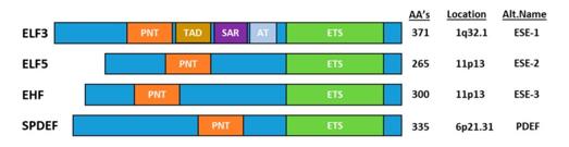 Fig. 1 Schematic representation of the epithelial-epecific ETS transcription factors.1
Key structural properties of ELF3:
- Conservative ETS domain
- Dual-module transcriptional activation region
- Functional regulation domain
Functions of ELF3
The main function of human ELF3 (E74/ESE-1) is to act as a specific transcription factor for epithelial tissue, regulating gene expression to maintain tissue homeostasis. However, it is also widely involved in various pathophysiological processes, including inflammatory responses, tissue repair and tumorigenesis.
| Function | Description |
| Maintenance of the epithelial barrier | By activating the expression of genes such as keratin and intercellular junction proteins, it maintains the physical barrier integrity of the mucous membranes of the skin, respiratory tract and digestive tract. |
| Inflammatory regulation | Under immune stimulation, the expression of chemokines (such as IL-8) and defencins can be induced, coordinating the innate immune response. However, excessive activation can lead to chronic inflammation. |
| Tissue repair and regeneration | It promotes the proliferation and migration of epithelial cells, accelerates the repair of mucosa after injury, and its expression level changes dynamically during the wound healing process. |
| Tumor-promoting effect | It is abnormally highly expressed in various epithelial cancers (such as breast cancer, lung cancer, and colon cancer), driving tumor progression by activating cell cycle and anti-apoptosis-related genes. |
| Regulation of cell differentiation | It participates in guiding the transformation of basal epithelial cells into terminally differentiated cells and affects the expression program of tissue-specific functional proteins. |
ELF3 directly binds to the promoter of the target gene through its conserved ETS domain, and its transcriptional activity is precisely regulated by signaling pathways such as MAPK/ERK. Under normal physiological conditions, it maintains tissue homeostasis. Under the stimulation of continuous inflammation or carcinogenic signals, its dysfunction will disrupt the balance and promote the development of the disease.
Applications of ELF3 and ELF3 Antibody in Literature
1. Yuan, Zengzhuang, et al. "Overexpression of ELF3 in the PTEN-deficient lung epithelium promotes lung cancer development by inhibiting ferroptosis." Cell Death & Disease 15.12 (2024): 897. https://doi.org/10.1038/s41419-024-07274-5
The article indicates that overexpression of ELF3 and deletion of PTEN synergistically drive the progression of lung cancer, mainly by regulating the ferroptosiia-related gene SLC7A11. Inhibiting SLC7A11 can induce ferroptosis and suppress tumor growth, and the expressions of both are associated with poor prognosis in patients.
2. Luk, Ian Y., Camilla M. Reehorst, and John M. Mariadason. "ELF3, ELF5, EHF and SPDEF transcription factors in tissue homeostasis and cancer." Molecules 23.9 (2018): 2191. https://doi.org/10.3390/molecules23092191
The article indicates that epithelial-specific ETS transcription factors (such as ELF3) maintain tissue homeostasis, and their dysfunction is associated with the progression of various epithelial carcinomas. This article reviews its normal functions, carcinogenic mechanisms, and potential strategies for targeted therapy.
3. Chen, Tianming, et al. "LncRNA ELF3‐AS1 is a prognostic biomarker and correlated with immune infiltrates in hepatocellular carcinoma." Canadian Journal of Gastroenterology and Hepatology 2021.1 (2021): 8323487. https://doi.org/10.1155/2021/8323487
The article indicates that the long non-coding RNA ELF3-AS1 is highly expressed in liver cancer and is significantly associated with the clinicopathological characteristics, immune infiltration and poor prognosis of patients, and can be used as a potential biomarker for the diagnosis and prognosis of liver cancer.
4. Sarmah, Swapnalee, et al. "Elf3 deficiency during zebrafish development alters extracellular matrix organization and disrupts tissue morphogenesis." PLoS One 17.11 (2022): e0276255. https://doi.org/10.1371/journal.pone.0276255
Research has found that during the embryonic development of zebrafish, the transcription factor ELF3 is widely expressed in various tissues, especially enriched at the tissue boundaries. The loss of its function leads to spinal curvature, cartilage deformity, neural defects and extracellular matrix disorder, indicating that ELF3 is indispensable in the development of the epidermis, interstitium and nerves.
5. Zhao, Qiaozhi, et al. "The ELF3-TRIM22-MAVS signaling axis regulates type I interferon and antiviral responses." Journal of Virology 99.5 (2025): e00004-25. https://doi.org/10.1128/jvi.00004-25
Research has found that TRIM22 activates the TBK1/IRF3 pathway by catalyzing the ubiquitination of the Lys63 position of MAVS and inhibits the MAVs-NLRX1 complex, thereby enhancing antiviral immunity. Viral infection upregulates TRIM22 by promoting the nuclear entry of the transcription factor ELF3, forming a positive regulatory axis.
Creative Biolabs: ELF3 Antibodies for Research
Creative Biolabs specializes in the production of high-quality ELF3 antibodies for research and industrial applications. Our portfolio includes monoclonal antibodies tailored for ELISA, Flow Cytometry, Western blot, immunohistochemistry, and other diagnostic methodologies.
- Custom ELF3 Antibody Development: Tailor-made solutions to meet specific research requirements.
- Bulk Production: Large-scale antibody manufacturing for industry partners.
- Technical Support: Expert consultation for protocol optimization and troubleshooting.
- Aliquoting Services: Conveniently sized aliquots for long-term storage and consistent experimental outcomes.
For more details on our ELF3 antibodies, custom preparations, or technical support, contact us at email.
Reference
- Luk, Ian Y., Camilla M. Reehorst, and John M. Mariadason. "ELF3, ELF5, EHF and SPDEF transcription factors in tissue homeostasis and cancer." Molecules 23.9 (2018): 2191. https://doi.org/10.3390/molecules23092191
Anti-ELF3 antibodies
 Loading...


Hot products 
-
Mouse Anti-ALB Recombinant Antibody (V2-55272) (CBMAB-H0819-FY)

-
Mouse Anti-CD19 Recombinant Antibody (CBXC-1224) (CBMAB-C1491-CQ)

-
Mouse Anti-BRD3 Recombinant Antibody (CBYY-0801) (CBMAB-0804-YY)

-
Mouse Anti-CSPG4 Recombinant Antibody (CBFYM-1050) (CBMAB-M1203-FY)

-
Mouse Anti-ACTN4 Recombinant Antibody (V2-6075) (CBMAB-0020CQ)

-
Mouse Anti-ENO2 Recombinant Antibody (H14) (CBMAB-E1341-FY)

-
Mouse Anti-FOSB Recombinant Antibody (CBXF-3593) (CBMAB-F2522-CQ)

-
Mouse Anti-FTH1 Recombinant Antibody (CBXF-1896) (CBMAB-F3426-CQ)

-
Mouse Anti-AKT1/AKT2/AKT3 (Phosphorylated T308, T309, T305) Recombinant Antibody (V2-443454) (PTM-CBMAB-0030YC)

-
Mouse Anti-2C TCR Recombinant Antibody (V2-1556) (CBMAB-0951-LY)

-
Mouse Anti-DMPK Recombinant Antibody (CBYCD-324) (CBMAB-D1200-YC)

-
Mouse Anti-CD33 Recombinant Antibody (P67.6) (CBMAB-C10189-LY)

-
Mouse Anti-ALB Recombinant Antibody (V2-180650) (CBMAB-A2186-YC)

-
Mouse Anti-CECR2 Recombinant Antibody (CBWJC-2465) (CBMAB-C3533WJ)

-
Human Anti-SARS-CoV-2 Spike Recombinant Antibody (CR3022) (CBMAB-CR014LY)

-
Mouse Anti-CAT Recombinant Antibody (724810) (CBMAB-C8431-LY)

-
Mouse Anti-ARID3A Antibody (A4) (CBMAB-0128-YC)

-
Mouse Anti-CASQ1 Recombinant Antibody (CBFYC-0863) (CBMAB-C0918-FY)

-
Mouse Anti-GDF5 Recombinant Antibody (1F4) (CBMAB-G2740-LY)

-
Mouse Anti-AFM Recombinant Antibody (V2-634159) (CBMAB-AP185LY)

- AActivation
- AGAgonist
- APApoptosis
- BBlocking
- BABioassay
- BIBioimaging
- CImmunohistochemistry-Frozen Sections
- CIChromatin Immunoprecipitation
- CTCytotoxicity
- CSCostimulation
- DDepletion
- DBDot Blot
- EELISA
- ECELISA(Cap)
- EDELISA(Det)
- ESELISpot
- EMElectron Microscopy
- FFlow Cytometry
- FNFunction Assay
- GSGel Supershift
- IInhibition
- IAEnzyme Immunoassay
- ICImmunocytochemistry
- IDImmunodiffusion
- IEImmunoelectrophoresis
- IFImmunofluorescence
- IGImmunochromatography
- IHImmunohistochemistry
- IMImmunomicroscopy
- IOImmunoassay
- IPImmunoprecipitation
- ISIntracellular Staining for Flow Cytometry
- LALuminex Assay
- LFLateral Flow Immunoassay
- MMicroarray
- MCMass Cytometry/CyTOF
- MDMeDIP
- MSElectrophoretic Mobility Shift Assay
- NNeutralization
- PImmunohistologyp-Paraffin Sections
- PAPeptide Array
- PEPeptide ELISA
- PLProximity Ligation Assay
- RRadioimmunoassay
- SStimulation
- SESandwich ELISA
- SHIn situ hybridization
- TCTissue Culture
- WBWestern Blot
