
EOMES Antibodies
Background
EOMES (also known as T-box brain protein 1) is a T-box transcription factor that plays a core role in the early embryonic development of vertebrates. This protein participates in key biological processes such as mesodermal differentiation, nervous system formation, and functional maturation of immune cells (such as CD8+ T cells) by regulating the expression of downstream target genes. Research has found that EOMES play a significant role in the trophoblast cell differentiation of placental mammals, and their abnormal expression is closely related to neurological developmental defects, immune dysfunction, and the progression of various tumors. As an important molecular marker in developmental biology, the research on the regulatory mechanism of EOMES has deepened people's understanding of the embryonic development program and cell fate determination, and provided potential targets for the diagnosis and treatment of related diseases.
Structure of EOMES
The molecular weight of the T-box transcription factor protein encoded by the EOMES gene is approximately 65-70 kDa, and its precise value fluctuates slightly due to differences in amino acid sequences among species and the presence of different transcripts.
| Species | Human | Mouse | Zebrafish | African clawed toad |
| Molecular Weight (kDa) | 70.2 | 69.8 | 68.5 | 67.1 |
| Primary Structural Differences | Domain contains T - box structure, the sequence is highly conserved | Very high homology with humans | The T-box domain is conservative, while the N/C ends are different | With typical structure in vertebrates |
This protein is composed of multiple functional domains, with its core being a highly conserved T-box domain (containing approximately 180 amino acids), which is responsible for specifically recognizing and binding to the target DNA sequence. This domain folds to form a unique three-dimensional conformation, with its secondary structure mainly composed of α -helices and β -folds, creating a binding interface that precisely interacts with the large grooves of DNA. Apart from the core structural domain, the regulatory region sequences at the amino and carboxyl ends vary among different species. These variations affect its transcriptional activity and the specificity of its interactions with other cofactors.
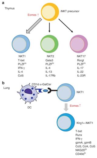 Fig. 1 The model depicting the role of Eomes during iNKT development in the thymus.1
Key structural properties of EOMES:
- Contains a highly conserved T-box domain
- This domain is composed of an α-helix and a β -fold to form the DNA binding interface
- Bind to the target sequence through specific DNA recognition helices
- Protein N and C regulatory regions influence the transcriptional activity and specificity
Functions of EOMES
The main function of the EOMES gene is to regulate cell fate determination as a transcription factor. However, it is also widely involved in various physiological and pathological processes, including immune responses and tumorigenesis.
| Function | Description |
| Regulation of cell differentiation | Activating mesoderm-specific gene expression in the early stage of embryonic development drives stem cells to differentiate into specific lineages. |
| Nervous system development | Regulating the proliferation and differentiation of neural precursor cells is crucial for the formation of the cerebral cortex and cerebellum. |
| Immune cell function | In adaptive immunity, it determines the effector functions of CD8+ T cells and NK cells and the formation of long-term memory. |
| Tumor-promoting effect | In a wide variety of cancer (such as liver cancer, colorectal cancer) in the abnormal high expression, through the regulation of epithelial - interstitial transformation to promote tumor invasion and metastasis. |
| Pathological protective effect | In certain circumstances can inhibit inflammation, and in the process of tissue repair regulating cell reprogramming. |
Compared with other synergistic transcription factors (such as T-bet), the regulation of target genes by EOMES usually shows more fundamental and earlier initiation characteristics, which is consistent with its "pioneer" role in cell fate determination.
Applications of EOMES and EOMES Antibody in Literature
1. Thelen, Benedikt, et al. "Eomes is sufficient to regulate IL-10 expression and cytotoxic effector molecules in murine CD4+ T cells." Frontiers in Immunology 14 (2023): 1058267. https://doi.org/10.3389/fimmu.2023.1058267
The article indicates that through a new genetic method, it was discovered that Eomes in CD4+ T cells can initiate a unique transcriptional program that partially overlaps with T-BET, which can not only induce the anti-inflammatory factor IL-10 but also work in synergy with T-BET to endow cytotoxic functions.
2. Wong, Pamela, et al. "T-BET and EOMES sustain mature human NK cell identity and antitumor function." The Journal of clinical investigation 133.13 (2023). https://doi.org/10.1172/JCI162530
Research reveals that EOMES and T-BET maintain the function of human NK cells through specific transcriptional programs. After deletion, the anti-tumor function, proliferation and cytokine response of NK cells are impaired, and there is a tendency to transform to other lineages such as ILC-3.
3. Llaó-Cid, Laura, et al. "EOMES is essential for antitumor activity of CD8+ T cells in chronic lymphocytic leukemia." Leukemia 35.11 (2021): 3152-3162. https://doi.org/10.1038/s41375-021-01198-1
Research has found that the EOMES gene variations associated with leukemia risk actually function by influencing CD8+ T cells. EOMES are crucial for the expansion of such cells, and their absence will directly weaken the body's immune ability to control leukemia.
4. Verma, Riva, et al. "Eomes expression defines group 1 innate lymphoid cells during metastasis in human and mouse." Frontiers in immunology 11 (2020): 1190. https://doi.org/10.3389/fimmu.2020.01190
Research has found that during the process of cancer metastasis, NK cells will be transformed into ILC1 along with the down-regulation of Eomes expression. This ILC1 with low expression of Eomes has weakened cytotoxicity, leading to a decline in herd immune surveillance function and thereby promoting disease progression.
5. Shimizu, Kanako, et al. "Eomes transcription factor is required for the development and differentiation of invariant NKT cells." Communications Biology 2.1 (2019): 150. https://doi.org/10.1038/s42003-019-0389-3
Research has found that the transcription factor Eomes not only regulates the differentiation of iNKT cells in the thymus into the NKT1 subtype, but also dominates their differentiation into KLRG1+ effector memory-like cells after peripheral activation, revealing its dual new role in iNKT cells.
Creative Biolabs: EOMES Antibodies for Research
Creative Biolabs specializes in the production of high-quality EOMES antibodies for research and industrial applications. Our portfolio includes monoclonal antibodies tailored for ELISA, Flow Cytometry, Western blot, immunohistochemistry, and other diagnostic methodologies.
- Custom EOMES Antibody Development: Tailor-made solutions to meet specific research requirements.
- Bulk Production: Large-scale antibody manufacturing for industry partners.
- Technical Support: Expert consultation for protocol optimization and troubleshooting.
- Aliquoting Services: Conveniently sized aliquots for long-term storage and consistent experimental outcomes.
For more details on our EOMES antibodies, custom preparations, or technical support, contact us at email.
Reference
- Shimizu, Kanako, et al. "Eomes transcription factor is required for the development and differentiation of invariant NKT cells." Communications Biology 2.1 (2019): 150. https://doi.org/10.1038/s42003-019-0389-3
Anti-EOMES antibodies
 Loading...
 Compare




Hot products 
-
Mouse Anti-DISP2 Monoclonal Antibody (F66A4B1) (CBMAB-1112CQ)

-
Mouse Anti-ESR1 Recombinant Antibody (Y31) (CBMAB-1208-YC)

-
Mouse Anti-BACE1 Recombinant Antibody (CBLNB-121) (CBMAB-1180-CN)

-
Mouse Anti-CARD11 Recombinant Antibody (CBFYC-0811) (CBMAB-C0866-FY)

-
Mouse Anti-CASP8 Recombinant Antibody (CBYY-C0987) (CBMAB-C2424-YY)

-
Mouse Anti-ASB9 Recombinant Antibody (1D8) (CBMAB-A0529-LY)

-
Mouse Anti-DES Monoclonal Antibody (440) (CBMAB-AP1857LY)

-
Mouse Anti-BCL6 Recombinant Antibody (CBYY-0442) (CBMAB-0445-YY)

-
Mouse Anti-ABIN2 Recombinant Antibody (V2-179106) (CBMAB-A0349-YC)

-
Mouse Anti-FN1 Monoclonal Antibody (D6) (CBMAB-1240CQ)

-
Mouse Anti-CCND2 Recombinant Antibody (DCS-3) (CBMAB-G1318-LY)

-
Mouse Anti-BSN Recombinant Antibody (219E1) (CBMAB-1228-CN)

-
Rat Anti-CD300A Recombinant Antibody (172224) (CBMAB-C0423-LY)

-
Mouse Anti-G6PD Recombinant Antibody (13B331) (CBMAB-G1553-LY)

-
Mouse Anti-ALOX5 Recombinant Antibody (33) (CBMAB-1890CQ)

-
Mouse Anti-Gro Monoclonal Antibody (1) (CBMAB-0482-LY)

-
Mouse Anti-CD2AP Recombinant Antibody (BR083) (CBMAB-BR083LY)

-
Mouse Anti-ALB Recombinant Antibody (V2-363290) (CBMAB-S0173-CQ)

-
Mouse Anti-ADGRE2 Recombinant Antibody (V2-261270) (CBMAB-C0813-LY)

-
Mouse Anti-AKT1 (Phosphorylated S473) Recombinant Antibody (V2-505430) (PTM-CBMAB-0067LY)

- AActivation
- AGAgonist
- APApoptosis
- BBlocking
- BABioassay
- BIBioimaging
- CImmunohistochemistry-Frozen Sections
- CIChromatin Immunoprecipitation
- CTCytotoxicity
- CSCostimulation
- DDepletion
- DBDot Blot
- EELISA
- ECELISA(Cap)
- EDELISA(Det)
- ESELISpot
- EMElectron Microscopy
- FFlow Cytometry
- FNFunction Assay
- GSGel Supershift
- IInhibition
- IAEnzyme Immunoassay
- ICImmunocytochemistry
- IDImmunodiffusion
- IEImmunoelectrophoresis
- IFImmunofluorescence
- IGImmunochromatography
- IHImmunohistochemistry
- IMImmunomicroscopy
- IOImmunoassay
- IPImmunoprecipitation
- ISIntracellular Staining for Flow Cytometry
- LALuminex Assay
- LFLateral Flow Immunoassay
- MMicroarray
- MCMass Cytometry/CyTOF
- MDMeDIP
- MSElectrophoretic Mobility Shift Assay
- NNeutralization
- PImmunohistologyp-Paraffin Sections
- PAPeptide Array
- PEPeptide ELISA
- PLProximity Ligation Assay
- RRadioimmunoassay
- SStimulation
- SESandwich ELISA
- SHIn situ hybridization
- TCTissue Culture
- WBWestern Blot
