GLO1 Antibodies
Background
The GLO1 gene encodes glyoxalase I, an important zinc metalloenzyme, which is mainly present in the cytoplasm. This enzyme uses glutathione as a cofactor to catalyze the conversion of toxic methylglyoxal into non-toxic D-lactic acid. This process is crucial for protecting cells from the formation of advanced glycation end products (AGEs) and the oxidative stress damage they cause. The expression and activity of the GLO1 gene are closely related to the pathophysiological mechanisms of various aging-related diseases, such as diabetic complications, neurodegenerative diseases and cardiovascular diseases. Since its function was discovered in the 20th century, it has been deeply studied through gene knockout and overexpression models, not only revealing its core position in the metabolic defense network, but also providing an important foundation for the research on the molecular mechanisms of related diseases and the development of potential therapeutic targets.
Structure of GLO1
GLO1 is an important cytoplasmic enzyme with a molecular weight of approximately 46 kDa. There are certain differences in molecular weight among different species, mainly due to minor changes in amino acid sequences.
| Species | Human | Mouse | Rat | Bovine |
| Molecular Weight (kDa) | 46 | 45.8 | 45.9 | 46.2 |
| Primary Structural Differences | Conservative active site | Sequence high homology | Very similar to the human structure | Individual amino acid substitution exists |
This protein is composed of approximately 185 amino acids, forming a typical double-domain folding conformation. Its primary structure folding generates two main domains: one for binding glutathione (GSH), and the other constitutes the catalytic active pocket. The active center contains a key zinc ion (Zn²⁺), which is stably fixed by coordinating with two glutamic acid residues and two histidine residues, which is crucial for catalyzing the toxic metabolite methylglyoxal (MG).
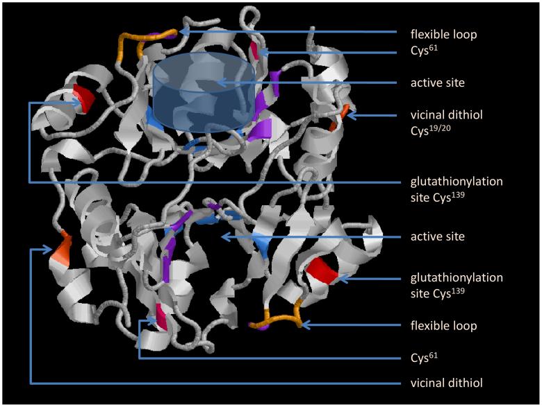 Fig. 1 3D structure of a Glo1 dimer .1
Key structural properties of GLO1:
- Typical double-domain folded conformation
- Active center has a conservative zinc ion binding sites
- Catalytic pockets relying on glutathione as a cofactor
Functions of GLO1
The core function of the GLO1 gene is to catalyze the detoxification of methylglyoxal. In addition, it is also involved in a variety of physiological and pathological processes, including anti-glycation stress and cellular metabolic regulation.
| Function | Description |
| Detoxification with methylglyoxal | As a key enzyme in the glyoxalase system, it catalyzes the conversion of the toxic metabolite methylglyoxal into the non-toxic D-lactic acid. |
| Anti-glycation stress | By eliminating methylglyoxal, the generation of advanced glycation end products can be effectively reduced, protecting protein function and cell health. |
| Maintenance of metabolic homeostasis | Regulating the levels of by-products of glucose metabolism indirectly affects energy metabolism pathways and overall cellular homeostasis. |
| Association with oxidative stress | The detoxification function helps reduce the oxidative damage caused by carbonyl stress, and oxidative stress pathways exist cross dialogue. |
| Disease risk regulation | The level of its activity is related to the susceptibility to various chronic diseases such as diabetic complications, neurodegenerative diseases and cancer. |
The catalytic mechanism of this enzyme relies on glutathione to form an efficient co-substrate circulation system, demonstrating its fundamental role in the cellular defense network.
Applications of GLO1 and GLO1 Antibody in Literature
1. Zhenhai, Zou, et al. "MiR-205-3p suppresses bladder cancer progression via GLO1 mediated P38/ERK activation." BMC cancer 23.1 (2023): 956. https://doi.org/10.1186/s12885-023-11175-9
This study reveals that miR-205-3p in bladder cancer inhibits the activation of the P38/ERK signaling pathway by targeting and inhibiting GLO1, ultimately suppressing the proliferation and metastasis of cancer cells. It indicates that GLO1 is a key downstream target in the tumor suppressor effect of miR-205-3p.
2. Zeng, Qiaoli, et al. "Association between GLO1 variants and gestational diabetes mellitus susceptibility in a Chinese population: A preliminary study." Frontiers in Endocrinology 14 (2023): 1235581. https://doi.org/10.3389/fendo.2023.1235581
This study is the first to explore the association between GLO1 gene polymorphism and the risk of gestational diabetes mellitus (GDM). Research has found that GLO1 rs1130534 increases susceptibility to GDM and is associated with higher blood glucose levels, while rs4746 reduces the risk of GDM. In addition, rs1781735 is associated with the accumulation of methylglyoxal in the mother and weight gain in newborns.
3. Cely, Ingrid, et al. "Glo1 reduction in mice results in age-and sex-dependent metabolic dysfunction." bioRxiv (2025). https://doi.org/10.1101/2025.01.24.634754
This study found through a Glo1 gene knockdown mouse model that reduced Glo1 expression can cause age - and gender-dependent metabolic disorders such as obesity and hyperglycemia, and this process is not dependent on advanced glycation end products (AGEs). Tissue-specific gene expression analysis suggests that alterations in transcription factors Hnf4a, Arntl, and methylglyoxal metabolites (such as pyruvate) may be the key mechanisms leading to metabolic abnormalities.
4. Šilhavý, Jan, et al. "Downregulation of the Glo1 gene is associated with reduced adiposity and ectopic fat accumulation in spontaneously hypertensive rats." Antioxidants 9.12 (2020): 1179. https://doi.org/10.3390/antiox9121179
This study found that down-regulating the Glo1 gene in spontaneously hypertensive rats (SHR), although it increased the level of methylglyoxal, unexpectedly reduced ectopic fat accumulation and decreased serum triglycerides and insulin. This protective effect may be related to the enhanced activity of AMPK in the heart.
5. Cheng, et al. "Protocatechualdehyde attenuates oxidative stress in diabetic cataract via GLO1-mediated inhibition of AGE/RAGE glycosylation." Frontiers in Pharmacology 16 (2025): 1586173. https://doi.org/10.3389/fphar.2025.1586173
This study reveals that protocatechualdehyde enhances the detoxification ability of the precursors of advanced glycation end products by up-regulating the expression of GLO1, thereby inhibiting the activation of the AGE-RAGE axis and oxidative stress, and thus delaying the progression of diabetic cataract.
Creative Biolabs: GLO1 Antibodies for Research
Creative Biolabs specializes in the production of high-quality GLO1 antibodies for research and industrial applications. Our portfolio includes monoclonal antibodies tailored for ELISA, Flow Cytometry, Western blot, immunohistochemistry, and other diagnostic methodologies.
- Custom GLO1 Antibody Development: Tailor-made solutions to meet specific research requirements.
- Bulk Production: Large-scale antibody manufacturing for industry partners.
- Technical Support: Expert consultation for protocol optimization and troubleshooting.
- Aliquoting Services: Conveniently sized aliquots for long-term storage and consistent experimental outcomes.
For more details on our GLO1 antibodies, custom preparations, or technical support, contact us at email.
Reference
- Birkenmeier, Gerd, et al. "Posttranslational modification of human glyoxalase 1 indicates redox-dependent regulation." PloS one 5.4 (2010): e10399. https://doi.org/10.1371/journal.pone.0010399
Anti-GLO1 antibodies
 Loading...


Hot products 
-
Rabbit Anti-ABL1 (Phosphorylated Y185) Recombinant Antibody (V2-443434) (PTM-CBMAB-0001YC)

-
Armenian hamster Anti-CD40 Recombinant Antibody (HM40-3) (CBMAB-C10365-LY)

-
Mouse Anti-DLG1 Monolconal Antibody (4F3) (CBMAB-0225-CN)

-
Mouse Anti-CCDC6 Recombinant Antibody (CBXC-0106) (CBMAB-C5397-CQ)

-
Mouse Anti-EIF4G1 Recombinant Antibody (2A9) (CBMAB-A2544-LY)

-
Mouse Anti-8-oxoguanine Recombinant Antibody (V2-7719) (CBMAB-1898CQ)

-
Mouse Anti-ACTN4 Recombinant Antibody (V2-6075) (CBMAB-0020CQ)

-
Mouse Anti-ACTB Recombinant Antibody (V2-179553) (CBMAB-A0870-YC)

-
Mouse Anti-BIRC5 Recombinant Antibody (6E4) (CBMAB-CP2646-LY)

-
Mouse Anti-BMI1 Recombinant Antibody (CBYC-P026) (CBMAB-P0108-YC)

-
Mouse Anti-BPGM Recombinant Antibody (CBYY-1806) (CBMAB-2155-YY)

-
Mouse Anti-C5B-9 Recombinant Antibody (CBFYA-0216) (CBMAB-X0304-FY)

-
Mouse Anti-CCDC25 Recombinant Antibody (CBLC132-LY) (CBMAB-C9786-LY)

-
Rat Anti-CD34 Recombinant Antibody (MEC 14.7) (CBMAB-C10196-LY)

-
Mouse Anti-BCL6 Recombinant Antibody (CBYY-0442) (CBMAB-0445-YY)

-
Mouse Anti-ATG5 Recombinant Antibody (9H197) (CBMAB-A3945-YC)

-
Rabbit Anti-CCN1 Recombinant Antibody (CBWJC-3580) (CBMAB-C4816WJ)

-
Rat Anti-ADAM10 Recombinant Antibody (V2-179741) (CBMAB-A1103-YC)

-
Mouse Anti-BLK Recombinant Antibody (CBYY-0618) (CBMAB-0621-YY)

-
Mouse Anti-FN1 Monoclonal Antibody (D6) (CBMAB-1240CQ)

- AActivation
- AGAgonist
- APApoptosis
- BBlocking
- BABioassay
- BIBioimaging
- CImmunohistochemistry-Frozen Sections
- CIChromatin Immunoprecipitation
- CTCytotoxicity
- CSCostimulation
- DDepletion
- DBDot Blot
- EELISA
- ECELISA(Cap)
- EDELISA(Det)
- ESELISpot
- EMElectron Microscopy
- FFlow Cytometry
- FNFunction Assay
- GSGel Supershift
- IInhibition
- IAEnzyme Immunoassay
- ICImmunocytochemistry
- IDImmunodiffusion
- IEImmunoelectrophoresis
- IFImmunofluorescence
- IGImmunochromatography
- IHImmunohistochemistry
- IMImmunomicroscopy
- IOImmunoassay
- IPImmunoprecipitation
- ISIntracellular Staining for Flow Cytometry
- LALuminex Assay
- LFLateral Flow Immunoassay
- MMicroarray
- MCMass Cytometry/CyTOF
- MDMeDIP
- MSElectrophoretic Mobility Shift Assay
- NNeutralization
- PImmunohistologyp-Paraffin Sections
- PAPeptide Array
- PEPeptide ELISA
- PLProximity Ligation Assay
- RRadioimmunoassay
- SStimulation
- SESandwich ELISA
- SHIn situ hybridization
- TCTissue Culture
- WBWestern Blot
