JUNB Antibodies
Background
The JUNB gene, as an important member of the transcription factor AP-1 family, is mainly expressed in immune cells of mammals and various other tissue cells. The protein encoded by this gene specifically binds to the TPA response elements in the promoter regions of target genes through homodimerization or heterodimerization, thereby precisely regulating the transcriptional activity of cytokines, chemokines, and immune regulatory molecules. The immune system relies on JUNB to maintain T cell differentiation and effector functions, and it plays a crucial role in the establishment of Th17 and follicular helper T cell lineages. Its expression imbalance is closely related to autoimmune diseases and chronic inflammation. Initially identified as an early response gene from the cDNA library of human lung fibroblasts, JUNB was later confirmed to be a molecular switch that regulates cell cycle exit and terminal differentiation. The structure of this gene contains a highly conserved bZIP domain, and its differential expression patterns with family members such as c-JUN and JUND together constitute the functional redundancy and specificity of the transcriptional regulatory network, providing an important entry point for understanding the molecular mechanisms of immune homeostasis maintenance, tumor immune escape, and inflammatory diseases.
Structure of JUNB
The protein encoded by the JUNB gene has a molecular weight of approximately 35 kDa. This protein contains 311 amino acids and its structure includes a highly conserved basic leucine zipper domain, which is responsible for dimerization and specific binding to DNA. JUNB forms heterodimers with FOS family proteins through its bZIP domain and recognizes the TPA response elements in the promoter regions of target genes. The basic regions in the protein structure mediate direct contact with the DNA backbone, while the leucine zipper structure is responsible for the stable binding between subunits. The transcriptional activity of JUNB is also precisely regulated by amino-terminal phosphorylation modifications, and different modification states affect its ability to bind to cofactors and the efficiency of transcriptional activation.
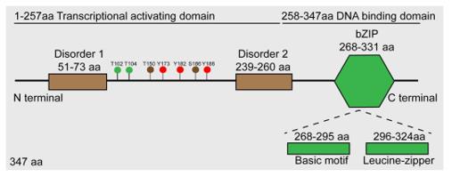 Fig. 1 Schematic diagram of JunB protein.1
Key structural properties of JUNB:
- Basic leucine zipper domains mediate dimerization
- DNA-binding regions are rich in positively charged amino acids
- The C-terminal transcriptional activation domain is in an unordered conformation
- Phosphorylation sites are distributed in the N-terminal regulatory region
Functions of JUNB
The core function of JUNB is to regulate the gene expression of immune cells, and it is involved in the cell cycle, differentiation and inflammatory responses.
| Function | Description |
| T cell differentiation | Regulates the specialization process of subgroups such as Th17 and Tfh, determining the direction of immune response. |
| Cytokine expression | Directly activates the transcription of effector molecules such as IL-4 and IL-17. |
| Proliferation and apoptosis | Inhibits p53 and regulates cyclins to affect cell survival. |
| Inflammation regulation | Mediates M2 polarization and inflammation resolution in macrophages. |
| Autoimmunity | Abnormal expression is related to diseases such as rheumatoid arthritis and asthma . |
JUNB has a similar structure to c-JUN but differs in function. The former is more related to maintaining immune homeostasis, while the latter is often associated with proliferative stress.
Applications of JUNB and JUNB Antibody in Literature
1. Ren, Fu-jia, et al. "JunB: a paradigm for Jun family in immune response and cancer." Frontiers in Cellular and Infection Microbiology 13 (2023): 1222265. https://doi.org/10.3389/fcimb.2023.1222265
The article indicates that JunB is a key member of the AP-1 complex, regulating the differentiation and function of immune cells, participating in both innate and adaptive immunity, and playing a significant role in tumor occurrence by influencing the proliferation, metastasis of tumor cells, and the tumor microenvironment.
2. Li, Guozheng, et al. "NAT10/ac4C/JunB facilitates TNBC malignant progression and immunosuppression by driving glycolysis addiction." Journal of Experimental & Clinical Cancer Research 43.1 (2024): 278. https://doi.org/10.1186/s13046-024-03200-x
The article indicates that in TNBC, NAT10 upregulates JunB through ac4C modification, thereby promoting LDHA expression and glycolysis, and forming an immunosuppressive microenvironment. Inhibiting NAT10 can activate T cells, and its inhibitor remodelin, when combined with CTLA-4 antibody, can enhance anti-tumor immunity.
3. Wang, Xiujian, et al. "MEK inhibition prevents CAR-T cell exhaustion and differentiation via downregulation of c-Fos and JunB." Signal Transduction and Targeted Therapy 9.1 (2024): 293. https://doi.org/10.1038/s41392-024-01986-y
This study reveals that MEK inhibitors can effectively alleviate the exhaustion and terminal differentiation of CAR-T cells and enhance their anti-tumor efficacy by down-regulating the c-Fos/JunB axis. This mechanism provides a new strategy for the combined application of MEK inhibitors to enhance the efficacy of CAR-T therapy.
4. Kalampounias, Georgios, Theodosia Androutsopoulou, and Panagiotis Katsoris. "JUNB and JUND in Urological Cancers: A Literature Review." Current Issues in Molecular Biology 47.9 (2025): 741. https://doi.org/10.3390/cimb47090741
This study reveals that JUNB and JUND are the key transcription factors of the AP-1 complex. In urinary system cancers, they exert both oncogenic and tumor-suppressive effects by regulating multiple processes such as proliferation and invasion. Their functions depend on the type and stage of the cancer and have the potential to become diagnostic and therapeutic targets.
5. Hsieh, Tsunghan, et al. "JunB is critical for survival of T helper cells." Frontiers in immunology 13 (2022): 901030. https://doi.org/10.3389/fimmu.2022.901030
This study reveals that JunB plays a crucial role in the proliferation of Th1, Th2 and Th17 cells by inhibiting the expression of apoptotic genes such as Bim. When JunB is lacking, antigen-activated CD4+ T cells are more prone to apoptosis, resulting in significant impairment of their accumulation within the body.
Creative Biolabs: JUNB Antibodies for Research
Creative Biolabs specializes in the production of high-quality JUNB antibodies for research and industrial applications. Our portfolio includes monoclonal and polyclonal antibodies tailored for ELISA, Flow Cytometry, Western blot, immunohistochemistry, and other diagnostic methodologies.
- Custom JUNB Antibody Development: Tailor-made solutions to meet specific research requirements.
- Bulk Production: Large-scale antibody manufacturing for industry partners.
- Technical Support: Expert consultation for protocol optimization and troubleshooting.
- Aliquoting Services: Conveniently sized aliquots for long-term storage and consistent experimental outcomes.
For more details on our JUNB antibodies, custom preparations, or technical support, contact us at info@creative-biolabs.com.
Reference
- Ren, Fu-jia, et al. "JunB: a paradigm for Jun family in immune response and cancer." Frontiers in Cellular and Infection Microbiology 13 (2023): 1222265. Distributed under Open Access license CC BY 4.0, without modification. https://doi.org/10.3389/fcimb.2023.1222265
Anti-JUNB antibodies
 Loading...


Hot products 
-
Mouse Anti-dsDNA Recombinant Antibody (22) (CBMAB-AP1954LY)

-
Mouse Anti-AQP2 Recombinant Antibody (E-2) (CBMAB-A3358-YC)

-
Rabbit Anti-ALOX5AP Recombinant Antibody (CBXF-1219) (CBMAB-F0750-CQ)

-
Mouse Anti-CD2AP Recombinant Antibody (BR083) (CBMAB-BR083LY)

-
Mouse Anti-CCDC25 Recombinant Antibody (CBLC132-LY) (CBMAB-C9786-LY)

-
Mouse Anti-BRCA2 Recombinant Antibody (CBYY-0790) (CBMAB-0793-YY)

-
Mouse Anti-HTLV-1 gp46 Recombinant Antibody (CBMW-H1006) (CBMAB-V208-1154-FY)

-
Mouse Anti-BrdU Recombinant Antibody (IIB5) (CBMAB-1038CQ)

-
Mouse Anti-BRD3 Recombinant Antibody (CBYY-0801) (CBMAB-0804-YY)

-
Rabbit Anti-DLK1 Recombinant Antibody (9D8) (CBMAB-D1061-YC)

-
Rabbit Anti-B2M Recombinant Antibody (CBYY-0059) (CBMAB-0059-YY)

-
Mouse Anti-BBS2 Recombinant Antibody (CBYY-0253) (CBMAB-0254-YY)

-
Mouse Anti-ASTN1 Recombinant Antibody (H-9) (CBMAB-1154-CN)

-
Mouse Anti-CCND2 Recombinant Antibody (DCS-3) (CBMAB-G1318-LY)

-
Mouse Anti-FYN Recombinant Antibody (10) (CBMAB-S6332-CQ)

-
Mouse Anti-C5AR1 Recombinant Antibody (R63) (CBMAB-C9553-LY)

-
Mouse Anti-ACLY Recombinant Antibody (V2-179314) (CBMAB-A0610-YC)

-
Mouse Anti-ASB9 Recombinant Antibody (1D8) (CBMAB-A0529-LY)

-
Mouse Anti-CORO1A Recombinant Antibody (4G10) (V2LY-1206-LY806)

-
Mouse Anti-G6PD Recombinant Antibody (13B331) (CBMAB-G1553-LY)

- AActivation
- AGAgonist
- APApoptosis
- BBlocking
- BABioassay
- BIBioimaging
- CImmunohistochemistry-Frozen Sections
- CIChromatin Immunoprecipitation
- CTCytotoxicity
- CSCostimulation
- DDepletion
- DBDot Blot
- EELISA
- ECELISA(Cap)
- EDELISA(Det)
- ESELISpot
- EMElectron Microscopy
- FFlow Cytometry
- FNFunction Assay
- GSGel Supershift
- IInhibition
- IAEnzyme Immunoassay
- ICImmunocytochemistry
- IDImmunodiffusion
- IEImmunoelectrophoresis
- IFImmunofluorescence
- IGImmunochromatography
- IHImmunohistochemistry
- IMImmunomicroscopy
- IOImmunoassay
- IPImmunoprecipitation
- ISIntracellular Staining for Flow Cytometry
- LALuminex Assay
- LFLateral Flow Immunoassay
- MMicroarray
- MCMass Cytometry/CyTOF
- MDMeDIP
- MSElectrophoretic Mobility Shift Assay
- NNeutralization
- PImmunohistologyp-Paraffin Sections
- PAPeptide Array
- PEPeptide ELISA
- PLProximity Ligation Assay
- RRadioimmunoassay
- SStimulation
- SESandwich ELISA
- SHIn situ hybridization
- TCTissue Culture
- WBWestern Blot
