
NGFR Antibodies
Background
NGFR (neurotrophic factor receptor) is a transmembrane glycoprotein located on the cell membrane, primarily functioning as a key receptor for neurotrophic factors. The protein encoded by this gene can specifically bind to ligands such as nerve growth factor (NGF), thereby mediating the survival, differentiation, and synaptic plasticity of neurons, and is crucial for the development and maintenance of the nervous system. Abnormalities in its function are closely related to neurodegenerative diseases, pain perception, and tumor progression. Since its identification in the 1980s, the study of the structure and signaling pathways of NGFR has greatly advanced the field of cell receptor biology, providing an important molecular basis for the development of targeted therapeutic strategies.
Structure of NGFR
NGFR is a type I transmembrane glycoprotein with a molecular weight of approximately 45 kDa. Its size varies slightly among different species due to differences in glycosylation. The primary structure of the NGFR protein consists of an extracellular domain, a transmembrane region, and an intracellular domain, each containing approximately 400 amino acids. The extracellular domain is rich in four conserved cysteine repeat sequences, forming a typical ligand-binding domain that specifically recognizes and binds neurotrophic factors. The secondary structure of this protein is mainly composed of β-sheets, collectively forming a stable ligand-binding interface. Although the intracellular domain lacks kinase activity, it recruits downstream signal adaptor proteins through its death domain, thereby initiating cell survival or apoptosis signaling pathways.
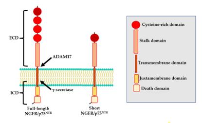 Fig. 1 Structure of NGFR/p75NTR isoforms.1
Key structural properties of NGFR:
- Type I transmembrane protein structure
- Extracellular ligand binding domains rich in cysteine repeat sequences
- Intracellular death domains lacking kinase activity
Functions of NGFR
The main function of the NGFR gene is to mediate the signal transduction of neurotrophic factors, which is crucial for the survival, differentiation and synaptic plasticity of neurons. At the same time, it is also involved in various pathological physiological processes.
| Function | Description |
| Neurotrophic Factor Signal Transduction | As receptors for factors such as NGF and BDNF, they activate downstream Trk or p75 signaling pathways to regulate cell growth and survival. |
| Neuron Survival and Differentiation | It mediates the survival, axonal guidance and phenotypic differentiation of neurons in the developing and adult nervous system. |
| Apoptosis Regulation | The p75 subtype can activate the cell apoptosis process by binding to neurotrophic factor precursors or ligands under specific conditions. |
| Pain Perception Regulation | It is involved in the transmission of pain signals at the peripheral and central nervous systems and the formation of chronic pain. |
| Impact of Tumor Progression | It is abnormally expressed in various cancers (such as melanoma, neuroblastoma), and is associated with tumor invasion, metastasis and drug resistance. |
Unlike typical receptors with a single high-affinity binding property, NGFR (especially p75NTR) can bind to multiple neurotrophic factors with lower affinity and often forms complexes with Trk receptors. Its signal output is highly context-dependent, capable of promoting survival as well as inducing apoptosis.
Applications of NGFR and NGFR Antibody in Literature
1. Bruno, Francesco, et al. "The nerve growth factor receptor (NGFR/p75NTR): A major player in Alzheimer's disease." International journal of molecular sciences 24.4 (2023): 3200. https://doi.org/10.3390/ijms24043200
The article indicates that NGFR/p75NTR, as a common receptor for neurotrophic factors and β-amyloid protein (Aβ), plays a crucial role in the pathological process, genetic mechanism, and diagnostic and therapeutic strategies of Alzheimer's disease. This article systematically reviews its multi-level functions in the disease.
2. Chen, Hao, et al. "NGFR increases the chemosensitivity of colorectal cancer cells by enhancing the apoptotic and autophagic effects of 5-fluorouracil via the activation of S100A9." Frontiers in Oncology 11 (2021): 652081. https://doi.org/10.3389/fonc.2021.652081
The article indicates that the high expression of nerve growth factor receptor (NGFR) in colorectal cancer can enhance the sensitivity of cancer cells to 5-FU. The mechanism may be related to the induction of apoptosis, autophagy, and regulation of pathways such as S100A9, suggesting that NGFR may serve as a potential biomarker for predicting the efficacy of chemotherapy.
3. Boshuizen, Julia, et al. "Reversal of pre-existing NGFR-driven tumor and immune therapy resistance." Nature communications 11.1 (2020): 3946. https://doi.org/10.1038/s41467-020-17739-8
This study has confirmed the existence of a pre-existing and stable subgroup with high expression of NGFR in melanoma. This subgroup is resistant to both T cells and targeted therapies, and predicts resistance to PD-1 therapy. Inhibiting NGFR can restore the tumor's sensitivity to T cells, providing a potential target for overcoming immune therapy resistance.
4. Xie, Ning, et al. "Efficient muscle regeneration by human PSC-derived CD82+ ERBB3+ NGFR+ skeletal myogenic progenitors." Cells 12.3 (2023): 362. https://doi.org/10.3390/cells12030362
The article indicates that by using the CD82, ERBB3 and NGFR markers, highly regenerative skeletal muscle progenitor cells can be efficiently isolated from teratoma derived from human stem cells. These cells can be expanded in vitro and, after transplantation, can efficiently regenerate muscle fibers and replenish the stem cell bank.
5. Lehmann, Julia, et al. "Escape from NK cell tumor surveillance by NGFR-induced lipid remodeling in melanoma." Science advances 9.2 (2023): eadc8825. https://doi.org/10.1126/sciadv.adc8825
The article indicates that the expression of NGFR enables melanoma cells to evade immune surveillance by natural killer cells. The mechanism involves down-regulating activating ligands and up-regulating the SCD enzyme. Inhibiting SCD can reverse the immune escape mediated by NGFR, providing a new target for the prevention and treatment of metastasis.
Creative Biolabs: NGFR Antibodies for Research
Creative Biolabs specializes in the production of high-quality NGFR antibodies for research and industrial applications. Our portfolio includes monoclonal and polyclonal antibodies tailored for ELISA, Flow Cytometry, Western blot, immunohistochemistry, and other diagnostic methodologies.
- Custom NGFR Antibody Development: Tailor-made solutions to meet specific research requirements.
- Bulk Production: Large-scale antibody manufacturing for industry partners.
- Technical Support: Expert consultation for protocol optimization and troubleshooting.
- Aliquoting Services: Conveniently sized aliquots for long-term storage and consistent experimental outcomes.
For more details on our NGFR antibodies, custom preparations, or technical support, contact us at email.
Reference
- Bruno, Francesco, et al. "The nerve growth factor receptor (NGFR/p75NTR): A major player in Alzheimer's disease." International journal of molecular sciences 24.4 (2023): 3200. Distributed under Open Access license CC BY 4.0, without modification. https://doi.org/10.3390/ijms24043200
Anti-NGFR antibodies
 Loading...
 Compare




Hot products 
-
Mouse Anti-C5b-9 Recombinant Antibody (aE11) (CBMAB-AO138LY)

-
Mouse Anti-CD24 Recombinant Antibody (ALB9) (CBMAB-0176CQ)

-
Mouse Anti-CRTAM Recombinant Antibody (CBFYC-2235) (CBMAB-C2305-FY)

-
Mouse Anti-DMPK Recombinant Antibody (CBYCD-324) (CBMAB-D1200-YC)

-
Mouse Anti-CCL18 Recombinant Antibody (64507) (CBMAB-C7910-LY)

-
Mouse Anti-AK4 Recombinant Antibody (V2-180419) (CBMAB-A1891-YC)

-
Mouse Anti-AKT1 (Phosphorylated S473) Recombinant Antibody (V2-505430) (PTM-CBMAB-0067LY)

-
Rabbit Anti-ADRA1A Recombinant Antibody (V2-12532) (CBMAB-1022-CN)

-
Mouse Anti-BRCA2 Recombinant Antibody (CBYY-0790) (CBMAB-0793-YY)

-
Mouse Anti-ARID1B Recombinant Antibody (KMN1) (CBMAB-A3546-YC)

-
Mouse Anti-FN1 Monoclonal Antibody (D6) (CBMAB-1240CQ)

-
Mouse Anti-ATG5 Recombinant Antibody (9H197) (CBMAB-A3945-YC)

-
Mouse Anti-AKT1 Recombinant Antibody (V2-180546) (CBMAB-A2070-YC)

-
Mouse Anti-ENO1 Recombinant Antibody (CBYC-A950) (CBMAB-A4388-YC)

-
Mouse Anti-ACLY Recombinant Antibody (V2-179314) (CBMAB-A0610-YC)

-
Mouse Anti-GDF5 Recombinant Antibody (1F4) (CBMAB-G2740-LY)

-
Mouse Anti-CD247 Recombinant Antibody (6B10.2) (CBMAB-C1583-YY)

-
Mouse Anti-APOE Recombinant Antibody (A1) (CBMAB-0078CQ)

-
Mouse Anti-CCDC25 Recombinant Antibody (CBLC132-LY) (CBMAB-C9786-LY)

-
Mouse Anti-A2M Recombinant Antibody (V2-178822) (CBMAB-A0036-YC)

- AActivation
- AGAgonist
- APApoptosis
- BBlocking
- BABioassay
- BIBioimaging
- CImmunohistochemistry-Frozen Sections
- CIChromatin Immunoprecipitation
- CTCytotoxicity
- CSCostimulation
- DDepletion
- DBDot Blot
- EELISA
- ECELISA(Cap)
- EDELISA(Det)
- ESELISpot
- EMElectron Microscopy
- FFlow Cytometry
- FNFunction Assay
- GSGel Supershift
- IInhibition
- IAEnzyme Immunoassay
- ICImmunocytochemistry
- IDImmunodiffusion
- IEImmunoelectrophoresis
- IFImmunofluorescence
- IGImmunochromatography
- IHImmunohistochemistry
- IMImmunomicroscopy
- IOImmunoassay
- IPImmunoprecipitation
- ISIntracellular Staining for Flow Cytometry
- LALuminex Assay
- LFLateral Flow Immunoassay
- MMicroarray
- MCMass Cytometry/CyTOF
- MDMeDIP
- MSElectrophoretic Mobility Shift Assay
- NNeutralization
- PImmunohistologyp-Paraffin Sections
- PAPeptide Array
- PEPeptide ELISA
- PLProximity Ligation Assay
- RRadioimmunoassay
- SStimulation
- SESandwich ELISA
- SHIn situ hybridization
- TCTissue Culture
- WBWestern Blot
