PON3 Antibodies
Background
The PON3 gene encodes phytate phosphatase 3, which is a calcium-dependent esterase mainly expressed in high-density lipoproteins. This protein plays antioxidant and anti-inflammatory roles in the body and can hydrolyze various substrates including lactate, aromatic esters, and oxidized lipids, thereby protecting cells and tissues from oxidative damage. Studies have shown that PON3 is highly active in the liver and macrophages, and it maintains cardiovascular health by regulating cholesterol metabolism and inhibiting the formation of atherosclerotic plaques. Since its gene was identified in 1996, PON3 has become a research focus due to its regulatory mechanisms in metabolic diseases and the aging process. The structural and functional studies of PON3 have provided an important model for understanding lipid protein metabolism and the defense system against oxidative stress.
Structure of PON3
PON3 is a serum glycoprotein with a molecular weight of approximately 40 kDa. There are slight interspecies differences in its weight among different mammals. As a member of the oxyphosphatase family, the active center of PON3 contains two calcium ions, forming a typical six-leaf β-spiral tertiary structure. This structure achieves the specific hydrolysis of substrates such as oxidized phospholipids through its hydrophobic channels and key amino acid residues around the active site (such as histidine and glutamate). The conserved "lid" region regulates the entry and exit of substrates into the active center, while the glycosylation modifications on the surface affect the stability and localization of it on high-density lipoprotein particles. Similar to PON1 and PON2, the catalytic mechanism of PON3 relies on calcium ion-mediated ester bond cleavage, but its substrate preference and tissue-specific expression enable it to play a unique role in the antioxidant defense.
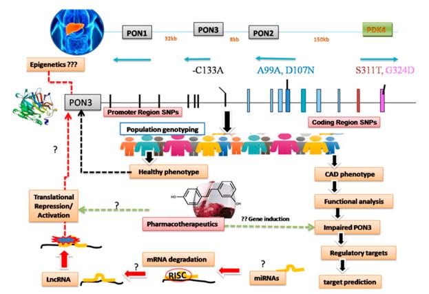 Fig. 1 Schematic of hidden links in CAD pathophysiology related to impaired paraoxonases, focusing on PON3.1
Key structural properties of PON3:
- Typical three-stage structure of six-bladed β-propeller
- Contains two active centers of calcium ions
- Conservative hydrophobic channels and catalytic sites
- Dependence on calcium ion-mediated esterase activity
- Key histidine and glutamic acid residues are involved in substrate recognition and hydrolysis
Functions of PON3
The PON3 gene encodes the peroxiredoxin 3 protein. Its primary function is to hydrolyze oxidized lipids and aromatic ester compounds, playing a crucial role in the body's metabolic defense. The specific physiological functions of this protein are as follows:
| Function | Description |
| Antioxidant Defense | Hydrolyze the oxidized phospholipids in oxidized low-density lipoprotein (ox-LDL), preventing their deposition on the arterial wall and slowing down the progression of atherosclerosis. |
| Anti-inflammatory Regulation | By reducing oxidative stress products and inhibiting the release of inflammatory factors, it regulates the inflammatory response of macrophages. |
| Lipid Metabolism Regulation | It functions on high-density lipoprotein (HDL) particles, influencing reverse cholesterol transport and lipid homeostasis. |
| Drug and Toxin Metabolism | It can hydrolyze certain endogenous esters and exogenous toxins (such as lactobacillus toxins), and has a certain detoxification function. |
| Cell Protection | Expressed in liver cells, endothelial cells, etc., it helps maintain the structural and functional integrity of cells by reducing oxidative damage. |
Compared with the homologous PON1, although PON3 lacks oxygen-phosphatase activity, it has a higher catalytic efficiency for aromatic esters and endogenous oxidized lipids. Moreover, its enzymatic activity is not affected by polymorphisms and plays a more stable protective role in maintaining the systemic oxidative balance.
Applications of PON3 and PON3 Antibody in Literature
1. Priyanka, Kumari, Surjit Singh, and Kirandip Gill. "Paraoxonase 3: Structure and its role in pathophysiology of coronary artery disease." Biomolecules 9.12 (2019): 817. https://doi.org/10.3390/biom9120817
The article indicates that the PON (phosphatidylcholine-oxidase) family is a potential therapeutic target for cardiovascular diseases, with insufficient research on PON3. This paper mainly explores the structure, function and genetic variations of PON3, providing new potential strategies for the prevention and treatment of coronary heart disease.
2. Riedmaier, Stephan, et al. "Paraoxonase (PON1 and PON3) polymorphisms: impact on liver expression and atorvastatin-lactone hydrolysis." Frontiers in Pharmacology 2 (2011): 41. https://doi.org/10.3389/fphar.2011.00041
The article indicates that in human liver microsomes, the protein levels of PON1 and PON3 are significantly correlated with the hydrolytic activity of atorvastatin δ-lactone. The study found that multiple polymorphic loci of the PON gene affect the expression of PON1, while non-genetic factors such as tumors and inflammation simultaneously affect the expression of PON1/PON3, which may be related to the efficacy or adverse reactions of statins.
3. Peng, Birong, et al. "Dnmt1 Alleviates S1PR1‐Mediated Pyroptosis after Spinal Cord Injury through Regulating Pon3 Expression." Advanced Science 12.42 (2025): e07330. https://doi.org/10.1002/advs.202507330
This study reveals that after spinal cord injury (SCI), the expression of Pon3 decreases while that of Dnmt1 increases. Overexpression of Pon3 can alleviate cell pyroptosis and improve the prognosis of neurological function by inhibiting the downstream target S1PR1 and promoting autophagy. Dnmt1 reduces pyroptosis damage after SCI by regulating the expression of Pon3.
4. Mohammed, Chrysan J., et al. "A PON for all seasons: comparing paraoxonase enzyme substrates, activity and action including the role of PON3 in health and disease." Antioxidants 11.3 (2022): 590. https://doi.org/10.3390/antiox11030590
This study reveals that as a member of the oxygen-phosphatase (PON) family, PON3 possesses the ability to hydrolyze lactones and has antioxidant potential, playing a significant role in anti-atherosclerosis. This article reviews the substrate characteristics, kinetic parameters of PON3, and the research progress on its association with various diseases such as cardiovascular diseases.
5. Bliźniewska-Kowalska, Katarzyna, et al. "Expression of PON1, PON2, PON3 and MPO genes in patients with depressive disorders." Journal of Clinical Medicine 11.12 (2022): 3321. https://doi.org/10.3390/jcm11123321
This study investigated the expression of antioxidant enzymes (including PON1, PON2, PON3 and MPO) in patients with depression. The results showed that the protein expression of PON2 and PON3 in the patients was significantly higher than that in the healthy controls, while the expression of MPO was significantly lower. The current study does not support the use of these enzymes as reliable biomarkers for depression.
Creative Biolabs: PON3 Antibodies for Research
Creative Biolabs specializes in the production of high-quality PON3 antibodies for research and industrial applications. Our portfolio includes monoclonal and polyclonal antibodies tailored for ELISA, Flow Cytometry, Western blot, immunohistochemistry, and other diagnostic methodologies.
- Custom PON3 Antibody Development: Tailor-made solutions to meet specific research requirements.
- Bulk Production: Large-scale antibody manufacturing for industry partners.
- Technical Support: Expert consultation for protocol optimization and troubleshooting.
- Aliquoting Services: Conveniently sized aliquots for long-term storage and consistent experimental outcomes.
For more details on our PON3 antibodies, custom preparations, or technical support, contact us at info@creative-biolabs.com.
Reference
- Priyanka, Kumari, Surjit Singh, and Kirandip Gill. "Paraoxonase 3: Structure and its role in pathophysiology of coronary artery disease." Biomolecules 9.12 (2019): 817. Distributed under Open Access license CC BY 4.0, without modification. https://doi.org/10.3390/biom9120817
Anti-PON3 antibodies
 Loading...


Hot products 
-
Mouse Anti-AK4 Recombinant Antibody (V2-180419) (CBMAB-A1891-YC)

-
Mouse Anti-ATG5 Recombinant Antibody (9H197) (CBMAB-A3945-YC)

-
Mouse Anti-CD83 Recombinant Antibody (HB15) (CBMAB-C1765-CQ)

-
Mouse Anti-CCDC6 Recombinant Antibody (CBXC-0106) (CBMAB-C5397-CQ)

-
Mouse Anti-ATM Recombinant Antibody (2C1) (CBMAB-A3970-YC)

-
Mouse Anti-APP Recombinant Antibody (5C2A1) (CBMAB-A3314-YC)

-
Mouse Anti-B2M Recombinant Antibody (CBYY-0050) (CBMAB-0050-YY)

-
Mouse Anti-CAPZB Recombinant Antibody (CBYY-C0944) (CBMAB-C2381-YY)

-
Mouse Anti-GFAP Recombinant Antibody (24) (CBMAB-G2927-LY)

-
Rat Anti-AChR Recombinant Antibody (V2-12500) (CBMAB-0990-CN)

-
Mouse Anti-AKR1B1 Antibody (V2-2449) (CBMAB-1001CQ)

-
Mouse Anti-CCDC25 Recombinant Antibody (CBLC132-LY) (CBMAB-C9786-LY)

-
Mouse Anti-CCND2 Recombinant Antibody (DCS-3) (CBMAB-G1318-LY)

-
Mouse Anti-COL1A2 Recombinant Antibody (CF108) (V2LY-1206-LY626)

-
Mouse Anti-ALB Recombinant Antibody (V2-55272) (CBMAB-H0819-FY)

-
Mouse Anti-NSUN6 Recombinant Antibody (D-5) (CBMAB-N3674-WJ)

-
Mouse Anti-AGK Recombinant Antibody (V2-258056) (CBMAB-M0989-FY)

-
Mouse Anti-CORO1A Recombinant Antibody (4G10) (V2LY-1206-LY806)

-
Mouse Anti-EMP3 Recombinant Antibody (CBFYE-0100) (CBMAB-E0207-FY)

-
Mouse Anti-ASB9 Recombinant Antibody (1D8) (CBMAB-A0529-LY)

- AActivation
- AGAgonist
- APApoptosis
- BBlocking
- BABioassay
- BIBioimaging
- CImmunohistochemistry-Frozen Sections
- CIChromatin Immunoprecipitation
- CTCytotoxicity
- CSCostimulation
- DDepletion
- DBDot Blot
- EELISA
- ECELISA(Cap)
- EDELISA(Det)
- ESELISpot
- EMElectron Microscopy
- FFlow Cytometry
- FNFunction Assay
- GSGel Supershift
- IInhibition
- IAEnzyme Immunoassay
- ICImmunocytochemistry
- IDImmunodiffusion
- IEImmunoelectrophoresis
- IFImmunofluorescence
- IGImmunochromatography
- IHImmunohistochemistry
- IMImmunomicroscopy
- IOImmunoassay
- IPImmunoprecipitation
- ISIntracellular Staining for Flow Cytometry
- LALuminex Assay
- LFLateral Flow Immunoassay
- MMicroarray
- MCMass Cytometry/CyTOF
- MDMeDIP
- MSElectrophoretic Mobility Shift Assay
- NNeutralization
- PImmunohistologyp-Paraffin Sections
- PAPeptide Array
- PEPeptide ELISA
- PLProximity Ligation Assay
- RRadioimmunoassay
- SStimulation
- SESandwich ELISA
- SHIn situ hybridization
- TCTissue Culture
- WBWestern Blot
