RAD1 Antibodies
Background
The RAD1 gene, as a key component of cell cycle checkpoint proteins, mainly participates in DNA damage repair and cell cycle regulation processes. The protein encoded by this gene forms a "9-1-1" complex with Hus1 and Rad9, which can recognize damage sites and activate checkpoint signaling pathways during DNA replication, thereby maintaining genomic stability. Researchers have found that the functional defect of RAD1 is closely related to cancer susceptibility and radiotherapy tolerance. Since its systematic identification in the 1990s, this gene has become a classic research model in the field of DNA damage response. The analysis of its conserved triasic ring structure provides a key framework for understanding the DNA surveillance mechanism of eukaryotes, continuously promoting the development of tumor therapeutic targets and genomic integrity research.
Structure of RAD1
RAD1 protein is a conserved nucleoprotein with a molecular weight of approximately 34 kDa. Its precise molecular weight varies slightly by about 1-2 kDa among different eukaryotes, mainly due to the substitution or insertion of a few amino acid residues among species.
| Species | Human | Mouse | Yeast | Arabidopsis thaliana |
| Molecular Weight (kDa) | 34.2 | 34.1 | 35.5 | 33.8 |
| Primary Structural Differences | PCNA sample containing typical structure domain, the C functional regulation area | Highly homologous to human RAD1 | As the earliest identification model, its structure is highly conservative | A plant specific n-terminal sequence |
RAD1 is composed of approximately 300 amino acids, and its core tertiary structure exhibits a typical circular folding of sliding clamp-loaded proteins (PCNA-like). This protein, through its conserved β -folded lamellae structure, jointly asseminates with Hus1 and Rad9 proteins to form a heterotrimer "9-1-1" complex, creating a closed loop surrounding DNA. The key hydrophobic core and interface residues (such as multiple conserved α -helical and cyclic regions) ensure the stability of the complex, while the positively charged amino acids located on the inner surface of the complex directly mediate the interaction with the damaged DNA skeleton, thereby precisely anchoring at the DNA damage site and initiating checkpoint signals.
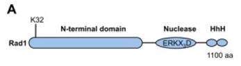 Fig. 1 Schematic depicting Rad1 domains and sumoylation site.1
Key structural properties of RAD1:
- Pcna-like cyclic domains
- Hydrophobic interface and bonding pocket
- DNA binding and damage recognition motifs
Functions of RAD1
The core function of the RAD1 protein is to act as a DNA damage checkpoint sensor and maintain genomic stability. In addition, it is also involved in regulating DNA replication and the cell cycle process.
| Function | Description |
| DNA damage identification | As a component of the "9-1-1" complex, it loads at DNA damage or replication fork arrest sites, recognizes abnormal structures and initiates checkpoint signals. |
| Checkpoint signal activation | Recruit and activate ATR kinase and its adaptor protein ATRIP, and then phosphorylate downstream effector factors (such as Chk1), thereby triggering cell cycle arrest. |
| Copy the pressure response | Under replication pressure, stably stagger the replication fork to prevent it from collapsing into harmful double-strand breaks and promote replication restart. |
| Regulation of DNA repair | It provides a platform for multiple repair pathways, such as nucleotide excision repair (NER) and homologous recombination (HR), and coordinates the localization and assembly of repair proteins. |
| Telomere maintenance | Participate in responding to the replication pressure at the telomere, assist in maintaining the integrity of telomere structure, and prevent telomere dysfunction. |
Unlike many DNA repair enzymes with catalytic activity, RAD1 itself does not have enzymatic activity. Its function mainly depends on the circular complex structure it forms as a scaffold protein. This structure binds to DNA with high affinity, providing a structural basis for its precise localization of damage signals in the genome and recruitment of downstream effector molecules.
Applications of RAD1 and RAD1 Antibody in Literature
- Seol, Ja-Hwan, et al. "Distinct roles of XPF-ERCC1 and Rad1-Rad10-Saw1 in replication-coupled and uncoupled inter-strand crosslink repair." Nature communications 9.1 (2018): 2025. https://doi.org/10.1038/s41467-018-04327-0
The article indicates that the RAD1-RAD10 complex repairs DNA cross-linking through different pathways: in the G1 phase, it relies on nucleotide excision for repair, while in the S/G2 phase, it collaborates with proteins such as Slx4 to independently complete replication coupling repair through the repair pathway.
- Sarangi, Prabha, et al. "Sumoylation of the Rad1 nuclease promotes DNA repair and regulates its DNA association." Nucleic acids research 42.10 (2014): 6393-6404. https://doi.org/10.1093/nar/gku300
The article indicates that the ubiquitination modification of Rad1 at DNA damage sites, especially at single lysine sites, will reduce the affinity of the RAD1-RAD10 complex for DNA, promoting its rapid dissociation after cleavage, thereby efficiently responding to high-load DNA damage.
- Wit, Niek, et al. "Lysine residue 185 of Rad1 is a topological but not a functional counterpart of lysine residue 164 of PCNA." Plos one 6.1 (2011): e16669. https://doi.org/10.1371/journal.pone.0016669
Research has found that the K185 site of the Rad1 protein in the 9-1-1 complex, although structurally corresponding to the K164 of PCNA, its monoubiquitination modification is not essential for key cellular activities in DNA damage responses and is not a functional corresponding site of PCNAK164.
- Davies, Adelina A., et al. "Role of the Rad1 and Rad10 Proteins in Nucleotide Excision Repair and Recombination (∗)." Journal of Biological Chemistry 270.42 (1995): 24638-24641. https://doi.org/10.1074/jbc.270.42.24638
Research has found that the Rad1-Rad10 complex can perform cleavage on the 5' side of the DNA bubble structure, and in collaboration with human XPG, it can simulate the double cleavage reaction of nucleotide excision repair. Its role in direct repeat sequence recombination may stem from the treatment of similar unpaired DNA intermediates.
- Hara, Kodai, et al. "The 9-1-1 DNA clamp subunit RAD1 forms specific interactions with clamp loader RAD17, revealing functional implications for binding-protein RHINO." Journal of Biological Chemistry 299.4 (2023). https://doi.org/10.1016/j.jbc.2023.103061
Research has found that the RAD17 protein specifically binds to the RAD1 subunit of the 9-1-1 complex through a motif at its N-terminal. This interaction is crucial for the loading of 9-1-1 and the subsequent activation of the ATR-CHK1 signaling pathway.
Creative Biolabs: RAD1 Antibodies for Research
Creative Biolabs specializes in the production of high-quality RAD1 antibodies for research and industrial applications. Our portfolio includes monoclonal antibodies tailored for ELISA, Flow Cytometry, Western blot, immunohistochemistry, and other diagnostic methodologies.
- Custom RAD1 Antibody Development: Tailor-made solutions to meet specific research requirements.
- Bulk Production: Large-scale antibody manufacturing for industry partners.
- Technical Support: Expert consultation for protocol optimization and troubleshooting.
- Aliquoting Services: Conveniently sized aliquots for long-term storage and consistent experimental outcomes.
For more details on our RAD1 antibodies, custom preparations, or technical support, contact us at email.
Reference
- Sarangi, Prabha, et al. "Sumoylation of the Rad1 nuclease promotes DNA repair and regulates its DNA association." Nucleic acids research 42.10 (2014): 6393-6404. https://doi.org/10.1093/nar/gku300
Anti-RAD1 antibodies
 Loading...


Hot products 
-
Mouse Anti-DHFR Recombinant Antibody (D0821) (CBMAB-D0821-YC)

-
Mouse Anti-14-3-3 Pan Recombinant Antibody (V2-9272) (CBMAB-1181-LY)

-
Mouse Anti-CRYAB Recombinant Antibody (A4345) (CBMAB-A4345-YC)

-
Mouse Anti-AGK Recombinant Antibody (V2-258056) (CBMAB-M0989-FY)

-
Mouse Anti-CD1C Recombinant Antibody (L161) (CBMAB-C2173-CQ)

-
Mouse Anti-CFL1 (Phospho-Ser3) Recombinant Antibody (CBFYC-1770) (CBMAB-C1832-FY)

-
Mouse Anti-C5B-9 Recombinant Antibody (CBFYA-0216) (CBMAB-X0304-FY)

-
Mouse Anti-DES Monoclonal Antibody (440) (CBMAB-AP1857LY)

-
Mouse Anti-APOH Recombinant Antibody (4D9A4) (CBMAB-A3249-YC)

-
Mouse Anti-FN1 Monoclonal Antibody (71) (CBMAB-1241CQ)

-
Mouse Anti-DDC Recombinant Antibody (8E8) (CBMAB-0992-YC)

-
Mouse Anti-CD247 Recombinant Antibody (6B10.2) (CBMAB-C1583-YY)

-
Human Anti-SARS-CoV-2 S1 Monoclonal Antibody (CBFYR-0120) (CBMAB-R0120-FY)

-
Rabbit Anti-ABL1 (Phosphorylated Y185) Recombinant Antibody (V2-443434) (PTM-CBMAB-0001YC)

-
Rabbit Anti-Acetyl-Histone H3 (Lys36) Recombinant Antibody (V2-623395) (CBMAB-CP0994-LY)

-
Mouse Anti-ARID3A Antibody (A4) (CBMAB-0128-YC)

-
Mouse Anti-FeLV g27 Recombinant Antibody (1) (CBMAB-V208-1714-FY)

-
Mouse Anti-AAV8 Recombinant Antibody (V2-634028) (CBMAB-AP022LY)

-
Mouse Anti-ELAVL4 Recombinant Antibody (6B9) (CBMAB-1132-YC)

-
Rabbit Anti-CCN1 Recombinant Antibody (CBWJC-3580) (CBMAB-C4816WJ)

- AActivation
- AGAgonist
- APApoptosis
- BBlocking
- BABioassay
- BIBioimaging
- CImmunohistochemistry-Frozen Sections
- CIChromatin Immunoprecipitation
- CTCytotoxicity
- CSCostimulation
- DDepletion
- DBDot Blot
- EELISA
- ECELISA(Cap)
- EDELISA(Det)
- ESELISpot
- EMElectron Microscopy
- FFlow Cytometry
- FNFunction Assay
- GSGel Supershift
- IInhibition
- IAEnzyme Immunoassay
- ICImmunocytochemistry
- IDImmunodiffusion
- IEImmunoelectrophoresis
- IFImmunofluorescence
- IGImmunochromatography
- IHImmunohistochemistry
- IMImmunomicroscopy
- IOImmunoassay
- IPImmunoprecipitation
- ISIntracellular Staining for Flow Cytometry
- LALuminex Assay
- LFLateral Flow Immunoassay
- MMicroarray
- MCMass Cytometry/CyTOF
- MDMeDIP
- MSElectrophoretic Mobility Shift Assay
- NNeutralization
- PImmunohistologyp-Paraffin Sections
- PAPeptide Array
- PEPeptide ELISA
- PLProximity Ligation Assay
- RRadioimmunoassay
- SStimulation
- SESandwich ELISA
- SHIn situ hybridization
- TCTissue Culture
- WBWestern Blot
