
Spectrin Antibodies
Background
Spectrin is a large cytoskeletal protein that mainly exists on the inner side of the cell membrane of eukaryotic cells and maintains cell morphology and mechanical stability by forming a reticular structure. This protein is assembled into a tetramer through heterodimerization of the α and β subunits, which then connects the actin microfilaments with the membrane protein complex, achieving the physical toughness and dynamic recombination ability of the cell membrane. The absence or mutation of Spectrin in the red blood cell membrane can lead to hemolytic anemia such as hereditary spherocytosis, highlighting its crucial role in the cell's resistance to shear force. First isolated and identified by Vincent Marchesi and others in the 1960s, Spectrin's research not only revealed the organizational principles of the cytoskeleton but also became a paradigm for the biomechanical study of membrane proteins, promoting a deeper understanding of cell deformation, adhesion and signal transduction mechanisms.
Structure of Spectrin
Spectrin is a large cytoskeletal protein, and its molecular weight varies significantly depending on the subtype. The common αII-spectrin is approximately 285 kDa, while βII-spectrin is about 274 kDa. This protein is composed of multiple repetitive domains, each containing approximately 106 amino acids, forming three helical bundles. Its main structure is highly conserved among different species, but there are specific functional domain differences among different subtypes (such as αI, αII, βI, etc.).
| Species | Human | Mouse | Chicken | Zebrafish |
| Molecular Weight (kDa) | αII: ~285, βII: ~274 | αII: ~284, βII: ~272 | αII: ~283, βII: ~273 | αII: ~282, βII: ~271 |
| Primary Structural Differences | Contains CH structural domain | Highly homologous to humans | Absence of erythrocyte profiling proteins | Embryo-specific subtypes exist |
Spectrin forms a flexible chain-like structure through its repeating units and binds to actin to form a reticular scaffold, maintaining cell morphology and membrane integrity. The CH domain at the N-terminal and the EF hand motif at the C-terminal respectively mediate the interaction with the cell membrane and calcium ions. This unique structure is the basis of its mechanical function.
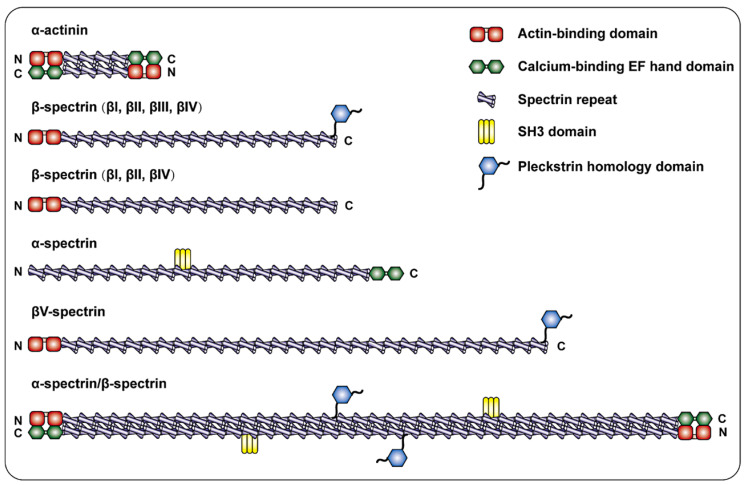 Fig. 1 Schematic diagram of the spectrin family and α-actinin.1
Key structural properties of Spectrin:
- The flexible chain consists of multiple repeated triple helical bundle domains connected in series
- Dimer and tetramer networks are formed, and the stability is maintained by hydrophobic interaction
- The CH domain and EF hand motif mediate the binding of proteins to membranes
Functions of Spectrin
The main function of Spectrin is to maintain cell morphology and membrane mechanical stability. In addition, it is also involved in various cellular processes, including cell division, cell adhesion and signal transduction.
| Function | Description |
| Maintenance of cell morphology | By forming a reticular skeleton to support the cell membrane, it endows the cell with a specific shape and toughness. |
| Mechanical stability | Disperse external mechanical stress to prevent cell membrane rupture, especially in red blood cells to cope with the shear force of blood circulation. |
| Cell division support | Helps establish and maintain cell polarity in mitosis and promotes symmetrical division. |
| Regulation of cell adhesion | By connecting membrane proteins to the intracellular skeleton, it affects the adhesion between cells and cells as well as between cells and matrix. |
| Signal transduction participation | As a scaffold protein, it recruits signaling molecules and participates in the regulation of important pathways such as apoptosis and differentiation. |
Spectrin, with its flexible repetitive unit structure and multivalent binding ability, can interact with multiple proteins such as actin and anchin simultaneously. This characteristic makes it a key integrator and functional coordinator at the cytoskeleton-membrane interface.
Applications of Spectrin and Spectrin Antibody in Literature
1. Yang, Panyu, et al. "βII spectrin (SPTBN1): biological function and clinical potential in cancer and other diseases." International journal of biological sciences 17.1 (2021): 32. https://doi.org/10.7150/ijbs.52375
The article indicates that βII shadow protein is a cytoskeletal protein widely present in nucleated cells. It not only participates in maintaining cell structure but also regulates multiple functions such as apoptosis and proliferation. Abnormal expression of it is closely related to developmental defects, cancer and other diseases, and may become an important target for future treatment.
2. Machnicka, B., et al. "Spectrin-based skeleton as an actor in cell signaling." Cellular and Molecular Life Sciences 69.2 (2012): 191-201. https://doi.org/10.1007/s00018-011-0804-5
The article indicates that spectrin is widely present in all metazoan cells, encoded by multiple genes to form various subtypes. It connects membrane proteins to the cytoskeleton through a heterotetramer structure and participates in maintaining membrane stability, signal transduction, and regulating cell morphology. Its tissue-specific expression is closely related to the developmental stage.
3. Perkins, Emma, Daumante Suminaite, and Mandy Jackson. "Cerebellar ataxias: β‐III spectrin's interactions suggest common pathogenic pathways." The Journal of physiology 594.16 (2016): 4661-4676. https://doi.org/10.1113/JP271195
The article indicates that β-III shadow protein plays a key role in maintaining the dendritic structure and membrane protein transport of cerebellar Purkinje cells. Mutations in its encoding gene SPTBN2 can lead to spinocerebellar ataxia type 5 (SCA5) and recessive ataxia SPARCA1, causing dendrite abnormalities, reduced sodium current and glutamate delivery disorders, ultimately resulting in cerebellar degeneration and functional disorders. This mechanism may be related to multiple SCA subtypes and aging-related cerebellar degeneration.
4. Liu, Cheng-Hsin, et al. "β spectrin-dependent and domain specific mechanisms for Na+ channel clustering." Elife 9 (2020): e56629. https://doi.org/10.7554/eLife.56629
Studies have shown that β4 spectrin is crucial for the aggregation and function of sodium ion channels in the axonal process initiation segment (AIS), and β1 spectrin cannot replace its function here. The simultaneous absence of β1 and β4 spectral proteins can exacerbate neurological dysfunction and lead to epileptic seizures, highlighting the crucial role of spectral proteins in maintaining the normal function of the nervous system.
5. Ibar, Consuelo, et al. "Competition between myosin II and βH-spectrin regulates cytoskeletal tension." Elife 12 (2023): RP84918. https://doi.org/10.7554/eLife.84918
Studies have shown that β H-shadow protein affects the Hippo signaling pathway by regulating cytoskeletal tension. Research has found that its function does not rely on α -shadowin but competes with myosin for binding to F-actin, thereby regulating myosin accumulation and cytoskeletal tension. This mechanism provides a new perspective for understanding cellular morphological changes.
Creative Biolabs: Spectrin Antibodies for Research
Creative Biolabs specializes in the production of high-quality Spectrin antibodies for research and industrial applications. Our portfolio includes monoclonal antibodies tailored for ELISA, Flow Cytometry, Western blot, immunohistochemistry, and other diagnostic methodologies.
- Custom Spectrin Antibody Development: Tailor-made solutions to meet specific research requirements.
- Bulk Production: Large-scale antibody manufacturing for industry partners.
- Technical Support: Expert consultation for protocol optimization and troubleshooting.
- Aliquoting Services: Conveniently sized aliquots for long-term storage and consistent experimental outcomes.
For more details on our Spectrin antibodies, custom preparations, or technical support, contact us at email.
Reference
- Yang, Panyu, et al. "βII spectrin (SPTBN1): biological function and clinical potential in cancer and other diseases." International journal of biological sciences 17.1 (2021): 32. https://doi.org/10.7150/ijbs.52375
Anti-Spectrin antibodies
 Loading...
 Compare




Hot products 
-
Mouse Anti-CRTAM Recombinant Antibody (CBFYC-2235) (CBMAB-C2305-FY)

-
Mouse Anti-CRYAB Recombinant Antibody (A4345) (CBMAB-A4345-YC)

-
Mouse Anti-ADGRE5 Recombinant Antibody (V2-360335) (CBMAB-C2088-CQ)

-
Mouse Anti-DMD Recombinant Antibody (D1190) (CBMAB-D1190-YC)

-
Mouse Anti-ABL2 Recombinant Antibody (V2-179121) (CBMAB-A0364-YC)

-
Mouse Anti-CA9 Recombinant Antibody (CBXC-2079) (CBMAB-C0131-CQ)

-
Mouse Anti-FLT1 Recombinant Antibody (11) (CBMAB-V0154-LY)

-
Mouse Anti-ALX1 Recombinant Antibody (96k) (CBMAB-C0616-FY)

-
Mouse Anti-ATP1B3 Recombinant Antibody (1E9) (CBMAB-A4021-YC)

-
Human Anti-SARS-CoV-2 S1 Monoclonal Antibody (CBFYR-0120) (CBMAB-R0120-FY)

-
Mouse Anti-GIPC2 Recombinant Antibody (10) (CBMAB-G0476-LY)

-
Mouse Anti-ALOX5 Recombinant Antibody (33) (CBMAB-1890CQ)

-
Mouse Anti-BAX Recombinant Antibody (CBYY-0216) (CBMAB-0217-YY)

-
Mouse Anti-GluRIIA Monoclonal Antibody (8B4D2) (CBMAB-0305-CN)

-
Mouse Anti-ATG5 Recombinant Antibody (9H197) (CBMAB-A3945-YC)

-
Mouse Anti-CIITA Recombinant Antibody (CBLC160-LY) (CBMAB-C10987-LY)

-
Mouse Anti-ASB9 Recombinant Antibody (1D8) (CBMAB-A0529-LY)

-
Mouse Anti-CHRNA9 Recombinant Antibody (8E4) (CBMAB-C9161-LY)

-
Rabbit Anti-AP2M1 (Phosphorylated T156) Recombinant Antibody (D4F3) (PTM-CBMAB-0610LY)

-
Mouse Anti-FPR2 Recombinant Antibody (1D6) (CBMAB-F2628-CQ)

- AActivation
- AGAgonist
- APApoptosis
- BBlocking
- BABioassay
- BIBioimaging
- CImmunohistochemistry-Frozen Sections
- CIChromatin Immunoprecipitation
- CTCytotoxicity
- CSCostimulation
- DDepletion
- DBDot Blot
- EELISA
- ECELISA(Cap)
- EDELISA(Det)
- ESELISpot
- EMElectron Microscopy
- FFlow Cytometry
- FNFunction Assay
- GSGel Supershift
- IInhibition
- IAEnzyme Immunoassay
- ICImmunocytochemistry
- IDImmunodiffusion
- IEImmunoelectrophoresis
- IFImmunofluorescence
- IGImmunochromatography
- IHImmunohistochemistry
- IMImmunomicroscopy
- IOImmunoassay
- IPImmunoprecipitation
- ISIntracellular Staining for Flow Cytometry
- LALuminex Assay
- LFLateral Flow Immunoassay
- MMicroarray
- MCMass Cytometry/CyTOF
- MDMeDIP
- MSElectrophoretic Mobility Shift Assay
- NNeutralization
- PImmunohistologyp-Paraffin Sections
- PAPeptide Array
- PEPeptide ELISA
- PLProximity Ligation Assay
- RRadioimmunoassay
- SStimulation
- SESandwich ELISA
- SHIn situ hybridization
- TCTissue Culture
- WBWestern Blot
