TGFA Antibodies
Background
The TGFA gene encodes a protein called transforming growth factor-α (TGF-α), which is mainly expressed in epithelial cells, neurons, and various tumor cells. This protein acts as a ligand for the epidermal growth factor receptor (EGFR), binding to the receptor and activating downstream signaling pathways, thereby promoting cell proliferation, differentiation, and migration. During embryonic development, TGF-α plays a crucial regulatory role in the formation of various organs and tissue remodeling. It was first identified in the 1980s by Rik Derynck and his colleagues through cloning from virus-transformed cells. TGF-α has attracted widespread attention due to its high expression in cancer cells and is regarded as one of the important regulatory factors in tumor occurrence and progression. Subsequent studies have revealed that this protein is also involved in normal physiological processes such as wound healing and tissue regeneration. The elucidation of its structure and function provides an important theoretical basis for understanding receptor-ligand interactions, cell signal transduction mechanisms, and the development of anti-cancer targeted therapies.
Structure of TGFA
The transforming growth factor-α encoded by the TGFA gene is a small molecule secreted protein with a molecular weight of approximately 5.5 kDa. Its precursor is a transmembrane glycoprotein, and the mature ligand is released after proteolytic cleavage.
| Species | Human | Mouse | Rat | Pig | Cow |
|---|---|---|---|---|---|
| Molecular Weight (kDa) | 5.5 | 5.5 | 5.4 | 5.6 | 5.5 |
| Primary Structural Differences | Contains six conserved cysteines forming three pairs of disulfide bonds | A few amino acids are replaced in the signal peptide region | Extracellular domain structure and human highly homologous | The hydrophobicity of the transmembrane region is slightly different | About 95% similarity to human sequence |
The precursor of transforming growth factor-α encoded by the TGFA gene consists of 160 amino acids and its structure includes four functional regions: the signal peptide, the extracellular domain, the transmembrane region, and the intracellular tail. The protein core contains a characteristic EGF-like domain, which forms a compact three-ring configuration through three pairs of highly conserved disulfide bonds (Cys16-Cys32, Cys21-Cys34, Cys28-Cys43). This structure is crucial for binding to EGFR. The membrane-anchored precursor can be cleaved by proteases such as TACE/ADAM17 in the extracellular near-membrane region, releasing the soluble mature ligand. The conserved zinc-binding motif in the extracellular domain participates in protein conformational stability, while the leucine zipper-like structure in the transmembrane region promotes homodimerization of the precursor on the membrane.
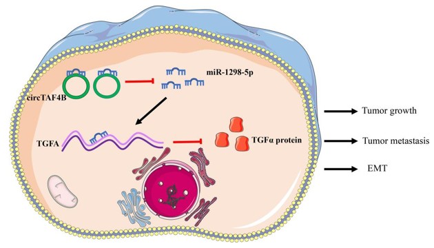 Fig. 1 Schematic diagram of the circTAF4B/miR-1298-5p/TGFA axis in the progression of BCa.1
Key structural properties of TGFA:
- Contains four domains: signal peptide, extracellular domain, transmembrane region, and intracellular tail
- Domain, EGF sample structure formed by three pairs of disulfide bond stability three-ring configurations
- Across the membrane area, hydrophobic sequence mediated anchor the precursor film
- The TACE protease cleavage site is located in the extracellular near membrane region
Functions of TGFA
The main function of the TGFA gene is to act as a ligand for the epidermal growth factor receptor (EGFR), regulating cell proliferation and differentiation. Additionally, it is involved in various physiological and pathological processes, including embryonic development, tissue repair, and tumor formation.
| Function | Description |
|---|---|
| Cell Proliferation Regulation | After binding with EGFR, TGFA activates downstream signaling pathways, promoting the division and proliferation of epithelial cells and fibroblasts. |
| Embryonic Development | During the embryonic development stage, it participates in the formation of organs, particularly in the development of epithelial tissues and the occurrence of branching morphogenesis. |
| Angiogenesis | By inducing the expression of vascular endothelial growth factor, it indirectly promotes the formation of new blood vessels. |
| Wound Repair | Released after injury by macrophages and keratinocytes, it stimulates the formation of granulation tissue and re-epithelialization. |
| Tumor Progression | Overexpressed in various cancer cells, it drives tumor growth and metastasis through autocrine or paracrine mechanisms. |
The expression regulation of the TGFA gene has a tissue-specific pattern, and its synergistic effect with EGFR exhibits a ligand-receptor dose-dependent characteristic, which is closely related to its dual role in embryonic development and tumor occurrence.
Applications of TGFA and TGFA Antibody in Literature
1. Zhang, Xiaoting, et al. "Circular RNA TAF4B promotes bladder cancer progression by sponging miR-1298-5p and regulating TGFA expression." Frontiers in Oncology 11 (2021): 643362. https://doi.org/10.3389/fonc.2021.643362
The study found that circTAF4B is highly expressed in bladder cancer. By adsorbing miR-1298-5p, it upregulates TGFA, promoting the proliferation, metastasis and epithelial-mesenchymal transition of cancer cells. CircTAF4B can serve as a new target for the diagnosis and treatment of bladder cancer.
2. Ma, Xiaoxuan, et al. "TGFA expression is associated with poor prognosis and promotes the development of cervical cancer." Journal of Cellular and Molecular Medicine 28.3 (2024): e18086. https://doi.org/10.1111/jcmm.18086
The research has found that TGFA is highly expressed in cervical cancer and promotes the proliferation and metastasis of cancer cells. Knocking down TGFA can significantly reduce the expression of IL and MMP family proteins. TGFA is expected to become a new target for the treatment of cervical cancer.
3. Feng, Cuijuan, et al. "Association between polymorphism of TGFA Taq I and cleft lip and/or palate: a meta-analysis." BMC oral health 14.1 (2014): 88. https://doi.org/10.1186/1472-6831-14-88
The study found that a meta-analysis indicated that the TGFA Taq I gene polymorphism was significantly associated with the risk of cleft lip and palate. Individuals carrying the allele C2 had a higher risk of the disease, and this association was particularly evident in the Caucasian population.
4. Piloto, Ana Margarida, et al. "Plastic antibodies tailored on quantum dots for an optical detection of myoglobin down to the femtomolar range." Scientific reports 8.1 (2018): 4944. https://doi.org/10.17305/bjbms.2020.4485
The study found that miR-376c targets and inhibits the expression of TGFA. Cisplatin inhibits the proliferation of osteosarcoma cells by up-regulating miR-376c and down-regulating TGFA. Overexpression of TGFA can reverse the anti-cancer effect of cisplatin.
5. Letra, Ariadne, et al. "Interaction between IRF6 and TGFA genes contribute to the risk of nonsyndromic cleft lip/palate." PLOS One (2012): e45441. https://doi.org/10.1371/journal.pone.0045441
The study found that there is a significant interaction between IRF6 and the TGFA gene, jointly increasing the risk of cleft lip and palate. In the Irf6 knockout mouse model, Tgfa is not expressed, further confirming the regulatory relationship between the two.
Creative Biolabs: TGFA Antibodies for Research
Creative Biolabs specializes in the production of high-quality TGFA antibodies for research and industrial applications. Our portfolio includes monoclonal and polyclonal antibodies tailored for ELISA, Flow Cytometry, Western blot, immunohistochemistry, and other diagnostic methodologies.
- Custom TGFA Antibody Development: Tailor-made solutions to meet specific research requirements.
- Bulk Production: Large-scale antibody manufacturing for industry partners.
- Technical Support: Expert consultation for protocol optimization and troubleshooting.
- Aliquoting Services: Conveniently sized aliquots for long-term storage and consistent experimental outcomes.
For more details on our TGFA antibodies, custom preparations, or technical support, contact us at email.
Reference
- Zhang, Xiaoting, et al. "Circular RNA TAF4B promotes bladder cancer progression by sponging miR-1298-5p and regulating TGFA expression." Frontiers in Oncology 11 (2021): 643362. Distributed under Open Access license CC BY 4.0, without modification. https://doi.org/10.3389/fonc.2021.643362
Anti-TGFA antibodies
 Loading...


Hot products 
-
Mouse Anti-BBS2 Recombinant Antibody (CBYY-0253) (CBMAB-0254-YY)

-
Mouse Anti-AOC3 Recombinant Antibody (CBYY-0014) (CBMAB-0014-YY)

-
Mouse Anti-ADAM12 Recombinant Antibody (V2-179752) (CBMAB-A1114-YC)

-
Mouse Anti-CALR Recombinant Antibody (CBFYC-0763) (CBMAB-C0818-FY)

-
Rat Anti-FABP3 Recombinant Antibody (CBXF-2299) (CBMAB-F1612-CQ)

-
Mouse Anti-ESR1 Recombinant Antibody (Y31) (CBMAB-1208-YC)

-
Mouse Anti-CGAS Recombinant Antibody (CBFYM-0995) (CBMAB-M1146-FY)

-
Rabbit Anti-AKT3 Recombinant Antibody (V2-12567) (CBMAB-1057-CN)

-
Mouse Anti-BACE1 Recombinant Antibody (61-3E7) (CBMAB-1183-CN)

-
Mouse Anti-CHRNA9 Recombinant Antibody (8E4) (CBMAB-C9161-LY)

-
Mouse Anti-ENO1 Recombinant Antibody (CBYC-A950) (CBMAB-A4388-YC)

-
Mouse Anti-BIRC3 Recombinant Antibody (315304) (CBMAB-1214-CN)

-
Rabbit Anti-AKT2 (Phosphorylated S474) Recombinant Antibody (V2-556130) (PTM-CBMAB-0605LY)

-
Rat Anti-EMCN Recombinant Antibody (28) (CBMAB-E0280-FY)

-
Mouse Anti-ATG5 Recombinant Antibody (9H197) (CBMAB-A3945-YC)

-
Mouse Anti-ADIPOR2 Recombinant Antibody (V2-179983) (CBMAB-A1369-YC)

-
Mouse Anti-ENPP1 Recombinant Antibody (CBFYE-0159) (CBMAB-E0375-FY)

-
Mouse Anti-ALX1 Recombinant Antibody (96k) (CBMAB-C0616-FY)

-
Rabbit Anti-CBL Recombinant Antibody (D4E10) (CBMAB-CP0149-LY)

-
Mouse Anti-Acetyl SMC3 (K105/K106) Recombinant Antibody (V2-634053) (CBMAB-AP052LY)

- AActivation
- AGAgonist
- APApoptosis
- BBlocking
- BABioassay
- BIBioimaging
- CImmunohistochemistry-Frozen Sections
- CIChromatin Immunoprecipitation
- CTCytotoxicity
- CSCostimulation
- DDepletion
- DBDot Blot
- EELISA
- ECELISA(Cap)
- EDELISA(Det)
- ESELISpot
- EMElectron Microscopy
- FFlow Cytometry
- FNFunction Assay
- GSGel Supershift
- IInhibition
- IAEnzyme Immunoassay
- ICImmunocytochemistry
- IDImmunodiffusion
- IEImmunoelectrophoresis
- IFImmunofluorescence
- IGImmunochromatography
- IHImmunohistochemistry
- IMImmunomicroscopy
- IOImmunoassay
- IPImmunoprecipitation
- ISIntracellular Staining for Flow Cytometry
- LALuminex Assay
- LFLateral Flow Immunoassay
- MMicroarray
- MCMass Cytometry/CyTOF
- MDMeDIP
- MSElectrophoretic Mobility Shift Assay
- NNeutralization
- PImmunohistologyp-Paraffin Sections
- PAPeptide Array
- PEPeptide ELISA
- PLProximity Ligation Assay
- RRadioimmunoassay
- SStimulation
- SESandwich ELISA
- SHIn situ hybridization
- TCTissue Culture
- WBWestern Blot
