TNFRSF4 Antibodies
Background
The TNFRSF4 gene encodes a transmembrane protein that is present on the surface of T cells and mainly participates in the immune co-stimulation signal transduction. This protein activates downstream signaling pathways such as NF-κB by binding to the ligand OX40L, thereby enhancing the proliferation, survival, and cytokine secretion of T cells, and playing a crucial role in adaptive immune responses and the formation of immune memory. Its dysfunction is closely related to autoimmune diseases, transplant rejection, and the regulation of the tumor immune microenvironment. This gene was first identified in the 1990s through studies on T cell activation, and its structure and signaling mechanism have become important research objects in the field of immunotherapy, providing a molecular basis for the development of tumor immunotherapy and targeted drugs for autoimmune diseases.
Structure of TNFRSF4
The protein encoded by the TNFRSF4 gene is a transmembrane receptor with a molecular weight of approximately 28-30 kDa. The molecular weight of this protein varies among different species, mainly due to glycosylation modifications and minor changes in the transmembrane region.
| Species | Human | Mouse | Rat |
| Molecular Weight (kDa) | ~30 | ~28 | ~29 |
| Primary Structural Differences | Cysteine-rich extracellular domains and conserved homologous regions of tumor necrosis factor receptors | Intracellular signal domain have species specificity | The amino acid sequence in the transmembrane region is highly conserved. |
This protein is composed of approximately 240 amino acids and includes a signal peptide, an extracellular ligand-binding domain rich in cysteines, a single transmembrane region, and a cytoplasmic tail. Its extracellular domain is composed of three conserved cysteine-rich domains (CRDs), forming a typical tumor necrosis factor receptor superfamily fold. The intracellular region lacks inherent enzymatic activity and mainly activates downstream NF-κB and MAPK signaling pathways by recruiting TRAF adaptor proteins. The ligand-binding pocket is formed by the CRD2 and CRD3 domains, and the key arginine residues are responsible for the specific recognition and binding to the ligand OX40L.
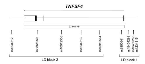 Fig. 1 The TNFSF4 gene as represented in the NCBI database.1
Key structural properties of TNFRSF4:
- Typical extracellular domains of the tumor necrosis factor receptor (TNFR) superfamily
- Fixed to the cell membrane by a single transmembrane region
- Intracellular area without catalytic function, but contains specific motif to raise a downstream signaling molecules
- Maintain structural stability through disulfide bonds and ensure specific binding with the ligand OX40L
Functions of TNFRSF4
The main function of the protein encoded by the TNFRSF4 gene is to regulate the activation and proliferation of T cells. However, it is also widely involved in various immune-related physiological and pathological processes, including the formation of immune memory and the regulation of autoimmunity.
| Function | Description |
| T cell co-stimulation | Expressed on the surface of T cells, upon binding to OX40L on antigen-presenting cells, it provides a crucial second activation signal, promoting the clonal proliferation and survival of T cells (especially effector T cells and memory T cells). |
| Enhancement of Immune Memory | The signaling pathway can inhibit activated-induced cell death (AICD) and upregulate the expression of anti-apoptotic proteins, thereby promoting the formation and maintenance of long-lasting memory T cell pools. |
| Regulatory T Cell (Treg) Function Regulation | It affects the stability and inhibitory function of Treg cells, participates in the balance of immune tolerance, and abnormal signaling can lead to autoimmune reactions. |
| Regulation of Tumor Immune Microenvironment | It is highly expressed in tumor-infiltrating lymphocytes, and its signals can enhance the anti-tumor immune response, but it may also be exploited by tumors to create an immunosuppressive environment. |
| Involvement of Inflammatory Diseases | In chronic inflammatory diseases such as rheumatoid arthritis and multiple sclerosis, their signaling pathways are abnormally activated, driving pathological immune responses. |
Unlike most receptors that require the aggregation of multiple ligands to be efficiently activated, TNFRSF4 has a high affinity binding with OX40L, which can rapidly initiate a strong and sustained signaling cascade. This highlights its crucial "amplifier" role in initiating and maintaining adaptive immune responses.
Applications of TNFRSF4 and TNFRSF4 Antibody in Literature
1. Ria, Massimiliano, et al. "A common polymorphism in the promoter region of the TNFSF4 gene is associated with lower allele-specific expression and risk of myocardial infarction." PLoS One 6.3 (2011): e17652. https://doi.org/10.1371/journal.pone.0017652
Studies have shown that the rs45454293 polymorphism in the promoter region of the TNFSF4 gene is related to its expression level. Carrying the T allele can reduce transcriptional activity and decrease the expression of TNFSF4, thereby increasing the risk of myocardial infarction in women. This variation works by influencing the binding of transcription factors.
2. Murphy, Catherine, et al. "In vivo cisplatin-resistant neuroblastoma metastatic model reveals tumour necrosis factor receptor superfamily member 4 (TNFRSF4) as an independent prognostic factor of survival in neuroblastoma." Plos one 19.5 (2024): e0303643. https://doi.org/10.1371/journal.pone.0303643
Studies have shown that in drug-resistant neuroblastoma models, low expression of TNFRSF4 is associated with enhanced metastasis ability of tumor cells and is an independent risk factor for poor prognosis in patients, which can affect event-free survival and overall survival.
3. Guo, Runqi, et al. "Microwave ablation triggers OX40L-mediated disruption of TNFRSF4+ Treg immunosuppressive activity." Frontiers in Immunology 16 (2025): 1637317. https://doi.org/10.3389/fimmu.2025.1637317
Research has found that microwave ablation can affect the tumor microenvironment by regulating the OX40L/TNFRSF4 axis: reducing the function of CTLA-4+ Tregs, enhancing the cytotoxicity of CD8+ T cells, and increasing the intracellular CD8+/Treg ratio, providing a new basis for combined immunotherapy.
4. Ma, Heng, et al. "Identification and validation of TNFRSF4 as a high-profile biomarker for prognosis and immunomodulation in endometrial carcinoma." BMC cancer 22.1 (2022): 543. https://doi.org/10.1186/s12885-022-09654-6
Studies have confirmed that TNFRSF4 can serve as a novel prognostic marker in endometrial cancer. Its expression level is positively correlated with CD4+, CD8+ T cells and Tregs infiltration within the tumor, and is significantly associated with the survival prognosis of patients.
5. Gu, Siyu, et al. "Elevated TNFRSF4 gene expression is a predictor of poor prognosis in non-M3 acute myeloid leukemia." Cancer Cell International 20.1 (2020): 146. https://doi.org/10.1186/s12935-020-01213-y
Studies have confirmed that in non-M3 type acute myeloid leukemia, high expression of TNFRSF4 is significantly associated with TP53, FLT3, NPM1 gene mutations and poor prognosis, and the overall survival of patients is significantly shortened, suggesting that it can serve as a potential prognostic biomarker.
Creative Biolabs: TNFRSF4 Antibodies for Research
Creative Biolabs specializes in the production of high-quality TNFRSF4 antibodies for research and industrial applications. Our portfolio includes monoclonal antibodies tailored for ELISA, Flow Cytometry, Western blot, immunohistochemistry, and other diagnostic methodologies.
- Custom TNFRSF4 Antibody Development: Tailor-made solutions to meet specific research requirements.
- Bulk Production: Large-scale antibody manufacturing for industry partners.
- Technical Support: Expert consultation for protocol optimization and troubleshooting.
- Aliquoting Services: Conveniently sized aliquots for long-term storage and consistent experimental outcomes.
For more details on our TNFRSF4 antibodies, custom preparations, or technical support, contact us at email.
Reference
- Ria, Massimiliano, et al. "A common polymorphism in the promoter region of the TNFSF4 gene is associated with lower allele-specific expression and risk of myocardial infarction." PLoS One 6.3 (2011): e17652. https://doi.org/10.1371/journal.pone.0017652
Anti-TNFRSF4 antibodies
 Loading...


Hot products 
-
Mouse Anti-AMIGO2 Recombinant Antibody (CBYY-C0756) (CBMAB-C2192-YY)

-
Mouse Anti-ACO2 Recombinant Antibody (V2-179329) (CBMAB-A0627-YC)

-
Mouse Anti-ALB Recombinant Antibody (V2-180650) (CBMAB-A2186-YC)

-
Rabbit Anti-AKT3 Recombinant Antibody (V2-12567) (CBMAB-1057-CN)

-
Mouse Anti-AKR1C3 Recombinant Antibody (V2-12560) (CBMAB-1050-CN)

-
Mouse Anti-EPO Recombinant Antibody (CBFYR0196) (CBMAB-R0196-FY)

-
Mouse Anti-FOXA3 Recombinant Antibody (2A9) (CBMAB-0377-YC)

-
Mouse Anti-CD83 Recombinant Antibody (HB15) (CBMAB-C1765-CQ)

-
Mouse Anti-CGAS Recombinant Antibody (CBFYM-0995) (CBMAB-M1146-FY)

-
Mouse Anti-CCT6A/B Recombinant Antibody (CBXC-0168) (CBMAB-C5570-CQ)

-
Mouse Anti-CD164 Recombinant Antibody (CBFYC-0077) (CBMAB-C0086-FY)

-
Mouse Anti-CAT Recombinant Antibody (724810) (CBMAB-C8431-LY)

-
Mouse Anti-AQP2 Recombinant Antibody (E-2) (CBMAB-A3358-YC)

-
Mouse Anti-FPR2 Recombinant Antibody (1D6) (CBMAB-F2628-CQ)

-
Mouse Anti-CFL1 (Phospho-Ser3) Recombinant Antibody (CBFYC-1770) (CBMAB-C1832-FY)

-
Mouse Anti-2C TCR Recombinant Antibody (V2-1556) (CBMAB-0951-LY)

-
Mouse Anti-AQP2 Recombinant Antibody (G-3) (CBMAB-A3359-YC)

-
Mouse Anti-CD1C Recombinant Antibody (L161) (CBMAB-C2173-CQ)

-
Mouse Anti-BCL6 Recombinant Antibody (CBYY-0442) (CBMAB-0445-YY)

-
Mouse Anti-ARID3A Antibody (A4) (CBMAB-0128-YC)

- AActivation
- AGAgonist
- APApoptosis
- BBlocking
- BABioassay
- BIBioimaging
- CImmunohistochemistry-Frozen Sections
- CIChromatin Immunoprecipitation
- CTCytotoxicity
- CSCostimulation
- DDepletion
- DBDot Blot
- EELISA
- ECELISA(Cap)
- EDELISA(Det)
- ESELISpot
- EMElectron Microscopy
- FFlow Cytometry
- FNFunction Assay
- GSGel Supershift
- IInhibition
- IAEnzyme Immunoassay
- ICImmunocytochemistry
- IDImmunodiffusion
- IEImmunoelectrophoresis
- IFImmunofluorescence
- IGImmunochromatography
- IHImmunohistochemistry
- IMImmunomicroscopy
- IOImmunoassay
- IPImmunoprecipitation
- ISIntracellular Staining for Flow Cytometry
- LALuminex Assay
- LFLateral Flow Immunoassay
- MMicroarray
- MCMass Cytometry/CyTOF
- MDMeDIP
- MSElectrophoretic Mobility Shift Assay
- NNeutralization
- PImmunohistologyp-Paraffin Sections
- PAPeptide Array
- PEPeptide ELISA
- PLProximity Ligation Assay
- RRadioimmunoassay
- SStimulation
- SESandwich ELISA
- SHIn situ hybridization
- TCTissue Culture
- WBWestern Blot
