Intracellular Staining Flow Cytometry
Besides using antibodies directed against surface markers, flow cytometry also allows simultaneous analysis of multiple intracellular targets using antibodies that detect signaling proteins, their post-translational modifications, epigenetic modifications, and downstream transcription factors. While techniques for cell surface staining are relatively standard, optimal staining for intracellular markers often depends on the biology of the target protein, such as the protein location and biochemistry of the target epitopes, association with other molecules, and its stability. For precisely this reason, cell preparation and staining methods are different for various target proteins. Refer to Cell Preparation for Flow Cytometry for different kinds of sample preparation. While detection of different intracellular proteins might require different conditions, the basic principles are the same.
Basic Principles of Intracellular Staining
Cells are fixed and permeabilized, stained intracellularly with fluorescent antibodies, and then analyzed by flow cytometry. For studies of secreted proteins, cells are first treated with the protein transport inhibitor to allow accumulation of the target protein inside the cell. When surface and intracellular staining is to be performed in the same sample, it is suggested that surface staining should be carried out first to avoid any potential effects of the intracellular staining protocol.
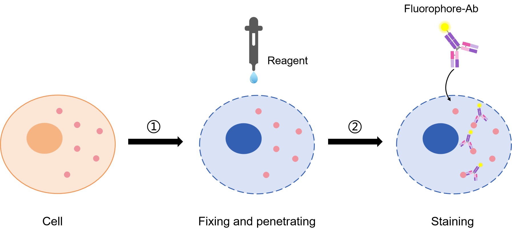 Fig.1 Intracellular staining procedure.
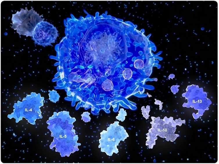
Nowadays, intracellular staining flow cytometry can be used to analyze various intracellular molecules including but not limited to phosphorylated signaling proteins, inflammatory mediators, transcription factors, and cytokines. To stain intracellular molecules, the cells need to be fixed in suspension and then permeabilized before the detection antibody is added. This fixation/permeabilization treatment allows the antibody to pass through the plasma membrane into the cell interior, while maintaining the morphological characteristics used to sort the cells. For instance, cytokines are typically secreted proteins. However, if they are trapped inside the cell, they can be stained as intracellular proteins using protein transport inhibitors. Transcription factors are often localized inside the nucleus and bound to DNA and other proteins. Phosphorylation of some proteins results in dimer formation that masks the phosphorylated epitope of interest. Also, intracellular phosphatases can quickly dephosphorylate these proteins. Therefore, after treatment, cells must be quickly fixed and subjected to stronger permeabilization conditions to allow the antibody to enter the nucleus and access the epitope within disrupted molecular complexes.
General Procedure of Intracellular Staining Flow Cytometry
- Sample preparation
- Intracellular staining
- Cell stimulation, fixing and permeabilization
- Data analysis
Advantages
- Concurrent detection of proteins associated with surface, cytosolic and nuclear compartments
- Direct correlation of phenotype with:
- Intracellular protein expression
- Functional output, such as cytokine release, production of transcription factors
- Study of signaling events on a single cell basis
- Study of the mechanism of action of pathway activation and/or inhibition for cells in suspension
- Quantitative assessment of intracellular protein expression
- Allows the detection of multiple proteins simultaneously at the level of individual cells
- Provides rich information concerning cellular function and signaling responses when combined with different kinds of markers
Protocol of Intracellular Staining Flow Cytometry is available for you. It mainly includes the following parts:
- Protocol of Direct Immunofluorescence Staining of Intracellular Cytokines in Blood
- Protocol of Direct Immunofluorescence Staining of Intracellular Antigens: Digitonin Method
- Protocol of Direct Immunofluorescence of Intracellular Antigens: Paraformaldehyde / Saponin Method
If you get in trouble, please refer to our Troubleshooting of Intracellular Staining Flow Cytometry.