CD5 Antibodies
Background
CD5 is a cell surface glycoprotein, mainly expressed on the surface of T lymphocytes and some B lymphocyte subsets (B-1 cells). As an immunomodulatory molecule, CD5 plays a significant role in the regulation of immune responses by participating in the signal transduction of T-cell receptors (TCR) and B-cell receptors (BCR). This gene was jointly identified by multiple research teams in 1986, and the protein it encodes belongs to the scavenger receptor cysteine-enriched (SRCR) superfamily. The unique structural features of CD5 include the presence of three extracellular SRCR domains. This special conformation enables it to interact with ligands such as CD72, thereby regulating lymphocyte activation and autoimmune tolerance. Recent studies have found that CD5 plays a dual role in the regulation of the tumor immune microenvironment. It can not only suppress excessive immune responses but also possibly affect anti-tumor immune responses, making it one of the important targets in cancer immunotherapy research.
Structure of CD5
CD5 is composed of three highly conserved extracellular immunoglobulin-like domains (V-type domains), and its amino acid sequence has approximately 60-70% homology among different mammals. The N-terminal of this protein contains multiple glycosylation sites, and these modifications are crucial for its immune function. The intracellular segment of CD5 contains multiple tyrosine residues, which can participate in signal transduction. Structurally, CD5 belongs to the scavenger receptor cysteine-enriched (SRCR) superfamily, and its extracellular region contains three consecutive SRCR domains, forming a typical β -folding structure. This special conformation enables it to specifically bind to ligands such as CD72. The transmembrane region of CD5 is composed of 23 hydrophobic amino acids, while the intracellular segment contains multiple conserved protein kinase action sites. These structural features jointly determine its key role in immune regulation.
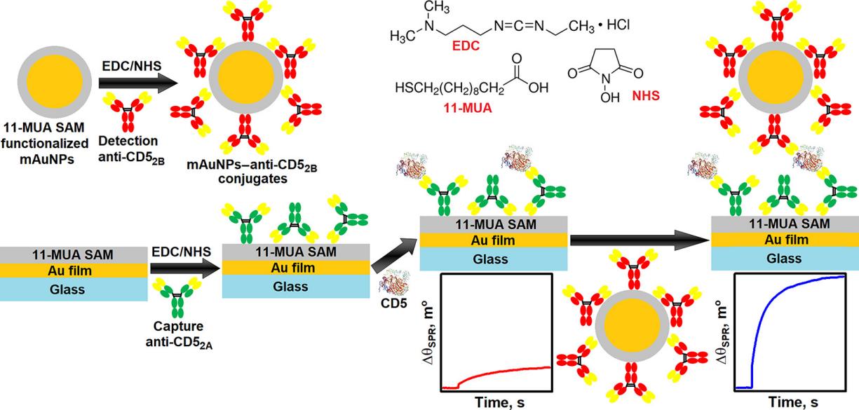 Fig. 1 Simplified schematic illustration of mAuNPs–anti-CD52B conjugate formation and the SPR-based direct and indirect sandwich immunoassay for CD5 detection.1
Key structural properties of CD5:
- Contains three highly conserved extracellular immunoglobulin-like domains (V-domain)
- Scavenger receptors that are rich in cysteine (SRCR) features of super family structure
- Across the membrane area consists of 23 hydrophobic amino acids
- Intracellular section contains multiple tyrosine phosphorylation site
Functions of CD5
The main function of the CD5 gene is to regulate the activation and signal transduction of immune cells, and it also plays a significant role in various pathophysiological processes.
| Function | Description |
| Immune regulation | As a co-receptor on the surface of T cells and B cells (B-1 subset), it regulates the signal transduction thresholds of TCR and BCR. |
| Maintenance of immune tolerance | By recruiting phosphatases such as SHP-1 to deliver inhibitory signals, autoimmune reactions can be prevented. |
| Tumor immunity | Regulating T cell function in the tumor microenvironment affects the anti-tumor immune response. |
| Inflammatory regulation | Participate in the development process of chronic inflammatory diseases, such as rheumatoid arthritis. |
| B-cell development | On b-1 cells play a key role to maintain development and function. |
The interaction between CD5 and its ligand CD72 exhibits specific binding characteristics, and its signal transduction is dose-dependent, indicating its precise regulatory role in immune balance. Unlike typical immune-activating receptors, CD5 mainly functions through inhibitory signaling pathways, a characteristic that makes it an important research target for autoimmune diseases and tumor immunotherapy.
Applications of CD5 and CD5 Antibody in Literature
1. Alotaibi, Faizah, et al. "Reduced CD5 on CD8+ T cells in tumors but not lymphoid organs is associated with increased activation and effector function." Frontiers in Immunology 11 (2021): 584937. https://doi.org/10.3389/fimmu.2020.584937
The article indicates that in the 4T1 breast cancer mouse model, the expression of CD5 in CD4+ T cells is higher than that in CD8+ T cells. The CD5 level of tumor-infiltrating T cells (TILs) is lower than that of peripheral organs, and CD8+ TILs with low CD5 exhibit stronger activation and effector functions. CD5 may be negatively correlated with the anti-tumor activity of T cells and may become a new target for immunotherapy.
2. Alisjahbana, Arlisa, et al. "CD5 surface expression marks intravascular human innate lymphoid cells that have a distinct ontogeny and migrate to the lung." Frontiers in Immunology 12 (2021): 752104. https://doi.org/10.3389/fimmu.2021.752104
Research has found that CD5+ innate lymphoid cells (ILCs) exist in humanized mouse models, mainly located in the pulmonary vascular and circulatory systems. Unlike conventional CD5-ILCs, CD5+ ILCs originate from the thymus/spleen and have a unique developmental pathway, containing mature ILC1s (producing IFNγ) and immature ILCs, or act as "blood sentinels" to participate in pulmonary immune defense.
3. Ma, Dongshen, et al. "Molecular subtyping of CD5+ diffuse large B-cell lymphoma based on DNA-targeted sequencing and Lymph2Cx." Frontiers in Oncology 12 (2022): 941347. https://doi.org/10.3389/fonc.2022.941347
Studies have found that CD5+ diffuse large B-cell lymphoma (DLBCL) has a poor prognosis, often with MCD subtypes (MYD88/CD79B mutations) and ABC origin. High-frequency mutations in PIM1, MYD88, and KMT2D, as well as double expression of MYC/BCL2, suggest a poor prognosis, while those with high expression of MME have a longer survival period.
4. Naso, Julia R., et al. "CD5 Immunoreactivity Is Associated With Longer Overall Survival in Thymic Carcinoma: A Brief Report." JTO Clinical and Research Reports 6.5 (2025): 100803.https://doi.org/10.1016/j.jtocrr.2025.100803
The research found that the prognosis of patients with CD5-positive thymic carcinoma (≥50% tumor cell staining) was significantly improved: the 3-year overall survival rate reached 100% (only 48% in the CD5-negative group). Multivariate analysis confirmed that CD5 is an independent prognostic factor (HR=0.18, p=0.005) and may become a novel prognostic marker.
5. Ma, Xiao‐bo, et al. "Coexpression of CD 5 and CD 43 predicts worse prognosis in diffuse large B‐cell lymphoma." Cancer Medicine 7.9 (2018): 4284-4295. https://doi.org/10.1002/cam4.1674
Research has found that patients with diffuse large B-cell lymphoma (DLBCL) co-expressing CD5/CD43 have the poorest prognosis: 5% of patients have co-expression, and their event-free survival (EFS) and overall survival (OS) are significantly lower than those with single expression (P<0.001), which is an independent adverse prognostic factor.
Creative Biolabs: CD5 Antibodies for Research
Creative Biolabs specializes in the production of high-quality CD5 antibodies for research and industrial applications. Our portfolio includes monoclonal antibodies tailored for ELISA, Flow Cytometry, Western blot, immunohistochemistry, and other diagnostic methodologies.
- Custom CD5 Antibody Development: Tailor-made solutions to meet specific research requirements.
- Bulk Production: Large-scale antibody manufacturing for industry partners.
- Technical Support: Expert consultation for protocol optimization and troubleshooting.
- Aliquoting Services: Conveniently sized aliquots for long-term storage and consistent experimental outcomes.
For more details on our CD5 antibodies, custom preparations, or technical support, contact us at email.
Reference
- Kausaite-Minkstimiene, Asta, Anton Popov, and Almira Ramanaviciene. "Surface plasmon resonance immunosensor with antibody-functionalized magnetoplasmonic nanoparticles for ultrasensitive quantification of the CD5 biomarker." ACS Applied Materials & Interfaces 14.18 (2022): 20720-20728.https://doi.org/10.1021/acsami.2c02936
Anti-CD5 antibodies
 Loading...


Hot products 
-
Mouse Anti-CD8 Recombinant Antibody (C1083) (CBMAB-C1083-LY)

-
Mouse Anti-CTCF Recombinant Antibody (CBFYC-2371) (CBMAB-C2443-FY)

-
Mouse Anti-EMP3 Recombinant Antibody (CBFYE-0100) (CBMAB-E0207-FY)

-
Mouse Anti-ENO1 Recombinant Antibody (CBYC-A950) (CBMAB-A4388-YC)

-
Mouse Anti-FYN Recombinant Antibody (10) (CBMAB-S6332-CQ)

-
Mouse Anti-CALR Recombinant Antibody (CBFYC-0763) (CBMAB-C0818-FY)

-
Rabbit Anti-CCN1 Recombinant Antibody (CBWJC-3580) (CBMAB-C4816WJ)

-
Mouse Anti-BAX Recombinant Antibody (CBYY-0216) (CBMAB-0217-YY)

-
Mouse Anti-BACE1 Recombinant Antibody (61-3E7) (CBMAB-1183-CN)

-
Mouse Anti-CCND2 Recombinant Antibody (DCS-3) (CBMAB-G1318-LY)

-
Mouse Anti-CGAS Recombinant Antibody (CBFYM-0995) (CBMAB-M1146-FY)

-
Mouse Anti-CEMIP Recombinant Antibody (3C12) (CBMAB-K0296-LY)

-
Mouse Anti-AFDN Recombinant Antibody (V2-58751) (CBMAB-L0408-YJ)

-
Mouse Anti-ALX1 Recombinant Antibody (96k) (CBMAB-C0616-FY)

-
Mouse Anti-CD59 Recombinant Antibody (CBXC-2097) (CBMAB-C4421-CQ)

-
Mouse Anti-BRCA2 Recombinant Antibody (CBYY-0790) (CBMAB-0793-YY)

-
Mouse Anti-CFL1 Recombinant Antibody (CBFYC-1771) (CBMAB-C1833-FY)

-
Rat Anti-4-1BB Recombinant Antibody (V2-1558) (CBMAB-0953-LY)

-
Mouse Anti-DLC1 Recombinant Antibody (D1009) (CBMAB-D1009-YC)

-
Rabbit Anti-ADRA1A Recombinant Antibody (V2-12532) (CBMAB-1022-CN)

- AActivation
- AGAgonist
- APApoptosis
- BBlocking
- BABioassay
- BIBioimaging
- CImmunohistochemistry-Frozen Sections
- CIChromatin Immunoprecipitation
- CTCytotoxicity
- CSCostimulation
- DDepletion
- DBDot Blot
- EELISA
- ECELISA(Cap)
- EDELISA(Det)
- ESELISpot
- EMElectron Microscopy
- FFlow Cytometry
- FNFunction Assay
- GSGel Supershift
- IInhibition
- IAEnzyme Immunoassay
- ICImmunocytochemistry
- IDImmunodiffusion
- IEImmunoelectrophoresis
- IFImmunofluorescence
- IGImmunochromatography
- IHImmunohistochemistry
- IMImmunomicroscopy
- IOImmunoassay
- IPImmunoprecipitation
- ISIntracellular Staining for Flow Cytometry
- LALuminex Assay
- LFLateral Flow Immunoassay
- MMicroarray
- MCMass Cytometry/CyTOF
- MDMeDIP
- MSElectrophoretic Mobility Shift Assay
- NNeutralization
- PImmunohistologyp-Paraffin Sections
- PAPeptide Array
- PEPeptide ELISA
- PLProximity Ligation Assay
- RRadioimmunoassay
- SStimulation
- SESandwich ELISA
- SHIn situ hybridization
- TCTissue Culture
- WBWestern Blot
