CDK2 Antibodies
Background
CDK2 belongs to the cyclin-dependent kinase family and is a serine/threonine kinase that plays a core regulatory role in the G1/S phase transition of the cell cycle. This protein forms a complex by binding to cyclin E and drives the initiation of DNA replication by phosphorylating substrates such as retinoblastoma protein, ensuring the orderly advancement of the cell cycle. In 1993, scientists first clarified the eutectic structure of CDK2 and cyclin A, revealing the conservation of the kinase active center and its regulatory mechanism. As an important model molecule in cell cycle research, the structural analysis of CDK2 not only promotes the understanding of cell division mechanisms but also provides a key template for the development of cancer-targeted drugs. The design of its inhibitors has become an important research direction in the field of tumor treatment.
Structure of CDK2
CDK2 is a serine/threonine protein kinase with a molecular weight of approximately 33.8 kDa. There are subtle differences in its molecular weight among different species, mainly due to minor variations in the gene coding sequence.
| Species | Human | Mouse | Rat | African clawed toad | Zebrafish |
| Molecular Weight (kDa) | 33.8 | 33.9 | 33.7 | 33.5 | 33.6 |
| Primary Structural Differences | Highly conserved structure of catalytic domain | Very high homology with humans | Kinase domains are similar | Cell cycle regulation functions are conserved | There are collateral homologous genes |
This protein is composed of 298 amino acids, and its spatial structure is formed by a typical kinase catalytic core. The active center of CDK2 is located in the fissure formed by two leaf-shaped domains, and its activation depends on binding to cyclin and phosphorylation at the T160 site. The key PSTAIRE helix undergoes conformational rearrangement when binding to cyclins, while the G loop (Glycine-rich loop) within the ATP-binding pocket is responsible for anchoring the phosphate group. The combined effect of the N-terminal and C-terminal leaf-shaped domains has accomplished the specific recognition and phosphorylation catalysis of the substrate protein.
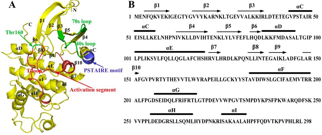 Fig. 1 The structure of monomeric CDK2.1
Key structural properties of CDK2:
- Typical bilobed structure of protein kinases
- ATP binding pockets are located in domain cracks
- Phosphorylation of the activation loop regulates kinase activity
Functions of CDK2
The core function of CDK2 is to drive the cell cycle process and DNA replication. In addition, this kinase is also involved in key life activities such as transcriptional regulation, cell differentiation and DNA damage response.
| Function | Description |
| Cell cycle advancement | By binding to cyclin E/A and phosphorylating substrates such as Rb, it promotes cells to transition from the G1 phase to the S phase, initiating DNA replication. |
| Transcriptional regulation | Phosphorylated transcription factors (such as NPAT and B-Myb) and the C-terminal domain of RNA polymerase II affect gene expression and histone biosynthesis. |
| Regulation of cell differentiation | In specific cell types (such as cardiomyocytes and neurons), the decline in CDK2 activity is closely related to the terminal differentiation process. |
| DNA damage checkpoint | Involved in cell cycle arrest after DNA damage, its activity by inhibiting factor of p21 gene regulation, provide time for DNA repair. |
| Induction of cellular senescence | Persistently highly active CDK2 can prompt cells to enter the state of replicative senescence prematurely, and this process is associated with telomere dysfunction. |
The activity regulation of CDK2 shows strict cycle-dependence. Its activation must rely on binding to cyclins and phosphorylation at the T160 site. This is both similar and specific to family members such as CDK1 and CDK4/6, reflecting its role in connecting the upper and lower parts of the cell cycle network.
Applications of CDK2 and CDK2 Antibody in Literature
1. Fagundes, Rafaela, and Leonardo K. Teixeira. "Cyclin E/CDK2: DNA replication, replication stress and genomic instability." Frontiers in cell and developmental biology 9 (2021): 774845. https://doi.org/10.3389/fcell.2021.774845
The article indicates that in normal cells, CDK2 binds to Cyclin E to regulate the G1/S phase transition and DNA replication. When it is abnormally activated, it can induce replication stress and DNA damage, which in turn leads to genomic instability and promotes the occurrence and development of human cancers.
2. Tyutyunyk-Massey, Liliya, et al. "CDK2 inhibition produces a persistent population of polyploid cancer cells." JCI insight 10.10 (2025): e189901. https://doi.org/10.1172/jci.insight.189901
The article indicates that in tumor treatment, inhibiting CDK2 can trigger abnormal chromosomal division and apoptosis, but it can also lead to the survival of polyploid cancer cells and the development of drug resistance. Studies have shown that the combined inhibition of CDK2 and CDK1 or KIF proteins can effectively eliminate such cells and enhance the anti-tumor effect.
3. Chen, Zibo, et al. "CDK2 inhibition disorders centrosome stoichiometry and alters cellular outcomes in aneuploid cancer cells." Cancer Biology & Therapy 24.1 (2023): 2279241. https://doi.org/10.1080/15384047.2023.2279241
The article indicates that CDK2 inhibition can prevent the normal clustering of excess centrosomes in cancer cells, disrupt their stoichiometry, lead to abnormal phenomena such as chromosomal loops and multipolar division, and thereby trigger apoptosis of cancer cells. This effect is significant in cancer cells but not obvious in normal alveolar epithelial cells, revealing its potential as an anti-cancer strategy.
4. Hydbring, Per, and Lars-Gunnar Larsson. "Cdk2: a key regulator of the senescence control function of Myc." Aging (Albany NY) 2.4 (2010): 244. https://doi.org/10.18632/aging.100140
Studies have shown that CDK2 can inhibit RAS-induced cellular senescence by phosphorylating the Myc protein, which is the key mechanism of the synergistic effect of Myc and Ras in carcinogenic transformation. Therefore, specific inhibition of CDK2 can force cancer cells into an senescent state, highlighting its significance as a potential therapeutic target for Myc or RAS-driven tumors.
5. Li, Yan, et al. "Insights on structural characteristics and ligand binding mechanisms of CDK2." International journal of molecular sciences 16.5 (2015): 9314-9340. https://doi.org/10.3390/ijms16059314
Studies have shown that the activity of CDK2 depends on its binding to cyclins or phosphorylation, a process that triggers changes in its three-dimensional conformation, particularly affecting the structure of the activation segment, and subsequently generating multiple inhibitor binding sites, including allosteric sites. This study systematically summarized the conformational characteristics of CDK2 and the changes in its binding mechanism with inhibitors, providing an important reference for understanding the molecular mechanism of this kinase.
Creative Biolabs: CDK2 Antibodies for Research
Creative Biolabs specializes in the production of high-quality CDK2 antibodies for research and industrial applications. Our portfolio includes monoclonal antibodies tailored for ELISA, Flow Cytometry, Western blot, immunohistochemistry, and other diagnostic methodologies.
- Custom CDK2 Antibody Development: Tailor-made solutions to meet specific research requirements.
- Bulk Production: Large-scale antibody manufacturing for industry partners.
- Technical Support: Expert consultation for protocol optimization and troubleshooting.
- Aliquoting Services: Conveniently sized aliquots for long-term storage and consistent experimental outcomes.
For more details on our CDK2 antibodies, custom preparations, or technical support, contact us at email.
Reference
- Li, Yan, et al. "Insights on structural characteristics and ligand binding mechanisms of CDK2." International journal of molecular sciences 16.5 (2015): 9314-9340. https://doi.org/10.3390/ijms16059314
Anti-CDK2 antibodies
 Loading...


Hot products 
-
Mouse Anti-CRYAB Recombinant Antibody (A4345) (CBMAB-A4345-YC)

-
Rabbit Anti-AKT3 Recombinant Antibody (V2-12567) (CBMAB-1057-CN)

-
Mouse Anti-ALX1 Recombinant Antibody (96k) (CBMAB-C0616-FY)

-
Mouse Anti-C5AR1 Recombinant Antibody (R63) (CBMAB-C9553-LY)

-
Rabbit Anti-ALOX5AP Recombinant Antibody (CBXF-1219) (CBMAB-F0750-CQ)

-
Mouse Anti-ACE2 Recombinant Antibody (V2-179293) (CBMAB-A0566-YC)

-
Mouse Anti-ATP1A2 Recombinant Antibody (M7-PB-E9) (CBMAB-A4013-YC)

-
Mouse Anti-EPO Recombinant Antibody (CBFYR0196) (CBMAB-R0196-FY)

-
Mouse Anti-ENO1 Recombinant Antibody (CBYC-A950) (CBMAB-A4388-YC)

-
Mouse Anti-ALPL Antibody (B4-78) (CBMAB-1009CQ)

-
Mouse Anti-GFAP Recombinant Antibody (5) (CBMAB-G0346-LY)

-
Mouse Anti-CGAS Recombinant Antibody (CBFYM-0995) (CBMAB-M1146-FY)

-
Mouse Anti-B2M Recombinant Antibody (CBYY-0050) (CBMAB-0050-YY)

-
Mouse Anti-AQP2 Recombinant Antibody (E-2) (CBMAB-A3358-YC)

-
Rat Anti-CD34 Recombinant Antibody (MEC 14.7) (CBMAB-C10196-LY)

-
Mouse Anti-CCN2 Recombinant Antibody (CBFYC-2383) (CBMAB-C2456-FY)

-
Mouse Anti-BMI1 Recombinant Antibody (CBYC-P026) (CBMAB-P0108-YC)

-
Mouse Anti-BrdU Recombinant Antibody (IIB5) (CBMAB-1038CQ)

-
Mouse Anti-AKT1/AKT2/AKT3 (Phosphorylated T308, T309, T305) Recombinant Antibody (V2-443454) (PTM-CBMAB-0030YC)

-
Mouse Anti-ALOX5 Recombinant Antibody (33) (CBMAB-1890CQ)

- AActivation
- AGAgonist
- APApoptosis
- BBlocking
- BABioassay
- BIBioimaging
- CImmunohistochemistry-Frozen Sections
- CIChromatin Immunoprecipitation
- CTCytotoxicity
- CSCostimulation
- DDepletion
- DBDot Blot
- EELISA
- ECELISA(Cap)
- EDELISA(Det)
- ESELISpot
- EMElectron Microscopy
- FFlow Cytometry
- FNFunction Assay
- GSGel Supershift
- IInhibition
- IAEnzyme Immunoassay
- ICImmunocytochemistry
- IDImmunodiffusion
- IEImmunoelectrophoresis
- IFImmunofluorescence
- IGImmunochromatography
- IHImmunohistochemistry
- IMImmunomicroscopy
- IOImmunoassay
- IPImmunoprecipitation
- ISIntracellular Staining for Flow Cytometry
- LALuminex Assay
- LFLateral Flow Immunoassay
- MMicroarray
- MCMass Cytometry/CyTOF
- MDMeDIP
- MSElectrophoretic Mobility Shift Assay
- NNeutralization
- PImmunohistologyp-Paraffin Sections
- PAPeptide Array
- PEPeptide ELISA
- PLProximity Ligation Assay
- RRadioimmunoassay
- SStimulation
- SESandwich ELISA
- SHIn situ hybridization
- TCTissue Culture
- WBWestern Blot
